. 2013 Oct;183(4):1209-1222.
doi: 10.1016/j.ajpath.2013.06.017. Epub 2013 Aug 3.

Galectin-3 modulates Th17 responses by regulating dendritic cell cytokines

Affiliations

Galectin-3 modulates Th17 responses by regulating dendritic cell cytokines

Agnes Fermin Lee et al. Am J Pathol. 2013 Oct.

Abstract

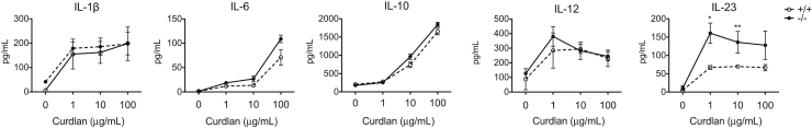
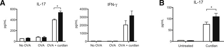
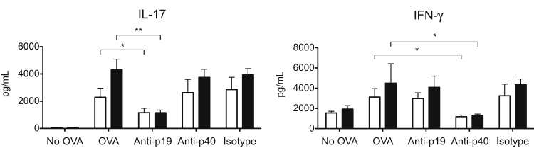
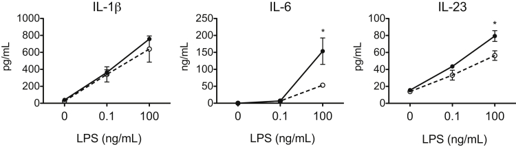
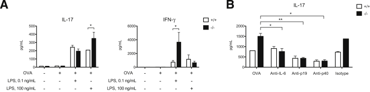
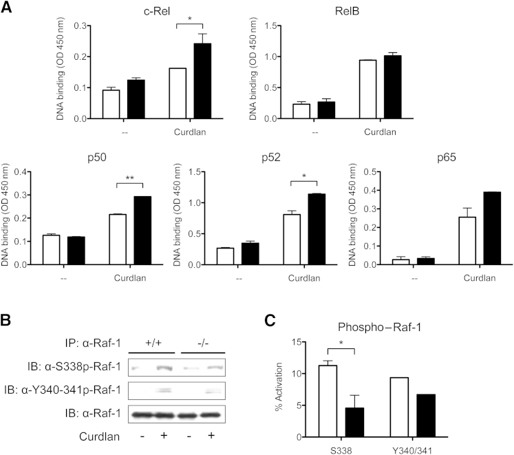
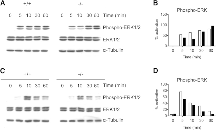
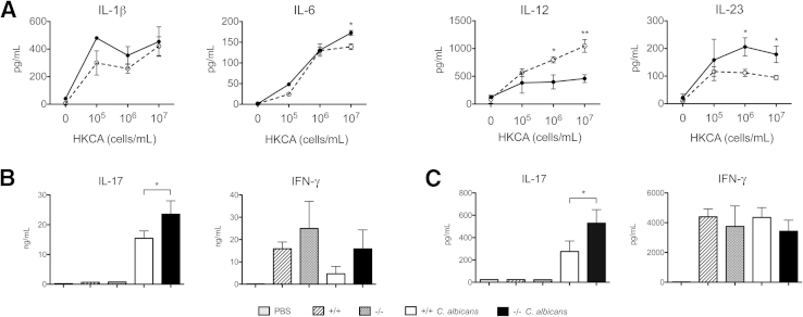
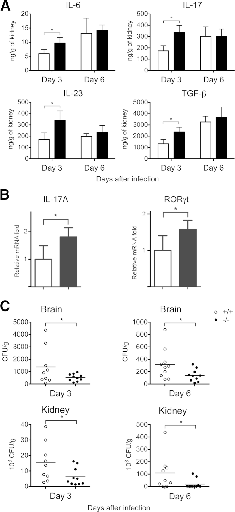
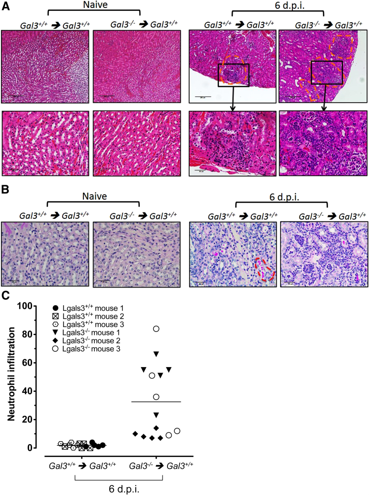
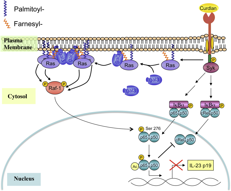
Galectin-3 is a β-galactoside-binding animal lectin with diverse functions, including regulation of T helper (Th) 1 and Th2 responses. Current data indicate that galectin-3 expressed in dendritic cells (DCs) may be contributory. Th17 cells have emerged as critical inducers of tissue inflammation in autoimmune disease and important mediators of host defense against fungal pathogens, although little is known about galectin-3 involvement in Th17 development. We investigated the role of galectin-3 in the induction of Th17 immunity in galectin-3-deficient (gal3(-/-)) and gal3(+/+) mouse bone marrow-derived DCs. We demonstrate that intracellular galectin-3 negatively regulates Th17 polarization in response to the dectin-1 agonist curdlan (a β-glucan present on the cell wall of fungal species) and lipopolysaccharide, agents that prime DCs for Th17 differentiation. On activation of dectin-1, gal3(-/-) DCs secreted higher levels of the Th17-axis cytokine IL-23 compared with gal3(+/+) DCs and contained higher levels of activated c-Rel, an NF-κB subunit that promotes IL-23 expression. Levels of active Raf-1, a kinase that participates in downstream inhibition of c-Rel binding to the IL23A promoter, were impaired in gal3(-/-) DCs. Modulation of Th17 by galectin-3 in DCs also occurred in vivo because adoptive transfer of gal3(-/-) DCs exposed to Candida albicans conferred higher Th17 responses and protection against fungal infection. We conclude that galectin-3 suppresses Th17 responses by regulating DC cytokine production.


Figures

Figure 1
Figure 1
Curdlan-stimulated gal3/ DCs produced higher levels of IL-23 compared with gal3+/+ DCs. ELISA of cytokines produced by gal3+/+ and gal3/ DCs after stimulation with various amounts of curdlan overnight. Data are given as means ± SD and are representative of at least four independent experiments. P < 0.05, ∗∗P < 0.01.
Figure 2
Figure 2
Galectin-3 in DCs negatively regulated Th17 responses. A: ELISA of cytokines in supernatants of CD4+ OT-II T cells that had been cultured with OVA-pulsed gal3+/+ (white bars) or gal3/ (black bars) DCs in the presence or absence of curdlan for 3 days. B: ELISA of IL-17 in supernatants of CD4+ cells cultured with curdlan-primed gal3+/+ (white bars) or gal3/ (black bars) DCs for 3 days in the presence of the superantigen Staphylococcus enterotoxin B. Data are given as means ± SD. Each experiment was performed at least two times. P < 0.05.
Figure 3
Figure 3
Differential IL-23 production by gal3/ (black bars) and gal3+/+ (white bars) DCs contributed to differences in Th17 induction. ELISA of cytokines in OVA-curdlan DC–T-cell (OT-II) co-cultures after the addition of neutralizing anti–IL-23p19, anti–IL-12/23p40, or isotype control antibodies on day 0. The co-cultures were maintained for 3 days. Data are given as means ± SD and are representative of at least four independent experiments. P < 0.05, ∗∗P < 0.01.
Figure 4
Figure 4
Gal3−/− (filled circles) DCs produced higher levels of the Th17-axis cytokines IL-6 and IL-23 compared with gal3+/+ (open circles) DCs after high-dose LPS stimulation. ELISA of cytokines produced by gal3+/+ and gal3/ DCs after stimulation with low- or high-dose LPS overnight. Data are given as means ± SD and are representative of at least three independent experiments. P < 0.05.
Figure 5
Figure 5
Gal3−/− DCs primed with high-dose LPS induced higher Th17 responses compared with gal3+/+ DCs. A: ELISA of cytokines in supernatants of CD4+ OT-II T cells that had been cultured with OVA-pulsed gal3+/+ or gal3/ DCs for 3 days in the presence of low-dose (0.1 ng/mL) or high-dose (100 ng/mL) LPS. B: ELISA of IL-17 in OVA-LPS (100 ng/mL) DC–T-cell (OT-II) co-cultures after the addition of anti–IL-6, anti–IL-23p19, anti–IL-12/23p40, or isotype control antibodies on day 0. The co-cultures were maintained for 3 days. Data are given as means ± SD and are representative of at least three independent experiments. ∗∗P < 0.01, ∗∗∗P < 0.001.
Figure 6
Figure 6
Galectin-3 negatively regulates c-Rel transcription factor activation through the Raf-1 signaling pathway. A: Nuclear extracts from unstimulated or curdlan-stimulated gal3+/+ (white bars) and gal3/ (black bars) DCs were added to plates coated with oligonucleotide containing an NF-κB consensus-binding site. Binding of activated NF-κB subunits was detected using antibodies against c-Rel, RelB, p50, p52, and p65. B: Raf-1 phosphorylation at Ser338 and Tyr340/341 as determined by immunoblot (IB) analysis in unstimulated and curdlan-stimulated gal3+/+ and gal3/ DCs. Data are representative of at least two independent experiments. IP, immunoprecipitate. C: Densitometry analysis of phosphorylated Raf-1. Percentage of activation was calculated as the ratio of phosphorylated Raf-1 to total Raf-1. Data are given as means ± SD and are representative of two independent experiments. P < 0.05, ∗∗P < 0.01.
Figure 7
Figure 7
Defective ERK signaling in gal3/ DCs stimulated with curdlan or LPS. A: Immunoblot analysis of ERK activation in gal3/ (black bars) and gal3+/+ (white bars) DCs stimulated with 10 μg/mL of curdlan. B: Densitometric analysis of ERK activation in curdlan-stimulated DCs. Data are representative of three experiments performed. C: Immunoblot analysis of ERK activation in gal3/ and gal3+/+ DCs stimulated with 100 ng/mL of LPS. D: Densitometric analysis of ERK activation in LPS-stimulated DCs. Data from one of two experiments are represented. Densitometric analyses were performed using ImageJ software version 1.45s (NIH, Bethesda, MD). Values shown are levels of phospho-ERK divided by total ERK.
Figure 8
Figure 8
Adoptive transfer of C. albicans–treated gal3/ DCs conferred higher Th17 responses. A: ELISA of cytokines produced by gal3+/+ (open circles) and gal3/ (closed circles) DCs after stimulation with HKCA overnight at various concentrations. Data are representative of at least three independent experiments. B: Untreated or HKCA-treated gal3/ or gal3+/+ DCs were s.c. injected into mice twice, 1 week apart. A week after the last DC transfer, splenocytes were harvested and evaluated for cytokine expression. Cytokines in culture supernatants of splenic CD4+ cells restimulated ex vivo with HKCA-treated DCs. C: Cytokines in culture supernatants of unfractionated splenocytes restimulated ex vivo with HKCA. All culture supernatants were harvested after 3 days. Data are representative of at least two independent experiments. Data are given as means ± SD. P < 0.05, ∗∗P < 0.01.
Figure 9
Figure 9
Galectin-3 in DCs regulated antifungal immunity in vivo. Gal3/ (black bars) or gal3+/+ (white bars) DCs mixed with live C. albicans were adoptively transferred into wild-type mice by i.v. injection. A: Kidneys of C. albicans–infected mice were collected on days 3 and 6 after adoptive transfer of gal3+/+ and gal3/ DCs and infection. Kidneys were homogenized, and cytokine levels in the supernatants of the homogenates were determined. The experiment was performed three times, with two to four mice included per group in each experiment. B: Real-time qPCR for indicated mRNAs in splenic CD4+ cells from mice that received C. albicans–exposed gal3/ or gal3+/+ DCs. The relative mRNA fold induction was calculated based on the fold induction of nontreated samples. Three to four mice were included in each group. The experiment was repeated three times. C: Fungal burdens [colony-forming units (CFUs) per gram of tissue] in brains and kidneys of mice that received DCs were collected on days 3 and 6 after infection. Nine to 10 mice were included in each group. Data are given as means ± SD. P < 0.05.
Figure 10
Figure 10
Renal histologic features in mice that received adoptively transferred gal3/ or gal3+/+ DCs exposed to C. albicans. gal3/ or gal3+/+ BMDCs were left untreated (naive) or were pulsed with C. albicans for 1 hour and then were adoptively transferred into gal3+/+ mice i.v. Six days after transfer (6 d.p.i.), kidneys were harvested and fixed in formalin. Tissue sections were stained with H&E (A) and PAS (B). A: Adoptive transfer of gal3/ DCs promoted increased levels of cellular infiltration into kidneys compared with gal3+/+ DCs after infection. Sites of cellular infiltration are indicated by orange dashed lines. Objectives: 10× (top); 40× (bottom). Scale bars: 250 μm (top); 50 μm (bottom). B: Higher C. albicans burden in mice that received adoptively transferred gal3+/+ DCs. Fungi are indicated by orange dashed lines.Objective, 40×. Scale bar = 50 μm. Representative images are shown. C: Higher levels of neutrophil infiltration in kidneys of mice receiving adoptively transferred gal3/ DCs. Neutrophils were identified by their polymorphic nuclei and cell size and were enumerated from five nonoverlapping high-power fields. Means are depicted by the horizontal lines.
Figure 11
Figure 11
Proposed model for galectin-3 in the regulation of cytokine expression in DCs. On ligand-mediated DC activation, galectin-3 in the cell cytosol may be recruited to membrane components, as previously demonstrated with Ras-GTP. At the membrane, galectin-3 may participate in the reorganization of membrane proteins or may act as a protein scaffold and subsequently influence downstream signal transduction. In association with Ras-GTP, galectin-3 aids in the formation of Ras nanoclusters, which promotes the efficient activation of Raf-1. By means of p65 phosphorylation, Raf-1 negatively regulates IL-23 expression.

Similar articles

Cited by

References

    1. Huang W., Na L., Fidel P.L., Schwarzenberger P. Requirement of interleukin-17A for systemic anti-Candida albicans host defense in mice. J Infect Dis. 2004;190:624–631. - PubMed
    1. Conti H.R., Shen F., Nayyar N., Stocum E., Sun J.N., Lindemann M.J., Ho A.W., Hai J.H., Yu J.J., Jung J.W., Filler S.G., Masso-Welch P., Edgerton M., Gaffen S.L. Th17 cells and IL-17 receptor signaling are essential for mucosal host defense against oral candidiasis. J Exp Med. 2009;206:299–311. - PMC - PubMed
    1. Cua D.J., Sherlock J., Chen Y., Murphy C.A., Joyce B., Seymour B., Lucian L., To W., Kwan S., Churakova T., Zurawski S., Wiekowski M., Lira S.A., Gorman D., Kastelein R.A., Sedgwick J.D. Interleukin-23 rather than interleukin-12 is the critical cytokine for autoimmune inflammation of the brain. Nature. 2003;421:744–748. - PubMed
    1. Langrish C.L., Chen Y., Blumenschein W.M., Mattson J., Basham B., Sedgwick J.D., McClanahan T., Kastelein R.A., Cua D.J. IL-23 drives a pathogenic T cell population that induces autoimmune inflammation. J Exp Med. 2005;201:233–240. - PMC - PubMed
    1. Bettelli E., Carrier Y., Gao W., Korn T., Strom T.B., Oukka M., Weiner H.L., Kuchroo V.K. Reciprocal developmental pathways for the generation of pathogenic effector TH17 and regulatory T cells. Nature. 2006;441:235–238. - PubMed

Publication types

MeSH terms