. 2013 Oct 4:312:48-55.
doi: 10.1016/j.tox.2013.07.015. Epub 2013 Jul 31.

Alterations to the circuitry of the frontal cortex following exposure to the polybrominated diphenyl ether mixture, DE-71

Affiliations

Alterations to the circuitry of the frontal cortex following exposure to the polybrominated diphenyl ether mixture, DE-71

Joshua M Bradner et al. Toxicology. .

Abstract

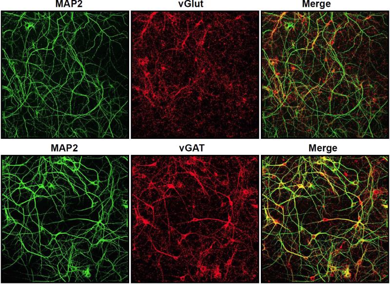
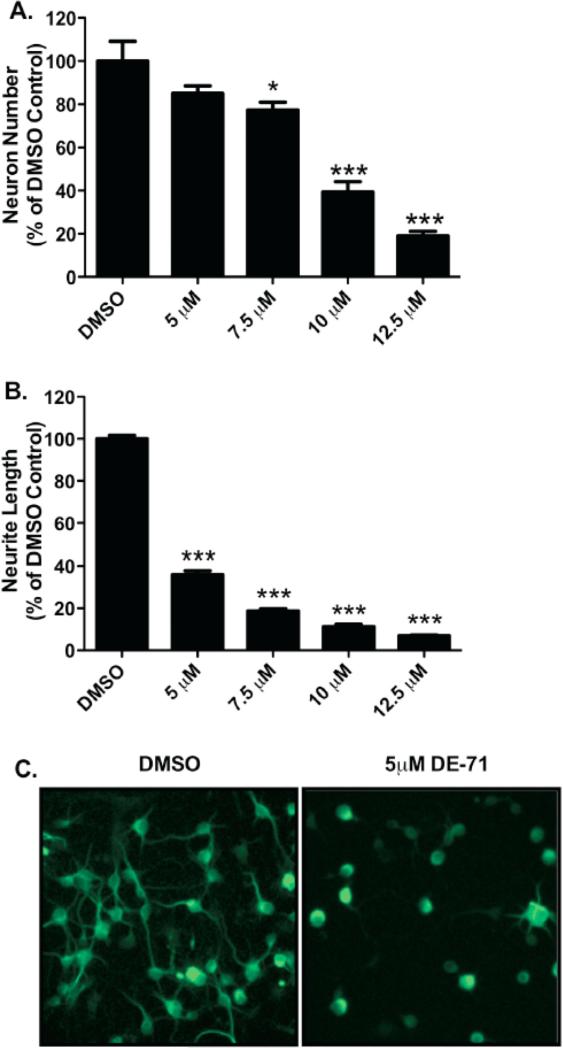
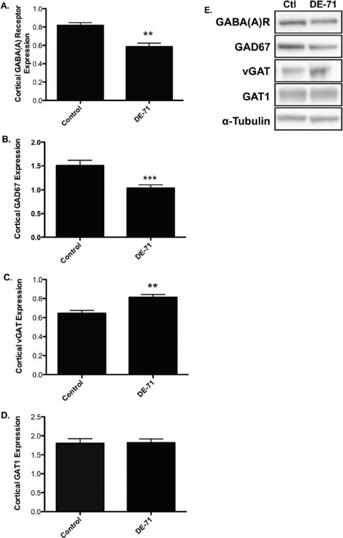
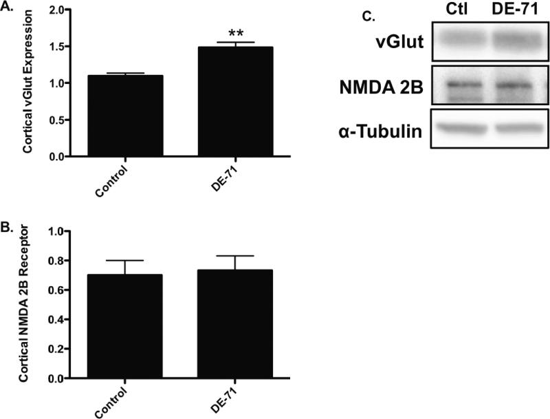
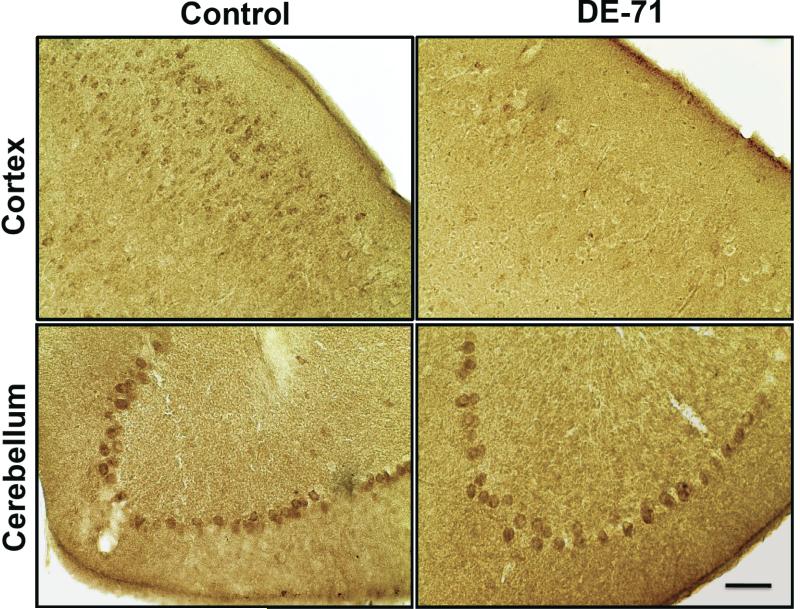
Recent studies have identified exposure to polybrominated diphenyl ethers (PBDEs) as a risk factor for deficits in cognitive functioning seen in children as well as adults. Additionally, similar alterations in learning and memory have also been observed in animal models of PBDE exposure. However, given these findings, the molecular alterations that may underlie these neurobehavioral endpoints have not been identified. As the frontal cortex is involved in modulating several cognitive functions, the purpose of our study was to investigate the possible changes to the GABAergic and glutamatergic neurotransmitter systems located in the frontal cortex following exposure to the PBDE mixture, DE-71. Primary cultured neurons isolated from the frontal cortex showed a dose-dependent reduction in neurons as well as neurite outgrowth. Furthermore, evaluation of DE-71 neurotoxicity in the frontal cortex using an in vivo model showed alterations to specific proteins involved in mediating GABA and glutamate neurotransmission, including GAD67, vGAT, vGlut, and GABA(A) 2α receptor subunit. Interestingly, these alterations appeared to be preferential for the GABA and glutamate systems located in the frontal cortex. These findings identify specific targets of PBDE neurotoxicity and provide a possible molecular mechanism for PBDE-mediated neurobehavioral deficits that arise from the frontal cortex.

Keywords: Cognition; DE-71; Frontal cortex; GABA; Glutamate; Polybrominated diphenyl ethers.


Figures

Figure 1
Figure 1
GABAergic and glutamatergic neuronal populations are highly enriched in primary cultured neurons isolated from the frontal cortex of postnatal day 1 (PND1) mice. Neurons were grown in culture for 7 days and then incubated with MAP2 to visualize all neurons and vGlut or vGAT to localize all glutamatergic or GABAergic neurons, respectively in culture.
Figure 2
Figure 2
Exposure of cortical primary cultures to DE-71 causes a loss of cortical neurons as well as reductions in neurite outgrowth. (A) Treatment of cortical cultures caused a significant reduction in the number of MAP2+ neurons, compared with DMSO control (52.3 ± 11.7) at 7.5 (40.5 ± 4.6), 10 (20.6 ± 6.0), and 12.5 μM (9.9 ± 2.8) DE-71. (B) Assessment of neurite length in these neurons demonstrated a greater loss of neurite outgrowth with a substantial reduction, compared with DMSO control (45.3 μm ± 1.8) at 5 μM (16.3 μm ± 2.0), 7.5 μM (8.5 μm ± 1.0), 10 μM (5.1 μm ± 1.4), and 12.5 μM (3.2 μm ± 0.4) DE-71. Columns represent the percent change from DMSO control for each genotype. Data represent the mean ± SEM of 4 experimental replicates per treatment group performed across 3 separate experiments. *Values for treatments significantly different from their respective genotype DMSO control (p < 0.05). ***Values significantly different from their respective genotype DMSO control (p < 0.001). (C) Representative cortical cultures stained for MAP2 and treated with DMSO or 5 μM DE-71.
Figure 3
Figure 3
Exposure of mice to 30 mg/kg DE-71 for 30 days show an alteration in the expression of pre- and postsynaptic GABAergic proteins in the frontal cortex. (A) Treatment with DE-71 significantly reduced cortical GABA(A) 2α receptor subunit expression compared with untreated mice. (B) Similarly, DE-71 treatment resulted in a significant reduction in cortical GAD67 expression compared with untreated animals. (C) In contrast, DE-71 caused an increase in cortical vGAT expression, relative to untreated mice. (D) Expression of GAT1 in the cortex was unchanged following DE-71 treatments. (E) Representative immunoblots for each protein examined. Data represent mean ± SEM (4-6 animals per treatment group). **Values for animals that are significantly different from controls (p < 0.01). ***Values for animals significantly different from controls (p < 0.001).
Figure 4
Figure 4
Exposure of mice to 30 mg/kg DE-71 for 30 days shows an alteration in the expression of a presynaptic glutamatergic protein in the frontal cortex. (A) Exposure to DE-71 caused a significant increase in expression of the cortical vGlut compared with untreated animals. (B) In contrast, DE-71 treatment did not have an effect on the expression of the NMDA 2B receptor in the frontal cortex. (C) Representative immunoblots for each protein examined. Data represent mean ± SEM (4-6 animals per treatment group). **Values for animals that are significantly different from controls (p < 0.01).
Figure 5
Figure 5
Immunohistochemical assessment of GABA(A) 2α receptor subunit expression in mice exposed to 30 mg/kg DE-71 for 30 days. Exposure to DE-71 caused a reduction in the expression of GABA(A) 2α receptor subunit in the frontal cortex, compared with untreated controls. In contrast, no change in GABA(A) 2α receptor subunit expression was observed in the Purkinje cells of the cerebellum of DE-71 exposed mice. Scale Bar: 100 μm.

References

    1. Alm H, Kultima K, Scholz B, Nilsson A, Andren PE, Fex-Svenningsen A, Dencker L, Stigson M. Exposure to brominated flame retardant PBDE-99 affects cytoskeletal protein expression in the neonatal mouse cerebral cortex. Neurotoxicology. 2008;29:628–637. - PubMed
    1. Alm H, Scholz B, Fischer C, Kultima K, Viberg H, Eriksson P, Dencker L, Stigson M. Proteomic evaluation of neonatal exposure to 2,2 ,4,4 ,5-pentabromodiphenyl ether. Environ Health Perspect. 2006;114:254–259. - PMC - PubMed
    1. Bradner JM, Suragh TA, Wilson WW, Lazo CR, Stout KA, Kim HM, Wang MZ, Walker DI, Pennell KD, Richardson JR, Miller GW, Caudle WM. Exposure to the Polybrominated Diphenyl Ether Mixture DE-71 Damages the Nigrostriatal Dopamine System: Role of Dopamine Handling in Neurotoxicity. Exp Neurol. 2012 - PMC - PubMed
    1. Caudle WM, Richardson JR, Delea KC, Guillot TS, Wang M, Pennell KD, Miller GW. Polychlorinated biphenyl-induced reduction of dopamine transporter expression as a precursor to Parkinson's disease-associated dopamine toxicity. Toxicol Sci. 2006;92:490–499. - PubMed
    1. Chao HR, Tsou TC, Huang HL, Chang-Chien GP. Levels of breast milk PBDEs from southern Taiwan and their potential impact on neurodevelopment. Pediatr Res. 2011;70:596–600. - PubMed

Publication types