Review
. 2014 Feb;24(2):90-9.
doi: 10.1016/j.tcb.2013.07.002. Epub 2013 Aug 1.

Molecular architecture of the mammalian circadian clock

Affiliations
Review

Molecular architecture of the mammalian circadian clock

Carrie L Partch et al. Trends Cell Biol. 2014 Feb.

Abstract

Circadian clocks coordinate physiology and behavior with the 24h solar day to provide temporal homeostasis with the external environment. The molecular clocks that drive these intrinsic rhythmic changes are based on interlocked transcription/translation feedback loops that integrate with diverse environmental and metabolic stimuli to generate internal 24h timing. In this review we highlight recent advances in our understanding of the core molecular clock and how it utilizes diverse transcriptional and post-transcriptional mechanisms to impart temporal control onto mammalian physiology. Understanding the way in which biological rhythms are generated throughout the body may provide avenues for temporally directed therapeutics to improve health and prevent disease.

Keywords: circadian; peripheral clock; post-transcription; transcription.


Figures

Figure 1
Figure 1
Temporal resolution of clock protein recruitment to genes that are transcriptionally regulated by the molecular circadian clock. (a) UCSC genome browser view of BMAL1 (blue), CLOCK (light green), NPAS2 (green), PER1 (orange), PER2 (gold), CRY1 (red) and CRY2 (purple) occupancy at the Dbp locus. Each track represents the normalized ChIP-seq read coverage (wiggle plot) at a single time point. For each transcription factor, six time points every 4 hr over a circadian cycle are shown beginning at CT0 and ending at CT20. Knockout (KO) mice were used as a negative control for each factor except NPAS2. The conservation track shows 30-Way Multiz Alignment & Conservation scores (PhastCons) provided by the UCSC genome browser. (b) Binding coverage profiles from ChIP-Seq experiments illustrate the orchestrated recruitment of core circadian proteins to over 1400 shared genomic sites and how the core circadian transcriptional regulatory complex remodels over the 24-hour day. The binding of individual clock proteins BMAL1 (blue), CLOCK (green), CRY1 (red), CRY2 (purple), PER1 (orange), and PER2 (brown) are shown from −260 to +260 base pairs surrounding the shared binding sites at different Circadian Times (CT) throughout the day in wild-type mice or in mice with a deletion of the indicated clock gene (KO). Reprinted with permission from [39].
Figure 2
Figure 2
Structural determinants of clock protein complex assembly and competition. (a) Ribbon diagram of the mouse CLOCK:BMAL1 heterodimer bHLH-PAS structure (Protein Data Bank (PDB) code 4F3L, taken from [59]) illustrating how the DNA-binding bHLH domain and tandem PAS domains each contribute to complex formation between CLOCK (green) and BMAL1 (blue). This structure lacks the C-terminal regions of each protein (471 of 855 residues in CLOCK and 179 of 626 residues in BMAL1) that lack ordered structure but are required for CLOCK:BMAL1 activity and clock function. Inset, a closer view of the CLOCK:BMAL1 bHLH domain bound to a canonical E-box element in DNA (PDB code 4H10 [60]). (b) A schematic representation of clock protein domain organization highlights regions on BMAL1, PER, and the E3 ubiquitin ligase SCFFBXL3 (blue) that compete for interaction with the conserved CC helix of cryptochromes (orange), as illustrated in the mouse CRY2: SCFFBXL3 complex structure (PDB code 4I6J [75]). The CC helix (orange) of CRY2 (red) serves as the primary docking site for the SCFFBXL3 E3 ubiquitin ligase (blue), which competes with PER2 for binding at this site [74, 75] to regulate the stability of cryptochromes.
Figure 3
Figure 3
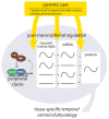
Schematic outline of the relationship between molecular mechanisms that contribute to the generation of circadian rhythms. Local control of circadian transcriptional and post-transcriptional mechanisms is integrated with systemic cues from the master clock in the SCN, as well as feeding and metabolism, to generate tissue-specific changes in transcription, mRNA accumulation and protein production. Different classes of clock-controlled genes are illustrated with either flat lines, to demonstrate constitutive expression at a given step (e.g. nascent transcript or mRNA), or sinusoidal lines, to demonstrate circadian regulation giving a once-per-day peak. Recent studies support the existence of these discrete classes [35, 39, 41], although the mechanisms used to discriminate between regulation of specific target genes are not well understood. The flexibility afforded by this complex, integrative approach likely allows for the maintenance of a core molecular clock in each cell (primarily driven by transcription) while allowing the tissue-specific control of clock-controlled genes necessary for temporal regulation of physiology by the clock.

References

    1. Lowrey P, Takahashi J. Mammalian circadian biology: elucidating genome-wide levels of temporal organization. Annual review of genomics and human genetics. 2004;5:407–441. - PMC - PubMed
    1. Kang TH, et al. Circadian oscillation of nucleotide excision repair in mammalian brain. Proc Natl Acad Sci U S A. 2009;106:2864–2867. - PMC - PubMed
    1. Kang TH, et al. Circadian control of XPA and excision repair of cisplatin-DNA damage by cryptochrome and HERC2 ubiquitin ligase. Proc Natl Acad Sci U S A. 2010;107:4890–4895. - PMC - PubMed
    1. Ko C, Takahashi J. Molecular components of the mammalian circadian clock. Human molecular genetics. 2006;15(Spec No 2):7. - PubMed
    1. Mohawk J, Takahashi J. Cell autonomy and synchrony of suprachiasmatic nucleus circadian oscillators. Trends in neurosciences 2011 - PMC - PubMed

Publication types