. 2013:4:2268.
doi: 10.1038/ncomms3268.

Prelamin A causes progeria through cell-extrinsic mechanisms and prevents cancer invasion

Affiliations

Prelamin A causes progeria through cell-extrinsic mechanisms and prevents cancer invasion

Jorge de la Rosa et al. Nat Commun. 2013.

Abstract

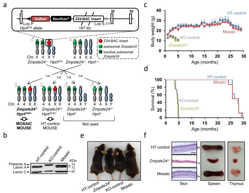
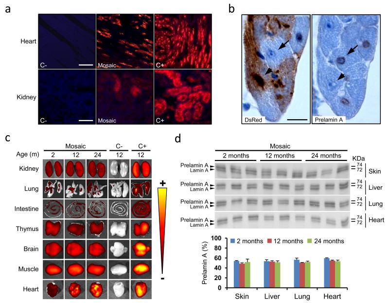
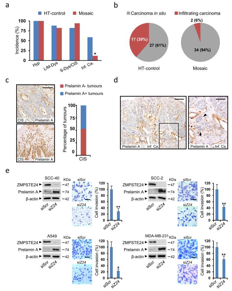
Defining the relationship between ageing and cancer is a crucial but challenging task. Mice deficient in Zmpste24, a metalloproteinase mutated in human progeria and involved in nuclear prelamin A maturation, recapitulate multiple features of ageing. However, their short lifespan and serious cell-intrinsic and cell-extrinsic alterations restrict the application and interpretation of carcinogenesis protocols. Here we present Zmpste24 mosaic mice that lack these limitations. Zmpste24 mosaic mice develop normally and keep similar proportions of Zmpste24-deficient (prelamin A-accumulating) and Zmpste24-proficient (mature lamin A-containing) cells throughout life, revealing that cell-extrinsic mechanisms are preeminent for progeria development. Moreover, prelamin A accumulation does not impair tumour initiation and growth, but it decreases the incidence of infiltrating oral carcinomas. Accordingly, silencing of ZMPSTE24 reduces human cancer cell invasiveness. Our results support the potential of cell-based and systemic therapies for progeria and highlight ZMPSTE24 as a new anticancer target.


Figures

Figure 1
Figure 1. Absence of progeroid phenotype in Zmpste24 mosaic mice
(a) Breeding scheme. Zmpste24−/−HprtZ24 males were bred with Zmpste24+/−Hprt+/+ females to yield female offspring that were either Zmpste24−/−HprtZ24/+ (mosaic) or Zmpste24+/−HprtZ24/+ (HT-control). (b) Western blot analysis of lamins A and C from control and mosaic neonatal fibroblasts. Original immunoblots images are shown in Supplementary Figure S7. (c) Body weight vs. age of HT-control, KO-control, Zmpste24−/− and mosaic mice (n=5 per genotype). Error bars represent s.e.m. (d) Kaplan-Meier survival plot of mice with genotypes as in (c) (n=8 per genotype). (e) Photographs of 4-month-old HT-control, Zmpste24−/−, KO-control, and mosaic mice. (f) Lack of progeroid features in skin, thymus and spleen from mosaic mice. Asterisk-labelled bars indicate the depth of the subcutaneous fat layer. Scale bar represents 500 μm.
Figure 2
Figure 2. Zmpste24-deficient cells are detected throughout lifespan of mosaic mice
(a) Fluorescence microscopy of DsRed-expressing cells in tissues from Zmpste24+/+Hprt+/+(C-), mosaic and Zmpste24+/−HprtZ24/Z24 (C+) mice. Scale bars represent 100 μm (heart) and 50 μm (kidney) (b) Immunohistochemical analysis of DsRed (left) and prelamin A (right) in mosaic hearts. Arrows correspond to a Zmpste24-deficient cell (DsRed-, prelamin A+), whereas arrowheads point to a Zmspte24-proficient cell (DsRed+, prelamin A-). Scale bar represents 10 μm. (c) DsRed fluorescent signal detection by an In Vivo Imaging System (IVIS) in whole organs from mosaic mice at different ages, compared to the same organs from 1-year-old Zmpste24+/+Hprt+/+ (C-) and Zmpste24+/−HprtZ24/Z24 (C+) mice. For comparison with DsRed signal in hearts from HT-control and Zmpste24+/−HprtΔZ24/+ mice see Supplementary Figure S2. (d) Western blot analysis of prelamin A and lamin A from skin, liver, lung and heart of 2-, 12- and 24- month-old mosaic mice. A densitometry analysis of the immunoblots is also shown. Error bars represent s.e.m. n=3.
Figure 3
Figure 3. Mosaic mice keep similar proportions of Zmpste24-deficient and - proficient cells at all ages
(a) Bisulfite pyrosequencing strategy to quantify the relative proportion of Zmpste24-deficient and -proficient cells. Four CpG sites around the Zmpste24 promoter were analyzed in DNA from Zmpste24-deficient adult fibroblasts isolated from mosaic mice (AF1, AF2) and in DNA from six wild type tissues (heart, liver, adipose tissue, kidney, lung and muscle). Western-blots show the progressive loss of mature lamin A and accumulation of prelamin A with serial passages of AF1 and AF2 cells, until only prelamin A is detected (dashed rectangles). The percent methylation of CpG sites is depicted for each of the cell populations. CpG 3 was subsequently used due to its low levels of methylation in wild-type tissues and consistent high values in the isolated fibroblasts. 1F and 1R indicate the positions of the oligonucleotides used (for sequences see Supplementary Table S4). TSS: Zmpste24 transcription start site. (b) Quantitative Zmpste24 methylation analysis by pyrosequencing in different organs from 2-, 12- and 24-month-old mosaic and HT-control mice (n≥3). Error bars represent s.e.m. (*), p < 0.05; (**), p < 0.01; two tailed Student’s t test.
Figure 4
Figure 4. Rescue of molecular and systemic progeria markers in mosaic mice
(a) Transcriptional qPCR analysis of p53-target genes (Cdkn1a, Igfbp3 and Atf3) in hearts from 6-month-old HT-control mice, 3-month-and 4-month-old Zmpste24−/− and 6-month-old mosaic mice. Data are shown as fold-change values as compared to HT-controls (n≥3). (b) Expression of Cdkn1a, Igfbp3 and Zmpste24 in microdissected DsRed fluorescent and negative cells from 6-month-old mosaic hearts (n=3). A fluorescence photomicrograph of DsRed positive (red) and negative (grey) heart areas, representative of those used for microdissection, is shown on the right. Scale bar corresponds to 100 μm. (c) Plasma levels of IGF1 and blood glucose concentration in HT-control, Zmpste24−/− and mosaic mice of the indicated age (n≥3). (m), months. In (a), (b) and (c) error bars represent s.e.m.; (*), p < 0.05; (**), p < 0.01; (***), p < 0.001; two tailed Student’s t-test. (d) Representative picture of SDS-PAGE analysis of MUPs content in urine from mice of each of the indicated genotypes.
Figure 5
Figure 5. Prelamin A accumulation prevents cancer invasion
(a) Percentage of HT-control (n=17) and mosaic (n=17) mice affected by oral lesions with the indicated histology: Hyp, hyperplasia; L/M-Dys, light/moderate dysplasia; S-Dys, severe dysplasia; CIS, carcinoma in situ; Inf. Ca., infiltrating carcinoma. (b) Number and proportion of in situ and infiltrating oral carcinomas in each group. (c) Representative photomicrographs of positive (bottom) and negative (top) prelamin A staining in oral CIS from mosaic mice. The percentage of prelamin A positive and negative CIS is plotted. A total of 17 CIS were analyzed by prelamin A immunohistochemistry. Scale bar represents 100 μm. (d) Representative photomicrographs of a prelamin A negative infiltrating carcinoma from a mosaic mouse. Arrowheads point at nests of invasive cancer cells. Scale bars represent 100 μm (left panel) and 50 μm (right panel). (e) Effect of ZMPSTE24 silencing on the invasiveness of SCC-40, SCC-2, A549 and MDA-MB-231 cancer cell lines. Left panels: Western blots showing the accumulation of prelamin A upon ZMPSTE24 (siZ24) vs. scrambled (siScr) silencing. Right panels: Representative pictures and average number of invasive ZMPSTE24-interfered and scrambled-treated cells. The assays were carried out at least in triplicate and results are expressed as mean ± s.e.m. (*), p < 0.05; (**), p < 0.01; Fisher’s exact test. Scale bars represent 100 μm.

References

    1. López-Otín C, Serrano M, Partridge L, Blasco MA, Kroemer G. The hallmarks of aging. Cell. 2013;153:1194–1217. - PMC - PubMed
    1. Scaffidi P, Misteli T. Lamin A-dependent nuclear defects in human aging. Science. 2006;312:1059–1063. - PMC - PubMed
    1. Burtner CR, Kennedy BK. Progeria syndromes and ageing: what is the connection? Nat. Rev. Mol. Cell. Biol. 2010;11:567–578. - PubMed
    1. Pendás AM, et al. Defective prelamin A processing and muscular and adipocyte alterations in Zmpste24 metalloproteinase-deficient mice. Nat. Genet. 2002;31:94–99. - PubMed
    1. De Sandre-Giovannoli A, et al. Lamin a truncation in Hutchinson-Gilford progeria. Science. 2003;300:2055. - PubMed

Publication types

MeSH terms