

Raccoon rabies virus variant transmission through solid organ transplantation
- PMID: 23917290
- PMCID: PMC7552820
- DOI: 10.1001/jama.2013.7986
Raccoon rabies virus variant transmission through solid organ transplantation
Abstract
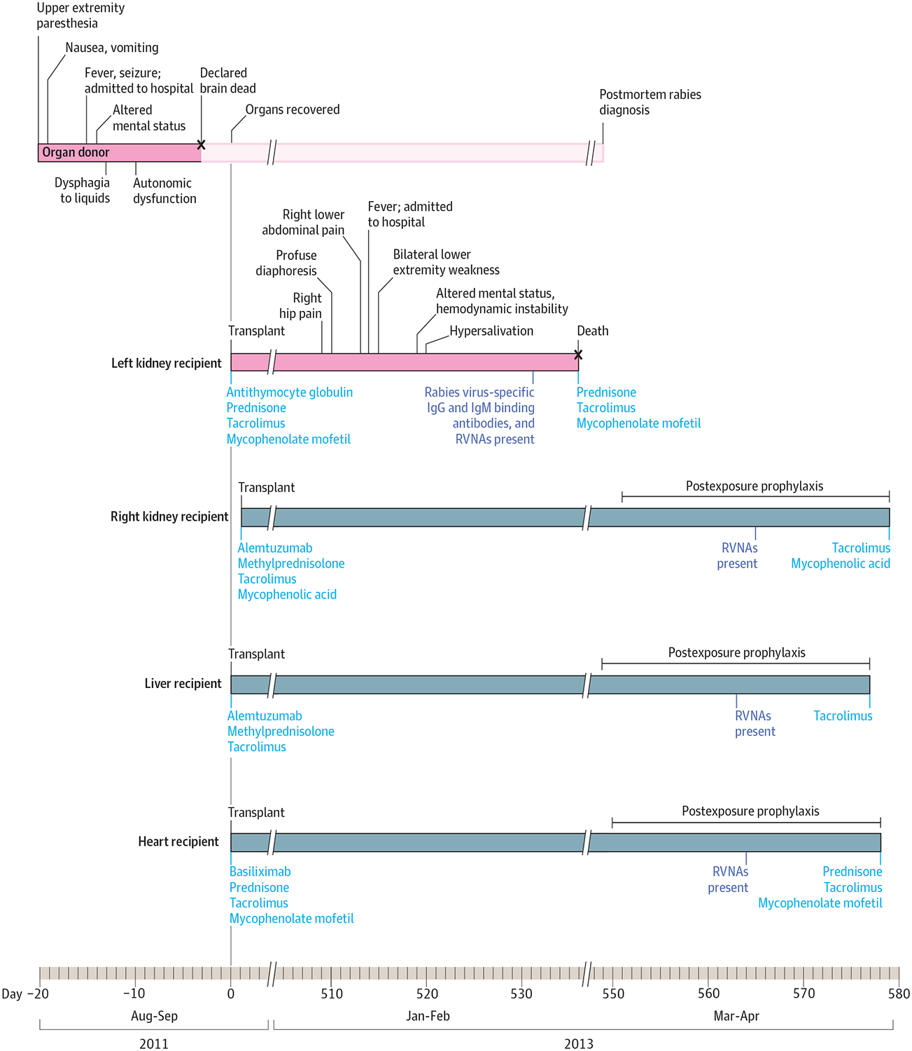
Importance: The rabies virus causes a fatal encephalitis and can be transmitted through tissue or organ transplantation. In February 2013, a kidney recipient with no reported exposures to potentially rabid animals died from rabies 18 months after transplantation.
Objectives: To investigate whether organ transplantation was the source of rabies virus exposure in the kidney recipient, and to evaluate for and prevent rabies in other transplant recipients from the same donor.
Design: Organ donor and all transplant recipient medical records were reviewed. Laboratory tests to detect rabies virus-specific binding antibodies, rabies virus neutralizing antibodies, and rabies virus antigens were conducted on available specimens, including serum, cerebrospinal fluid, and tissues from the donor and the recipients. Viral ribonucleic acid was extracted from tissues and amplified for nucleoprotein gene sequencing for phylogenetic comparisons.
Main outcomes and measures: Determination of whether the donor died from undiagnosed rabies and whether other organ recipients developed rabies.
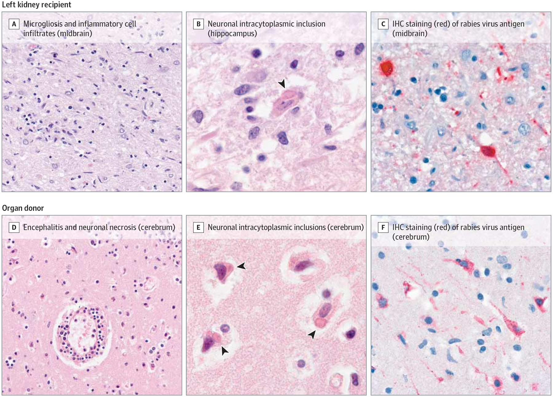
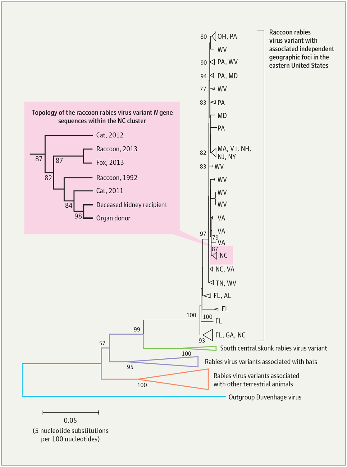
Results: In retrospect, the donor's clinical presentation (which began with vomiting and upper extremity paresthesias and progressed to fever, seizures, dysphagia, autonomic dysfunction, and brain death) was consistent with rabies. Rabies virus antigen was detected in archived autopsy brain tissue collected from the donor. The rabies viruses infecting the donor and the deceased kidney recipient were consistent with the raccoon rabies virus variant and were more than 99.9% identical across the entire N gene (1349/1350 nucleotides), thus confirming organ transplantation as the route of transmission. The 3 other organ recipients remained asymptomatic, with rabies virus neutralizing antibodies detected in their serum after completion of postexposure prophylaxis (range, 0.3-40.8 IU/mL).
Conclusions and relevance: Unlike the 2 previous clusters of rabies virus transmission through solid organ transplantation, there was a long incubation period in the recipient who developed rabies, and survival of 3 other recipients without pretransplant rabies vaccination. Rabies should be considered in patients with acute progressive encephalitis of unexplained etiology, especially for potential organ donors. A standard evaluation of potential donors who meet screening criteria for infectious encephalitis should be considered, and risks and benefits for recipients of organs from these donors should be evaluated.
Conflict of interest statement
Figures
Comment in
-
Donor-derived infections with central nervous system pathogens after solid organ transplantation.JAMA. 2013 Jul 24;310(4):378-9. doi: 10.1001/jama.2013.7989. JAMA. 2013. PMID: 23917287 No abstract available.
References
-
- Hemachudha T, Laothamatas J, Rupprecht CE. Human rabies: a disease of complex neuropathogenetic mechanisms and diagnostic challenges. Lancet Neurol. 2002;1(2):101–109. - PubMed
-
- Petersen B, Rupprecht C. Chapter 11: Human rabies epidemiology and diagnosis In: Tkachev S, ed. Non-Flavivirus Encephalitis. http://www.intechopen.com/books/non-flavivirus-encephalitis. Accessed July 8, 2013.
-
- Centers for Disease Control and Prevention (CDC). First human death associated with raccoon rabies: Virginia, 2003. MMWR Morb Mortal Wkly Rep. 2003;52(45):1102–1103. - PubMed
Publication types
MeSH terms
Substances
Grants and funding
LinkOut - more resources
Full Text Sources
Other Literature Sources
Medical
