. 2013 May;14(5):458-65.
doi: 10.4161/cbt.24424.

PARP and CHK inhibitors interact to cause DNA damage and cell death in mammary carcinoma cells

Affiliations

PARP and CHK inhibitors interact to cause DNA damage and cell death in mammary carcinoma cells

Laurence Booth et al. Cancer Biol Ther. 2013 May.

Abstract

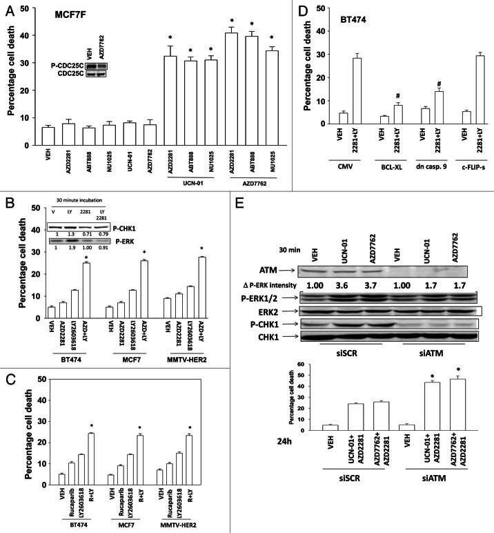
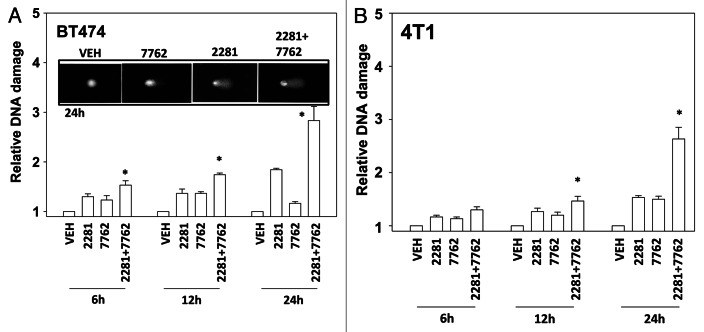
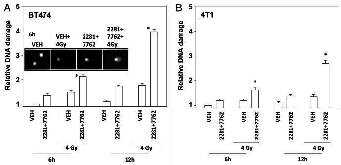
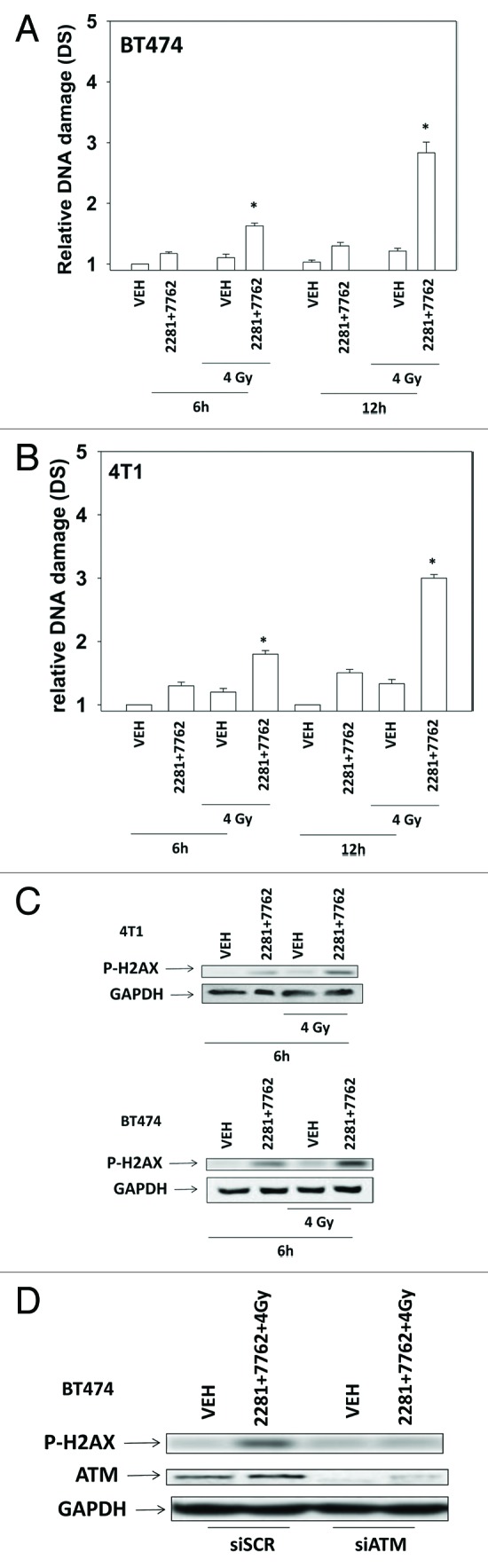
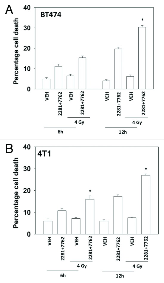
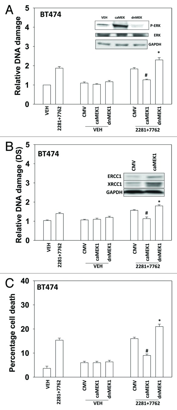
The present studies examined viability and DNA damage levels in mammary carcinoma cells following PARP1 and CHK1 inhibitor drug combination exposure. PARP1 inhibitors [AZD2281 ; ABT888 ; NU1025 ; AG014699] interacted with CHK1 inhibitors [UCN-01 ; AZD7762 ; LY2603618] to kill mammary carcinoma cells. PARP1 and CHK1 inhibitors interacted to increase both single strand and double strand DNA breaks that correlated with increased γH2AX phosphorylation. Treatment of cells with CHK1 inhibitors increased the phosphorylation of CHK1 and ERK1/2. Knock down of ATM suppressed the drug-induced increases in CHK1 and ERK1/2 phosphorylation and enhanced tumor cell killing by PARP1 and CHK1 inhibitors. Expression of dominant negative MEK1 enhanced drug-induced DNA damage whereas expression of activated MEK1 suppressed both the DNA damage response and tumor cell killing. Collectively our data demonstrate that PARP1 and CHK1 inhibitors interact to kill mammary carcinoma cells and that increased DNA damage is a surrogate marker for the response of cells to this drug combination.

Keywords: ATM; CHK1; DNA damage; PARP1; apoptosis; comet; kinase.


Figures

None
Figure 1. PARP1 and CHK1 inhibitors interact to kill breast cancer cells. (A) Fulvestrant resistant MCF7F cells were treated with AZD2281 (1 μM), ABT888 (1 μM), NU1025 (10 μM), UCN-01 (50 nM) and AZD7762 (25 nM), and these agents in combination as presented in the panel. Cells were isolated 48h after exposure, and viability was determined using trypan blue exclusion. (n = 3 ± SEM) * p < 0.05 value greater than corresponding vehicle control. Inset blot: Cells were treated with vehicle (DMSO) or AZD7762 (25 nM); cells were isolated after 60 min and the phosphorylation of CDC25C determined. (B) BT474, MCF7 and MMTV-HER2 cells were treated with AZD2281 (1 μM), LY2603618 (1 μM) or the drugs in combination. Cells were isolated 24h after exposure, and viability was determined using trypan blue exclusion. (n = 3 ± SEM) * p < 0.05 value greater than corresponding vehicle control. Inset blot: BT474 cells were treated with AZD2281 (1 μM), LY2603618 (1 μM) or the drugs in combination. Cells were isolated 30 min after exposure and blotting performed to determine P-ERK and P-CHK1 levels. The fold change in P-ERK to total ERK and P-CHK1 to total CHK1 levels is presented. (C) BT474, MCF7 and MMTV-HER2 cells were treated with Rucaparib (1 μM), LY2603618 (1 μM) or the drugs in combination. Cells were isolated 24h after exposure, and viability was determined using trypan blue exclusion. (n = 3 ± SEM). * p < 0.05 value greater than corresponding vehicle control. (D) BT474 cells were infected with an empty vector virus (CMV) or viruses to express BCL-XL, dominant negative caspase 9 or c-FLIP-s. Twenty four h after infection cells are treated with vehicle (DMSO) or AZD2281 (1 μM) and LY2603618 (1 μM). Cells were isolated 24h after exposure, and viability was determined using trypan blue exclusion. (n = 3 ± SEM). # p < 0.05 value less than corresponding virus control. (E) BT474 cells were transfected with scrambled siRNA (siSCR, 20 nM) or an siRNA to knock down ATM expression. Lower Graph: 24h after transfection cells were treated with vehicle (DMSO) or with [UCN-01, 50 nM + AZD2281, 1 μM] or [AZD7762, 25 nM + AZD2281 1 μM]. Cells were isolated 48h after exposure, and viability was determined using trypan blue exclusion. (n = 3 +/− SEM). * p < 0.05 value greater than corresponding siSCR control. Upper blots: 24h after transfection cells were treated with vehicle (DMSO) or with [UCN-01, 50 nM] or [AZD7762, 25 nM]. Cells were isolated 30 min after exposure and blotting performed to determine P-ERK and P-CHK1 levels.
None
Figure 2. CHK1 and PARP1 inhibitors interact to cause DNA damage. (A) BT474, (B) 4T1 cells were treated with AZD2281 (1 μM), AZD7762 (25 nM) or the drug combination for the indicated times. Cells were isolated and subjected to alkaline comet assay. The length of the tail being scored 1–5 (n = 3 ± SEM) * p < 0.05 value greater than corresponding vehicle control.
None
Figure 3. CHK1 and PARP1 inhibitors and ionizing radiation interact to cause DNA damage. (A) BT474, (B) 4T1 cells were treated with AZD2281 (1 μM) and AZD7762 (25 nM) in combination for the indicated times. Cells were irradiated (4 Gy) 30 min after drug exposure. Cells were isolated and subjected to alkaline comet assay. The length of the tail being scored 1–5 (n = 3 ± SEM). * p < 0.05 value greater than corresponding vehicle control.
None
Figure 4. CHK1 and PARP1 inhibitors interact to cause double stranded DNA damage. (A) BT474, (B) 4T1 cells were treated with AZD2281 (1 μM) and AZD7762 (25 nM) in combination for the indicated times. Cells were irradiated (4 Gy) 30 min after drug exposure. Cells were isolated and subjected to neutral comet assay. The length of the tail being scored 1–5 (n = 3 ± SEM). * p < 0.05 value greater than corresponding vehicle control. (C) BT474 and 4T1 cells were treated with AZD2281 (1 μM) and AZD7762 (25 nM) in combination for 6h. As indicated cells were irradiated (4 Gy) 30 min after drug exposure. Blotting was performed to determine P-H2AX levels. (D) BT474 cells were transfected with scrambled siRNA (siSCR, 20 nM) or an siRNA to knock down ATM expression. Twenty four h after transfection cells were treated with AZD2281 (1 μM) and AZD7762 (25 nM) in combination for 6h. As indicated cells were irradiated (4 Gy) 30 min after drug exposure. Blotting was performed to determine P-H2AX levels.
None
Figure 5. PARP1 and CHK1 inhibitors radiosensitize mammary carcinoma cells. (A) BT474, (B) 4T1 cells were treated with AZD2281 (1 μM) and AZD7762 (25 nM) in combination for 6h. As indicated cells were irradiated (4 Gy) 30 min after drug exposure. Cells were isolated 6h and 12h after exposure, and viability was determined using trypan blue exclusion. (n = 3 ± SEM). * p < 0.05 value greater than corresponding vehicle control.
None
Figure 6. MER-ERK signaling regulates the DNA damage response following PARP1 and CHK1 inhibitor treatment. (A) BT474 cells were infected with empty vector adenovirus (CMV) or viruses to express dominant negative MEK1 (dnMEK1) or activated MEK1 (caMEK1). Twenty four h after infection cells were treated with AZD2281 (1 μM) and AZD7762 (25 nM) in combination for 24h. Cells were isolated and subjected to alkaline comet assay. The length of the tail being scored 1–5 (n = 3 ± SEM). * p < 0.05 value greater than corresponding vehicle control; # p < 0.05 value less than corresponding vehicle control. Inset blot: the levels of ERK1/2 phosphorylation in cells expressing caMEK1 and dnMEK1. (B) BT474 cells were infected with empty vector adenovirus (CMV) or viruses to express dominant negative MEK1 (dnMEK1) or activated MEK1 (caMEK1). Twenty four h after infection cells were treated with AZD2281 (1 μM) and AZD7762 (25 nM) in combination for 24h. Cells were isolated and subjected to neutral comet assay. The length of the tail being scored 1–5 (n = 3 ± SEM). * p < 0.05 value greater than corresponding vehicle control; # p < 0.05 value less than corresponding vehicle control. Inset blot: the levels of ERCC1 and XRCC1 in cells expressing caMEK1. (C) BT474 cells were infected with empty vector adenovirus (CMV) or viruses to express dominant negative MEK1 (dnMEK1) or activated MEK1 (caMEK1). Twenty four h after infection cells were treated with AZD2281 (1 μM) and AZD7762 (25 nM) in combination. Cells were isolated 24h after exposure, and viability was determined using trypan blue exclusion. (n = 3 ± SEM). * p < 0.05 value greater than corresponding vehicle control; # p < 0.05 value less than corresponding vehicle control.

References

    1. Lukas C, Falck J, Bartkova J, Bartek J, Lukas J. Distinct spatiotemporal dynamics of mammalian checkpoint regulators induced by DNA damage. Nat Cell Biol. 2003;5:255–60. doi: 10.1038/ncb945. - DOI - PubMed
    1. Fernandez-Capetillo O, Lee A, Nussenzweig M, Nussenzweig A. H2AX: the histone guardian of the genome. DNA Repair (Amst) 2004;3:959–67. doi: 10.1016/j.dnarep.2004.03.024. - DOI - PubMed
    1. Lee JH, Paull TT. Activation and regulation of ATM kinase activity in response to DNA double-strand breaks. Oncogene. 2007;26:7741–8. doi: 10.1038/sj.onc.1210872. - DOI - PubMed
    1. Bartek J, Lukas J. Chk1 and Chk2 kinases in checkpoint control and cancer. Cancer Cell. 2003;3:421–9. doi: 10.1016/S1535-6108(03)00110-7. - DOI - PubMed
    1. Morgan MA, Parsels LA, Zhao L, Parsels JD, Davis MA, Hassan MC, et al. Mechanism of radiosensitization by the Chk1/2 inhibitor AZD7762 involves abrogation of the G2 checkpoint and inhibition of homologous recombinational DNA repair. Cancer Res. 2010;70:4972–81. doi: 10.1158/0008-5472.CAN-09-3573. - DOI - PMC - PubMed

Publication types

MeSH terms