. 2014 Jan;44(1):64-72.
doi: 10.1007/s00247-013-2752-8. Epub 2013 Aug 6.

Pediatric neuroimaging using magnetic resonance imaging during non-sedated sleep

Affiliations

Pediatric neuroimaging using magnetic resonance imaging during non-sedated sleep

Douglas C Dean 3rd et al. Pediatr Radiol. 2014 Jan.

Abstract

Background: Etiological studies of many neurological and psychiatric disorders are increasingly turning toward longitudinal investigations of infant brain development in order to discern predisposing structural and/or functional differences prior to the onset of overt clinical symptoms. While MRI provides a noninvasive window into the developing brain, MRI of infants and toddlers is challenging due to the modality's extreme motion sensitivity and children's difficulty in remaining still during image acquisition.

Objective: Here, we outline a broad research protocol for successful MRI of children under 4 years of age during natural, non-sedated sleep.

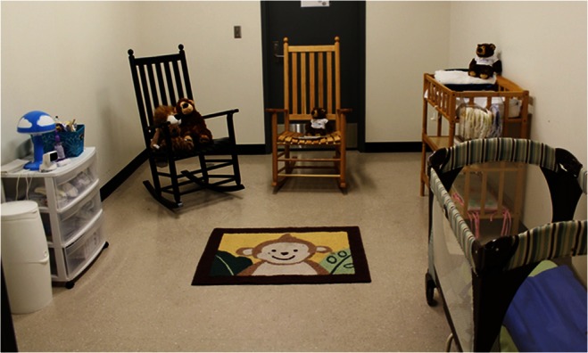
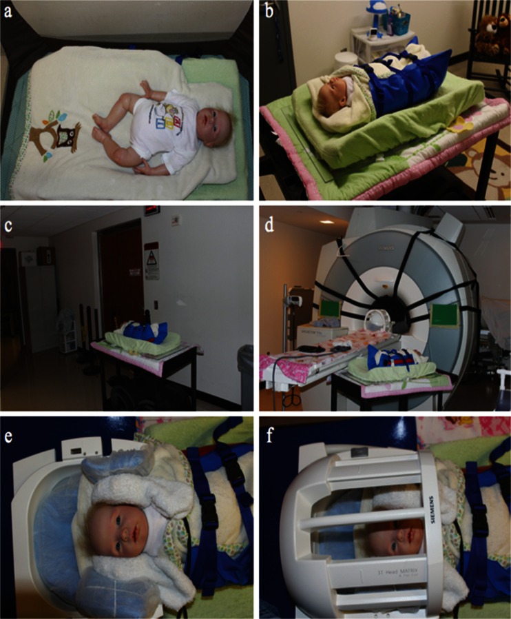
Materials and methods: All children were imaged during natural, non-sedated sleep. Active and passive measures to reduce acoustic noise were implemented to reduce the likelihood of the children waking up during acquisition. Foam cushions and vacuum immobilizers were used to limit intra-scan motion artifacts.

Results: More than 380 MRI datasets have been successfully acquired from 220 children younger than 4 years of age within the past 39 months. Implemented measures permitted children to remain asleep for the duration of the scan and allowed the data to be acquired with an overall 97% success rate.

Conclusion: The proposed method greatly advances current pediatric imaging techniques and may be readily implemented in other research and clinical settings to facilitate and further improve pediatric neuroimaging.


Figures

Fig. 1
Fig. 1
Private sleeping room setup for an infant. Rooms are equipped with a crib (or bed in the case of an older child), rocking chairs, video baby monitor and snacks. Normal bedtime routines could be practiced so that children did not feel uncomfortable sleeping in a new environment
Fig. 2
Fig. 2
MRI scanner with sound-insulating foam insert (Ultra Barrier HD Composite; American Micro Industries, Chambersburg, PA, USA) being installed into the bore of the scanner. Straps are used to hold the foam insert tight to the bore of the scanner. Noise reductions of up to 20 dB can be achieved
Fig. 3
Fig. 3
Logistics of setting up for scanning of a sleeping child. a Children fall asleep either in crib or on bed. b Once asleep, children are buckled into a MedVac immobilizer and transferred to a MRI compatible cart. c Children are then wheeled into the MRI suite (d) and moved to the MRI scanner’s bed. e Electrodynamic headphones are carefully positioned onto children’s ears and held in place using memory foam cushions. f Children are then landmarked and moved to the center of the bore for scanning
Fig. 4
Fig. 4
Anatomical T1-weighted and derived T1, T2, and myelin water fraction (MWF) maps from a representative 21-month-old depicting the quality of the acquired and calculated quantitative images. T2 values were calculated in voxels with T1 values below 3,500 ms
Fig. 5
Fig. 5
Example of an inadequate scan that would necessitate either repeating or having the child return for a second visit. T1‐weighted SPGR images were acquired from a 3‐month‐old boy
Fig. 6
Fig. 6
Attrition rate and number of active study subjects enrolled in the longitudinal study. The number of active subjects included those whose MRI data has been acquired and subjects who are scheduled for a follow-up MRI scan. Attrition rates correspond to the attrition between follow-up visits

References

    1. Giedd JN. Structural magnetic resonance imaging of the adolescent brain. Ann N Y Acad Sci. 2004;1021:77–85. doi: 10.1196/annals.1308.009. - DOI - PubMed
    1. Edwards AD, Arthurs OJ. Paediatric MRI under sedation: is it necessary? What is the evidence for the alternatives? Pediatr Radiol. 2011;41:1353–1364. doi: 10.1007/s00247-011-2147-7. - DOI - PubMed
    1. de Bie HMA, Boersma M, Wattjes MP, et al. Preparing children with a mock scanner training protocol results in high quality structural and functional MRI scans. Eur J Pediatr. 2010;169:1079–1085. doi: 10.1007/s00431-010-1181-z. - DOI - PMC - PubMed
    1. Raschle NM, Lee M, Buechler R, et al. Making MR imaging child’s play—pediatric neuroimaging protocol. Guidelines and Procedure. J Vis Exp. 2009;29:e1309. - PMC - PubMed
    1. Woods-Frohlich L, Martin T. Training children to reduce motion and increase success of MRI scanning. Curr Med Imaging Rev. 2010;6:165–170. doi: 10.2174/157340510791636255. - DOI

Publication types

MeSH terms