

Reviewing subchondral cartilage surgery: considerations for standardised and outcome predictable cartilage remodelling: a technical note
- PMID: 23917852
- PMCID: PMC3824892
- DOI: 10.1007/s00264-013-2025-z
Reviewing subchondral cartilage surgery: considerations for standardised and outcome predictable cartilage remodelling: a technical note
Abstract
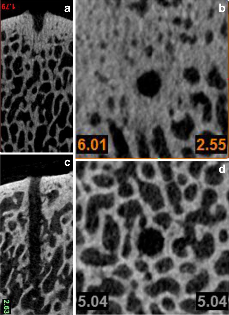
Purpose: The potential of subchondral mesenchymal stem cell stimulation (MSS) for cartilage repair has led to the widespread use of microfracture as a first line treatment for full thickness articular cartilage defects. Recent focus on the effects of subchondral bone during cartilage injury and repair has expanded the understanding of the strengths and limitations in MSS and opened new pathways for potential improvement. Comparative studies have shown that bone marrow access has positive implications for pluripotential cell recruitment, repair quality and quantity, i.e. deeper channels elicited better cartilage fill, more hyaline cartilage character with higher type II collagen content and lower type I collagen content compared to shallow marrow access.
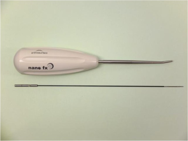
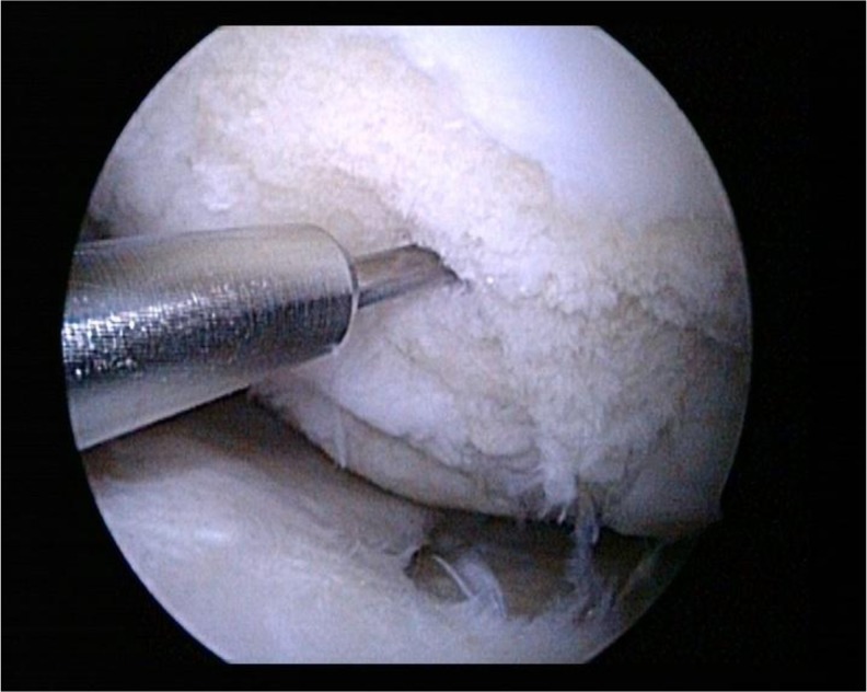
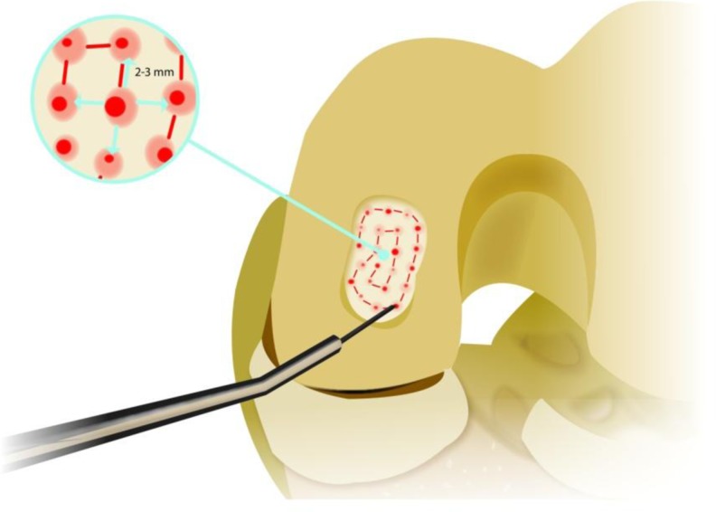
Methods: A subchondral needling procedure using standardised and thin subchondral perforations deep into the subarticular bone marrow making the MSS more consistent with the latest developments in subchondral cartilage remodelling is proposed.
Results: As this is a novel method clinical studies have been initiated to evaluate the procedure especially compared to microfracturing. However, the first case studies and follow-ups indicate that specific drills facilitate reaching the subchondral bone marrow while the needle size makes perforation of the subchondral bone easier and more predictable. Clinical results of the first group of patients seem to compare well to microfracturing.
Conclusion: The authors suggest a new method for a standardised procedure using a new perforating device. Advances in MSS by subchondral bone marrow perforation are discussed. It remains to be determined by clinical studies how this method compares to microfracturing. The subchondral needling offers the surgeon and the investigator a method that facilitates comparison studies because of its defined depth of subchondral penetration and needle size.
Figures

References
-
- Pridie KH. A method of resurfacing osteoarthritic knee joints. J Bone Joint Surg. 1959;41B:618–619.
-
- Steadman JR, Rodkey WG, Singleton SB, Briggs KK. Microfracture technique for full thickness chondral defects: technique and clinical results. Oper Tech Orthop. 1997;7:300–304. doi: 10.1016/S1048-6666(97)80033-X. - DOI
MeSH terms
LinkOut - more resources
Full Text Sources
Other Literature Sources
Medical
Research Materials
