Review
. 2013 Oct;61(10):1571-81.
doi: 10.1002/glia.22541. Epub 2013 Aug 5.

Communication between neuronal somata and satellite glial cells in sensory ganglia

Affiliations
Review

Communication between neuronal somata and satellite glial cells in sensory ganglia

Li-Yen M Huang et al. Glia. 2013 Oct.

Abstract

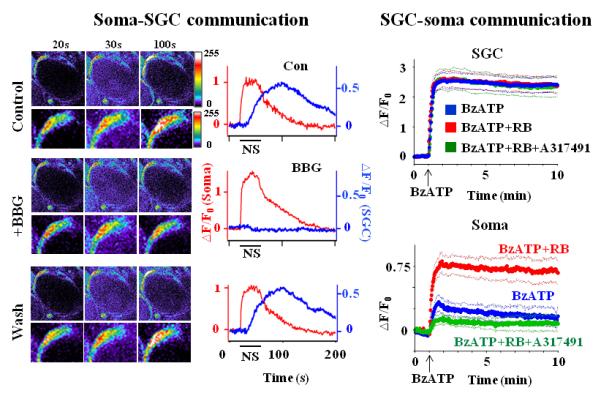
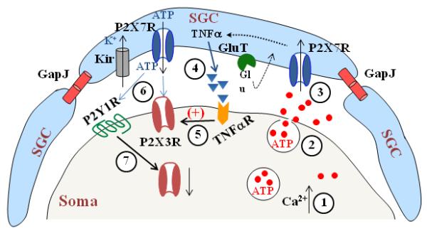
Studies of the structural organization and functions of the cell body of a neuron (soma) and its surrounding satellite glial cells (SGCs) in sensory ganglia have led to the realization that SGCs actively participate in the information processing of sensory signals from afferent terminals to the spinal cord. SGCs use a variety ways to communicate with each other and with their enwrapped soma. Changes in this communication under injurious conditions often lead to abnormal pain conditions. "What are the mechanisms underlying the neuronal soma and SGC communication in sensory ganglia?" and "how do tissue or nerve injuries affect the communication?" are the main questions addressed in this review.

Keywords: cytokine; gap junction; pain; pannexin; purinergic receptor.


Figures

Fig. 1
Fig. 1. Structural characteristics of dorsal root ganglia
(Left) Locations of L4 and L5 DRGs in the spinal column. (Upper right) A micrograph showing a neuron being tightly wrapped by SGCs. (Lower right) An enlarged view of the pseudo-unipolar structure of a DRG neuron.
Fig. 2
Fig. 2. Dependence of neuronal soma-SGC-soma communication on P2X7R activation
(Left) Soma-SGC communication. Images of Ca2+-dependent fluorescence changes in a neuronal soma and SGCs in response to a 20 Hz, 30 sec nerve stimulus are shown on the left. The number above each column is the time that the images were taken. Enlarged views of the SGC are shown directly below each panel. The time courses of the fluorescence change following nerve stimulation (NS) are given on the right. The NS caused an increase in the fluorescence in the neuronal soma ((F-F0)/F0 =△F/F0 (Soma)) and an increase in the SGC, i.e., (△F/F0 (SGC)) with a delay. F0 is the basal fluorescence in either the soma or in the SGC before NS. The △F/F0 in the soma was not affected by the application of the reversible P2X7R antagonist, BBG (1 μM) while △F/F0 in SGCs was inhibited by BBG. Thus, NS induced ATP release from the neuronal soma to activate P2X7Rs in SGCs. (Right) SGC-soma communication. Application of the P2X7R agonist, BzATP (100 μM) (arrows), evoked a large increase in △F/F0 in SGCs but a relatively small increase in somata. F0 is the basal fluorescence before the BzATP application. Preincubation of DRGs with the P2YR antagonist, RB (1 μM), had no effect on the BzATP-induced △F/F0 change in SGCs, but enhanced the △F/F0 change in somata. The enhancement was inhibited when P2X3R activity in somata was blocked by the P2X3R antagonist, A31749 (60 μM). Thus, stimulation of P2X7Rs in SGCs affects the activity of P2YRs and P2X3Rs in neuronal somata. The thick lines are the average fluorescences; the thin lines are the standard errors of the average values. Adapted from Zhang et al (2007) and Chen et al (2008)(copyright #2007 and #2008 by National Academy of Sciences, USA).
Fig. 3
Fig. 3. Mechanisms involved in Neuronal soma - SGC- soma communication in the DRG
A schematic drawing to illustrate that gap junctions (GapJ) are involved in SGC--SGC communication, GluT (glutamate transporter) and Kir (inwardly rectifying K+ channels) regulate glutamate and K+ concentration surrounding the soma respectively and P2X7Rs in SGCs and P2Y1Rs and P2X3Rs in the soma participate in neuron---> SGC--> soma communication. Nerve stimulation evokes Ca2+-dependent ATP release from the soma (1, 2) to activate P2X7Rs in SGCs (3). P2X7R activation promotes the cytokine, e.g. TNFα, release from SGCs (4) to increase activity of P2X3Rs in the soma (5). ATP release resulting from P2X7R activation increases P2X3R activity (6). In addition, ATP also activates P2Y1Rs (6), which in turn down-regulate P2X3R expression (7).

References

    1. Amir R, Devor M. Electrical excitability of the soma of sensory neurons is required for spike invasion of the soma, but not for through-conduction. Biophys J. 2003a;84:2181–91. - PMC - PubMed
    1. Amir R, Devor M. Extra spike formation in sensory neurons and the disruption of afferent spike patterning. Biophys J. 2003b;84:2700–8. - PMC - PubMed
    1. Anderson CM, Swanson RA. Astrocyte glutamate transport: review of properties, regulation, and physiological functions. Glia. 2000;32:1–14. - PubMed
    1. Bak LK, Schousboe A, Waagepetersen HS. The glutamate/GABA-glutamine cycle: aspects of transport, neurotransmitter homeostasis and ammonia transfer. Journal of neurochemistry. 2006;98:641–53. - PubMed
    1. Bao L, Locovei S, Dahl G. Pannexin membrane channels are mechanosensitive conduits for ATP. FEBS Lett. 2004;572:65–8. - PubMed

Publication types