. 2013 Oct;61(10):1687-97.
doi: 10.1002/glia.22549. Epub 2013 Aug 5.

Retinal regeneration in adult zebrafish requires regulation of TGFβ signaling

Affiliations

Retinal regeneration in adult zebrafish requires regulation of TGFβ signaling

Jenny R Lenkowski et al. Glia. 2013 Oct.

Abstract

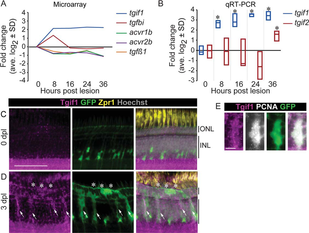
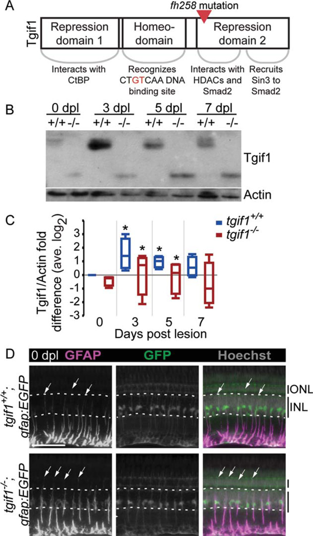
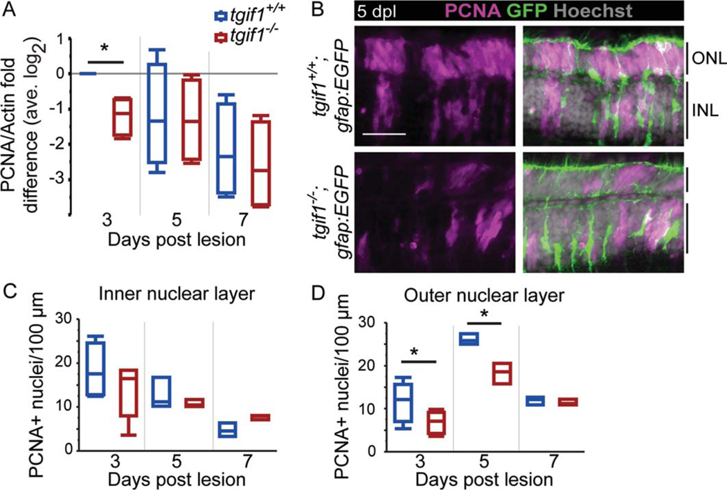
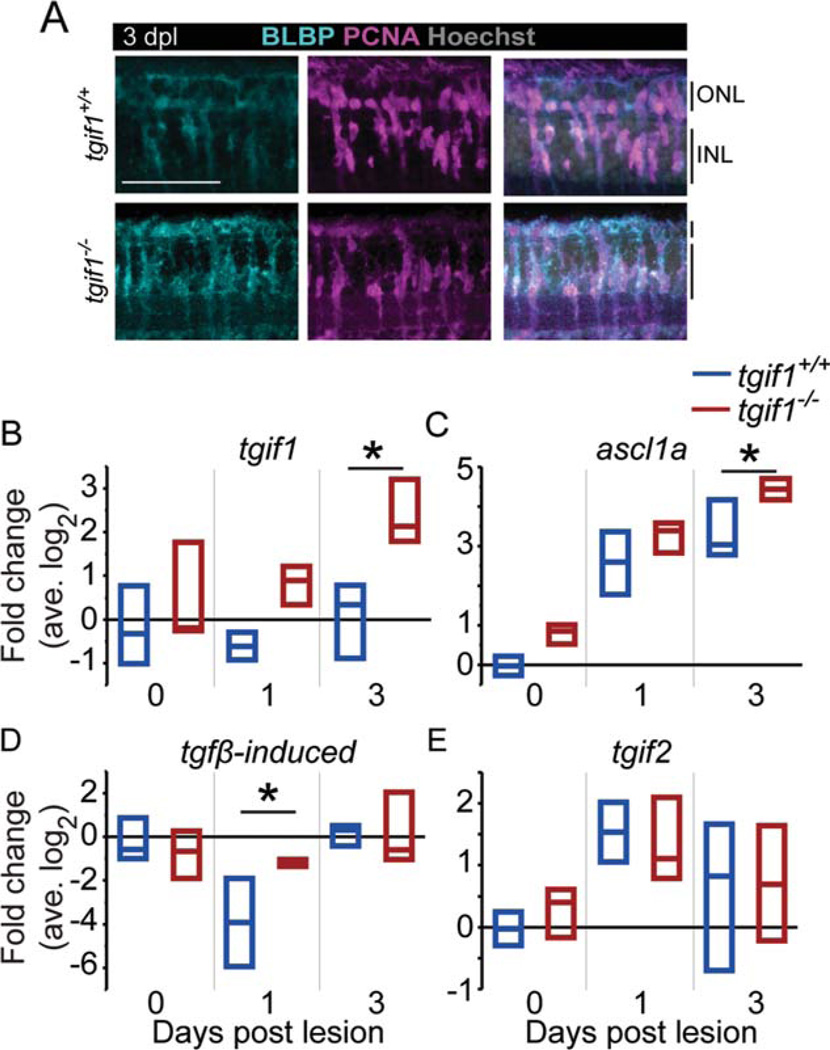
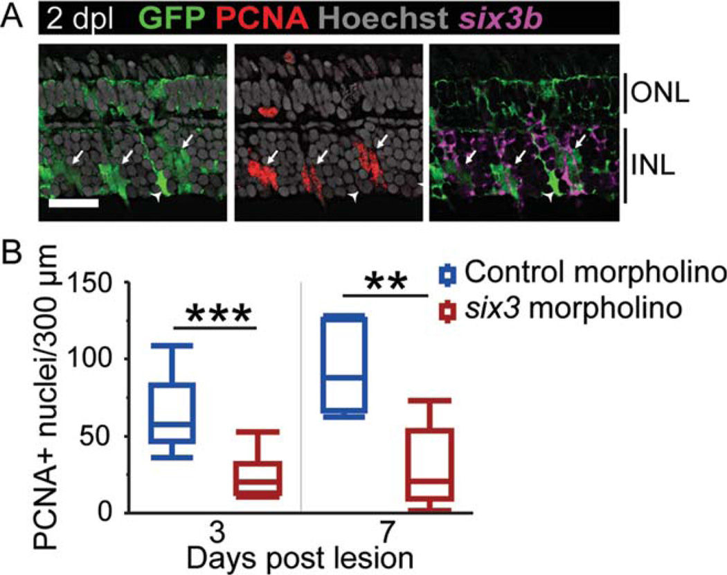
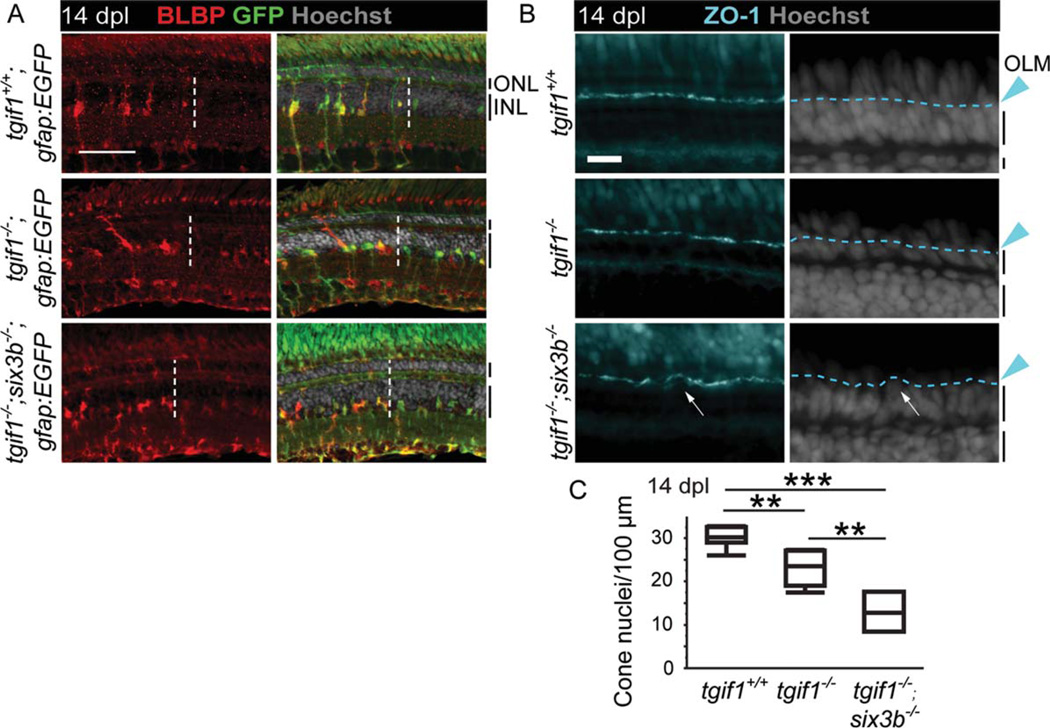
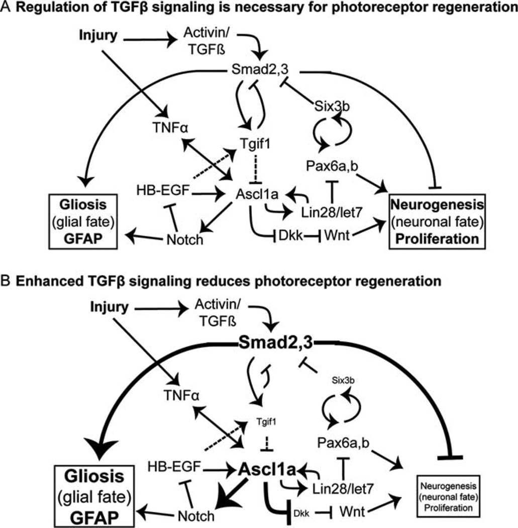
Müller glia are the resident radial glia in the vertebrate retina. The response of mammalian Müller glia to retinal damage often results in a glial scar and no functional replacement of lost neurons. Adult zebrafish Müller glia, in contrast, are considered tissue-specific stem cells that can self-renew and generate neurogenic progenitors to regenerate all retinal neurons after damage. Here, we demonstrate that regulation of TGFβ signaling by the corepressors Tgif1 and Six3b is critical for the proliferative response to photoreceptor destruction in the adult zebrafish retina. When function of these corepressors is disrupted, Müller glia and their progeny proliferate less, leading to a significant reduction in photoreceptor regeneration. Tgif1 expression and regulation of TGFβ signaling are implicated in the function of several types of stem cells, but this is the first demonstration that this regulatory network is necessary for regeneration of neurons.

Keywords: Müller glia; photoreceptor; six3b; stem cell; tgif1.


Figures

FIGURE 1
FIGURE 1
Smad2/3 signaling is downregulated and the corepressor Tgif1 upregulated after photoreceptors destruction. (A) Changes in expression of Smad2/3 signaling pathway in isolated Müller glia after acute light lesion, compared with uninjured retina, by microarray analysis (GEO GSE14495; Qin et al., 2009). (B) Changes in expression of tgif1 and tgif2 in isolated Müller glia measured by qRT-PCR. * P<0.05 relative to 0 hpl, Student t-test. Tgif1 immunoreactivity in an undamaged retina (C) and at 3 dpl (D) in gfap:EGFP fish that have GFP in the Müller glia and progenitors (arrows = Müller glia; *= lesion). (E) Colocalization of Tgif1 and PCNA in GFP-positive Müller glia in gfap:EGFP fish at 3 dpl. Scale bars = 50 µm in (C,D); 10 µm in (E). ONL = outer nuclear layer; INL = inner nuclear layer; hpl = hours post lesion; dpl 5 days post lesion.
FIGURE 2
FIGURE 2
Fish homozygous for the fh258 allele of tgif1 express a truncated Tgif1 and GFAP localization in Müller glia is altered. (A) Schematic of the Tgif1 protein and the point mutation in the fh258 allele. (B) Representative Tgif1 Western blot of whole retina extract from tgif1+/+ and tgif1−/− fish. (C) Densitometric analysis of Tgif1 protein in control and light lesioned retinas from tgif1+/+ and tgif1−/− fish normalized to actin and relative to 0 dpl tgif1+/+ (retinas pooled from two fish, n = 4 replicates). * P<0.05 relative to 0 dpl of the same genotype, Student t-test. (D) GFAP immunolocalization (first column) in the unlesioned retina of tgif1+/+;gfap:EGFP (top) and tgif1−/−;gfap:EGFP (bottom) fish expressing GFP only in the Müller glia. We used the same exposure time and did not alter the GFAP images postcapture. Arrows = top of GFAP distribution in individual Müller glia; dotted white lines delineate the base and top of the INL. Abbreviations as in Fig. 1. Scale bar = 50µm.
FIGURE 3
FIGURE 3
Proliferation is reduced in tgif1−/− fish compared with tgif1+/+ fish. (A) Densitometric analysis of PCNA in Western blots normalized to actin and relative to 3 dpl tgif1+/+ (retinas pooled from two fish, n = 4 replicates). (B) Representative images of PCNA immu-noreactivity at 5 dpl in tgif1+/+ and tgif1−/− fish with the gfap:EGFP transgene. The average number of PCNA-positive nuclei in the INL (C) and ONL (D) in tgif1+/+ and tgif1−/− fish; 8–17 sections per fish; n = 5 at 3 dpl; n = 3 at 5 and 7 dpl. Scale bars=25 µm. * P<0.05, Student t-test comparing genotypes at the same lesion interval. Abbreviations as in Fig. 1.
FIGURE 4
FIGURE 4
After acute light lesion, Müller glia in tgif1−/− fish express a dedifferentiation marker similar to tgif1+/+ but misre-gulate Smad2/3 target genes. (A) BLBP and PCNA immunoreac-tivity in tgif1+/+ and tgif1−/− fish at 3 dpl in the lesioned area, as determined by the absence of cone nuclei. (B–E) qRT-PCR of whole retina extracts (retinas pooled from individual fish, n=3 replicates). We normalized gene expression to gpia and calculated fold change relative to the average tgif1+/+ 0 dpl level. * P<0.05, Student t-test comparing genotypes at the same lesion interval. Abbreviations as in Fig. 1.
FIGURE 5
FIGURE 5
Repression of the Smad2/3 corepressor six3 inhibits proliferation after photoreceptor lesion. (A) six3b in situ hybridization and PCNA immunoreactivity in GFP-positive Müller glia and their progeny (arrows) at 2 dpl in gfap:EGFP fish. The arrowhead indicates a PCNA-negative Müller glial cell that is not expressing six3b. (B) The number of PCNA-positive nuclei at 3 and 7 dpl after electroporation of morpholinos targeting six3a/b. n=11 for control morpholino, n=10 for six3 morpholino at 3 dpl, and n=5 at 7 dpl. Scale bar=20 µm. ** P<0.01; *** P<0.0001, Student t-test comparing genotypes at the same lesion interval. Abbreviations as in Fig. 1.
FIGURE 6
FIGURE 6
Regeneration is significantly impaired in fish homozygous for tgif1−/− or both tgif1−/− and six3b−/− relative to tgif1+/+ fish. (A) BLBP immunoreactivity at 14 dpl in tgif1+/+;gfap:EGFPtgif1−/−;gfap:EGFP, and tgif1−/−;six3b−/−;gfap:EGFP fish expressing GFP in the Müller glia (lesioned area is left of dotted line). (B) ZO-1 immunolocalization at 14 dpl (panels at left) to visualize the OLM (dashed line in panels at right) of tgif1+/+tgif1−/−, and tgif1−/−;six3b−/− fish. (C) The number of photoreceptor nuclei in the regenerated area at 14 dpl. Scale bars=50 mm in (A), 10 µm in (B). ** P<0.005, *** P<0.0001, Student t-test pairwise comparisons of genotypes. OLM=outer limiting membrane. Other abbreviations as in Fig. 1.
FIGURE 7
FIGURE 7
Signaling pathways that regulate Müller glial-derived regeneration of retinal neurons. Signaling pathways implicated in Müller glial response to retinal injury and retinal regeneration based on the present work and published studies (Bernardos et al., 2005; Ikeda and Puro, 1995; Inbal et al., 2007; Liu et al., 2006; Liu et al., 2011; Meyers et al., 2012; Nelson et al., 2012, 2013; Ramachandran et al., 2010; Robel et al., 2011; Thummel et al., 2010; Wan et al., 2012). Regeneration requires a dynamically balanced regulation of signals and transcriptional regulators promoting proliferation and differentiation, as well as cell fate choices between a glial fate (gliosis) and a neuronal fate (neurogenesis). See text for details.

References

    1. Aigner L, Bogdahn U. TGF-beta in neural stem cells and in tumors of the central nervous system. Cell Tissue Res. 2008;331:225–241. - PubMed
    1. Ando H, Kobayashi M, Tsubokawa T, Uyemura K, Furuta T, Okamoto H. Lhx2 mediates the activity of Six3 in zebrafish forebrain growth. Dev Biol. 2005;287:456–468. - PubMed
    1. Barrios-Rodiles M, Brown KR, Ozdamar B, Bose R, Liu Z, Donovan RS, Shinjo F, Liu Y, Dembowy J, Taylor IW, Luga V, Przulj N, Robinson M, Suzuki H, Hayashizaki Y, Jurisica I, Wrana JL. High-throughput mapping of a dynamic signaling network in mammalian cells. Science. 2005;307:1621–1625. - PubMed
    1. Bartholin L, Powers SE, Melhuish TA, Lasse S, Weinstein M, Wotton D. TGIF inhibits retinoid signaling. Mol Cell Biol. 2006;26:990–1001. - PMC - PubMed
    1. Bernardos RL, Barthel LK, Meyers JR, Raymond PA. Late-stage neuronal progenitors in the retina are radial Müller glia that function as retina stem cells. J Neurosci. 2007;27:7028–7040. - PMC - PubMed

Publication types

MeSH terms