. 2013 Aug 20;110(34):13869-74.
doi: 10.1073/pnas.1222341110. Epub 2013 Aug 5.

Auditory ganglion source of Sonic hedgehog regulates timing of cell cycle exit and differentiation of mammalian cochlear hair cells

Affiliations

Auditory ganglion source of Sonic hedgehog regulates timing of cell cycle exit and differentiation of mammalian cochlear hair cells

Jinwoong Bok et al. Proc Natl Acad Sci U S A. .

Abstract

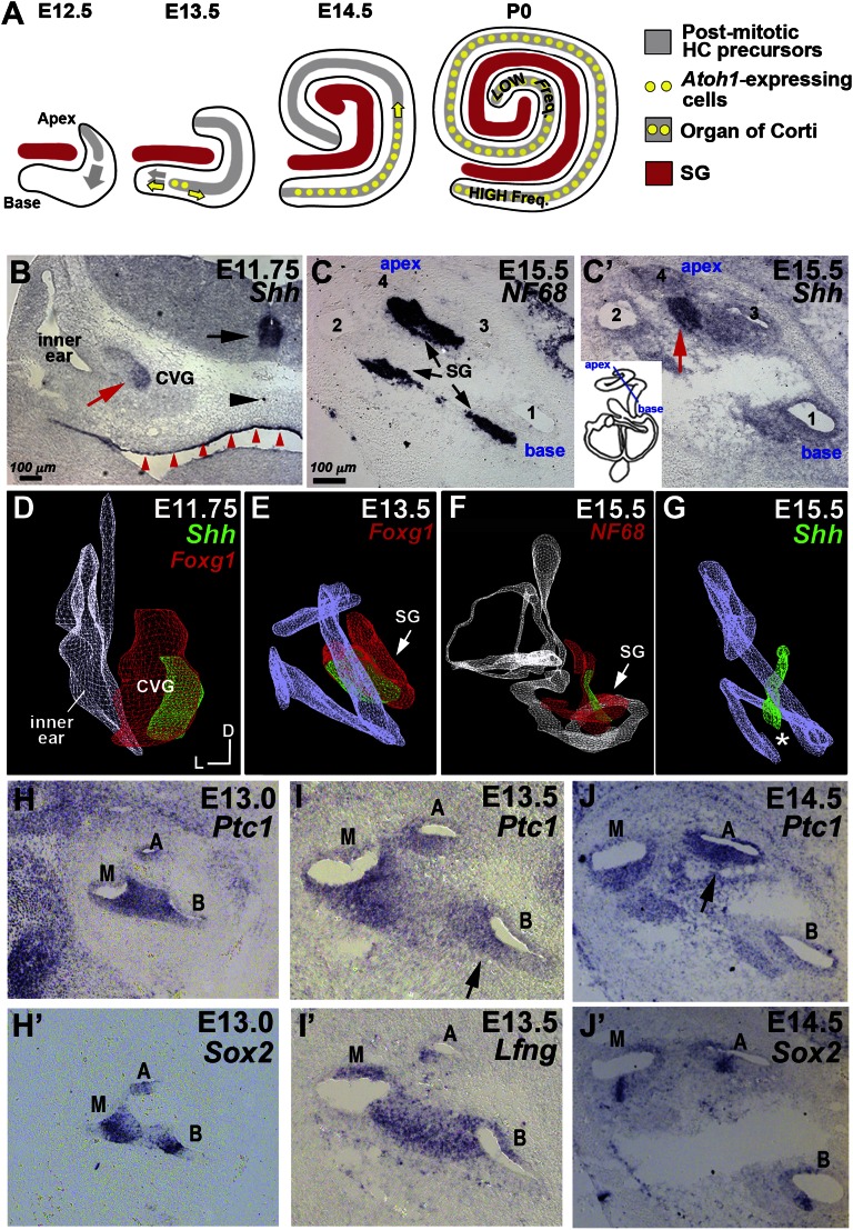
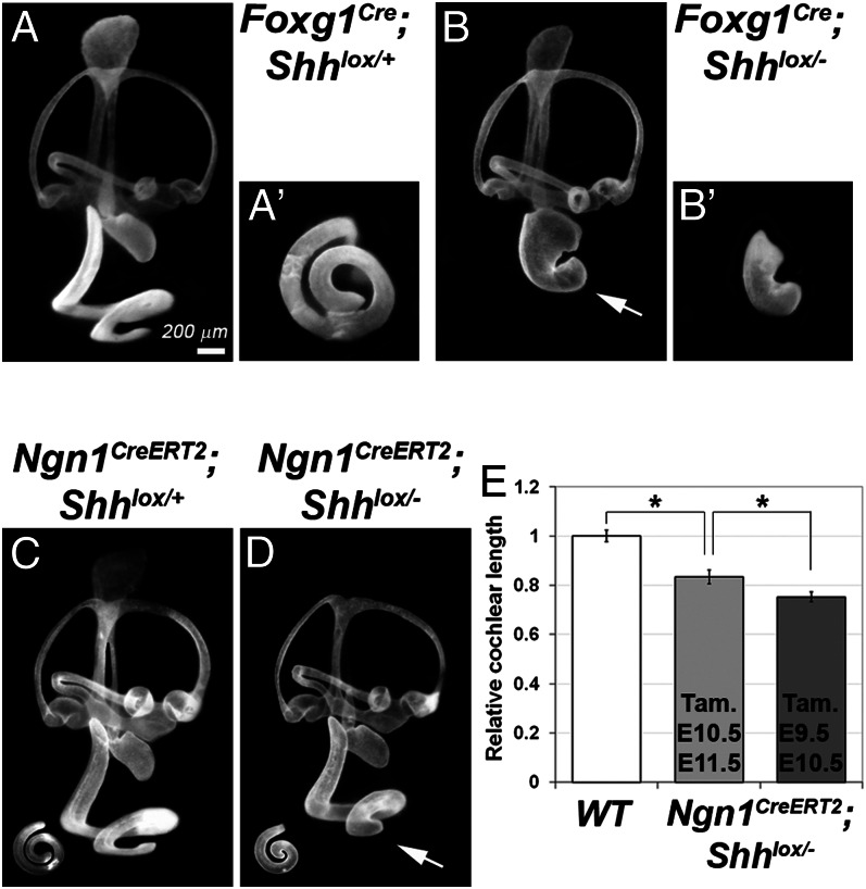
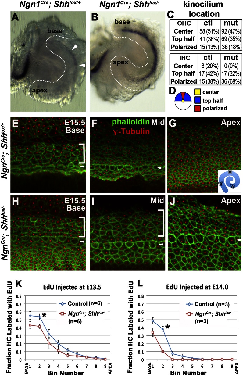
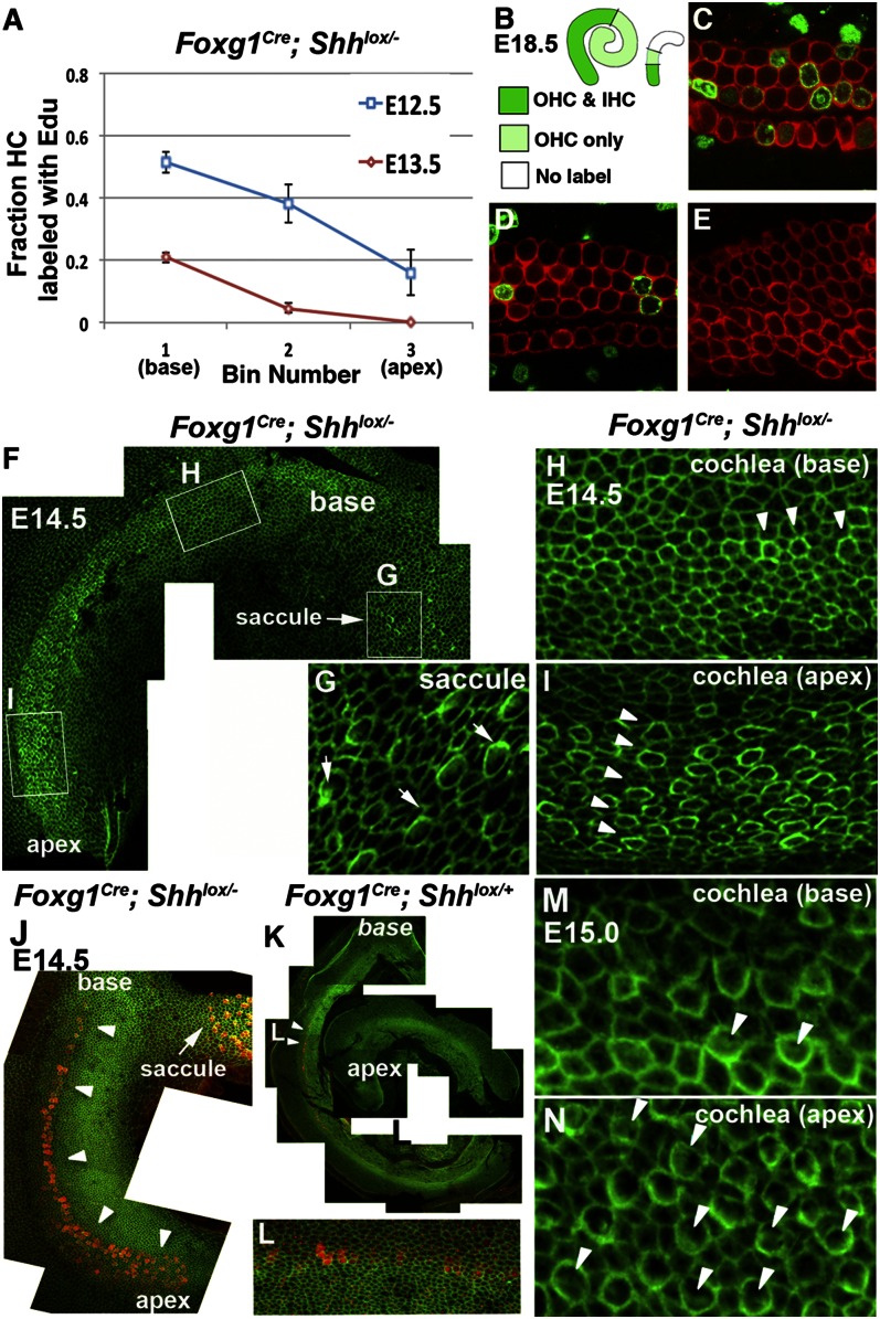
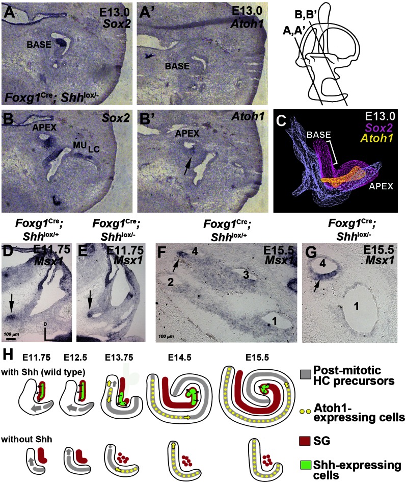
Neural precursor cells of the central nervous system undergo successive temporal waves of terminal division, each of which is soon followed by the onset of cell differentiation. The organ of Corti in the mammalian cochlea develops differently, such that precursors at the apex are the first to exit from the cell cycle but the last to begin differentiating as mechanosensory hair cells. Using a tissue-specific knockout approach in mice, we show that this unique temporal pattern of sensory cell development requires that the adjacent auditory (spiral) ganglion serve as a source of the signaling molecule Sonic hedgehog (Shh). In the absence of this signaling, the cochlear duct is shortened, sensory hair cell precursors exit from the cell cycle prematurely, and hair cell differentiation closely follows cell cycle exit in a similar apical-to-basal direction. The dynamic relationship between the restriction of Shh expression in the developing spiral ganglion and its proximity to regions of the growing cochlear duct dictates the timing of terminal mitosis of hair cell precursors and their subsequent differentiation.

Keywords: Atoh1; morphogenesis; tonotopy.


Conflict of interest statement

The authors declare no conflict of interest.

Figures

Fig. 1.
Fig. 1.
Expression of Shh in the developing spiral ganglion and Ptc1 in the cochlear duct. (A) In mouse cochlea, HC precursors at the apex start exiting from the cell cycle at E12.5, whereas HC differentiation starts at the midbase at E13.5 and progresses bidirectionally along the duct. (B) Shh is expressed in the CVG (red arrow), floor plate (black arrow), notochord (black arrowhead), and pharyngeal endoderm (red arrowheads). (C and C′) Adjacent cochlear sections showing Shh expression in a subset of neurofilament-positive (NF68) spiral ganglion (SG) neurons (black arrows) associated with the apical turn of the cochlear duct (red arrow). (D–G) Posterior views of 3D images of inner ears (D and F) and cochleae (E and G). Shh expression (green) in the Foxg1+ (D and E) or NF68+ (F) spiral ganglion (red) was gradually restricted by the apex of the cochlear duct over time (asterisk). (H–J′) Ptc1 expression (n = 10) is weak in the apex of the Sox2+ prosensory domain at E13 (H and H′), reduced at the basal prosensory region at E13.5 (I and I′, arrow), and strongest at the apex (arrow) by E14.5 (J and J′, arrow). A, apex; B, base; M, mid, D, dorsal; L, lateral.
Fig. 2.
Fig. 2.
Shortened cochlea in Foxg1Cre; Shhlox/ and Ngn1CreERT2; Shhlox/ mutants. (A–D) Paint-filled inner ears of E15.5 Foxg1Cre; Shhlox/+ (A), Foxg1Cre; Shhlox/ (B), Ngn1CreER; Shhlox/+ (C), and Ngn1CreER; Shhlox/ (D) embryos. (A′ and B′) Ventral view of the cochlea. In B and D, the arrow points to the shortened cochlea. (E) The length of the Ngn1CreER; Shhlox/- cochlear duct is dependent on whether tamoxifen (Tam) is administered at E10.5 and E11.5 (light gray) or at E9.5 and E10.5 (dark gray). *P < 0.0001. The scale bar in A applies to B–D as well.
Fig. 3.
Fig. 3.
Premature Atoh1 expression and HC development but mild premature CCE in Ngn1Cre; Shhlox/ cochlea. (A and B) Whole-mount in situ hybridization of Ngn1Cre; Shhlox/+ (A) and Ngn1Cre; Shhlox/ (B) cochlea at E13.5. Atoh1 is expressed at the midbase of the Ngn1Cre; Shhlox/+ cochlea (arrowheads), whereas its expression is strongly up-regulated in the Ngn1Cre; Shhlox/ cochlea (n = 4). (C) Table presenting the number and percentages (brackets) of HCs with kinocilium (anti–γ-tubulin antibody) in indicated locations (defined in D) of control (E) and mutant (H) basal cochleae (bin 2). Nascent HCs initially display a centrally located kinocilium (yellow) that later migrates through the top half of the cell (blue) before arriving at its final position at the lateral edge (red). More HCs with polarized kinocilia are seen in Ngn1Cre; Shhlox/ cochleae (H) compared with control cochleae (E) (OHC, P = 0.015; IHC, P = 0.002; χ2 test with Yates’ correction). (E–J) Luminal surface of Ngn1Cre; Shhlox/+ (E–G) and Ngn1Cre; Shhlox/ (H–J) cochleae at the basal (E and H), mid (F and I), and apical (G and J) regions of the organ of Corti showing stereocilia labeled with phalloidin (green) and basal bodies labeled with anti–γ-tubulin (red; n = 3). Arrowheads and brackets point to cortical condensation of IHC and OHC, respectively. (K and L) Comparison of fraction of EdU-labeled HCs in Ngn1Cre; Shhlox/+ and Ngn1Cre; Shhlox/ cochleae that were injected with EdU at E13.5 (K) and E14 (L). *P < 0.05. Error bars represent SEM.
Fig. 4.
Fig. 4.
EdU analysis and HC maturity of Foxg1Cre; Shhlox/ cochlea. (A) Comparison of fraction of HCs labeled with EdU at the base, mid, and apex of E18.5 Foxg1Cre; Shhlox/ cochleae that were injected with EdU at E12.5 and E13.5. (B) Traced confocal images of EdU labeling of control (Left, mean length 3,517 ± 262.89 μm; n = 7) and Foxg1Cre; Shhlox/ (Right, mean length 678 ± 318.11 μm; n = 4) E18.5 cochlea after EdU injection at E13.5. Dark-green, light-green, and white regions represent EdU labeling in both IHCs and OHCs (C), in OHCs only (D), and in neither type of HCs (E), respectively. (F–I) Whole-mount preparation of an E14.5 Foxg1Cre; Shhlox/ mutant cochlea labeled with phalloidin (green) showing more HCs with stronger actin condensation in the apex (I) than the base (H, arrowheads; n = 4). Also shown is the saccule displaying many distinct stereocilia-containing HCs (G, arrows). (M and N) More mature HCs with stereocilia are observed in the apex (N) than in the base (M) of Foxg1Cre; Shhlox/ cochleae at E15.5 (arrowheads; n = 2). (J–L) Double-labeling of E14.5 Foxg1Cre; Shhlox/ cochlea (J) with phalloidin (green) and anti-myosin VI antibodies (red) shows stronger myosin VI staining at the apex than at the base (arrowheads), whereas only weak myosin VI immunoreactivity is detectable at the midbase of control cochlea (n = 2). A similar pattern is observed with anti-myosin VII staining.
Fig. 5.
Fig. 5.
Expression patterns of Atoh1 and Msx1 in Foxg1Cre; Shhlox/ cochlea. (A–C) Atoh1 expression of Foxg1Cre; Shhlox/ cochlea at E13. Adjacent sections were probed for Sox2 (A and B) and Atoh1 (A′ and B′) at the base (A and A′) and apex (B and B′) of the cochlea. Atoh1 expression was detected only at the apex within the Sox2+ prosensory domain (B′, arrow). LC, lateral crista; MU, macula utricle. (C) A 3D image of Sox2 (pink) and Atoh1 (yellow) expression domains in Foxg1Cre; Shhlox/ cochlea, showing a lack of Atoh1 expression at the base of the Sox2+ prosensory domain (bracket). (D–G) Msx1 expression in the ventral tip of the inner ear at E11.75 (D and E, arrow) and apex of the cochlear duct at E15.5 (F and G, arrow) of control (D and F) and Foxg1Cre; Shhlox/− mutant (E and G). (H) Summary diagram of WT cochlea (Upper) showing that Shh, produced in a subpopulation (green) of the spiral ganglion (red), promotes growth of the cochlear duct and inhibition of HC differentiation. As the tip of the cochlear duct extends beyond the reach of Shh, prosensory cells start to exit from cell cycle in that location, after which CCE progresses toward the base. As the cochlear duct continues to grow, Shh expression and signaling become restricted toward the apex of the cochlear duct, which allows initiation of HC differentiation at the midbasal region. The lack of Shh causes poor spiral ganglion development and premature CCE, followed promptly by HC differentiation (Lower).

References

    1. Corwin JT, Warchol ME. Auditory hair cells: Structure, function, development, and regeneration. Annu Rev Neurosci. 1991;14:301–333. - PubMed
    1. Lim DJ. Cochlear anatomy related to cochlear micromechanics: A review. J Acoust Soc Am. 1980;67(5):1686–1695. - PubMed
    1. Mann ZF, Kelley MW. Development of tonotopy in the auditory periphery. Hear Res. 2011;276(1-2):2–15. - PubMed
    1. Lee YS, Liu F, Segil N. A morphogenetic wave of p27Kip1 transcription directs cell cycle exit during organ of Corti development. Development. 2006;133(15):2817–2826. - PubMed
    1. Ruben RJ. Development of the inner ear of the mouse: A radioautographic study of terminal mitoses. Acta Otolaryngol. 1967;220 220, 1–44. - PubMed

Publication types

MeSH terms