. 2013 Sep 13;288(37):26775-84.
doi: 10.1074/jbc.M113.483743. Epub 2013 Aug 5.

Protein phosphatase 2A enables expression of interleukin 17 (IL-17) through chromatin remodeling

Affiliations

Protein phosphatase 2A enables expression of interleukin 17 (IL-17) through chromatin remodeling

Sokratis A Apostolidis et al. J Biol Chem. .

Abstract

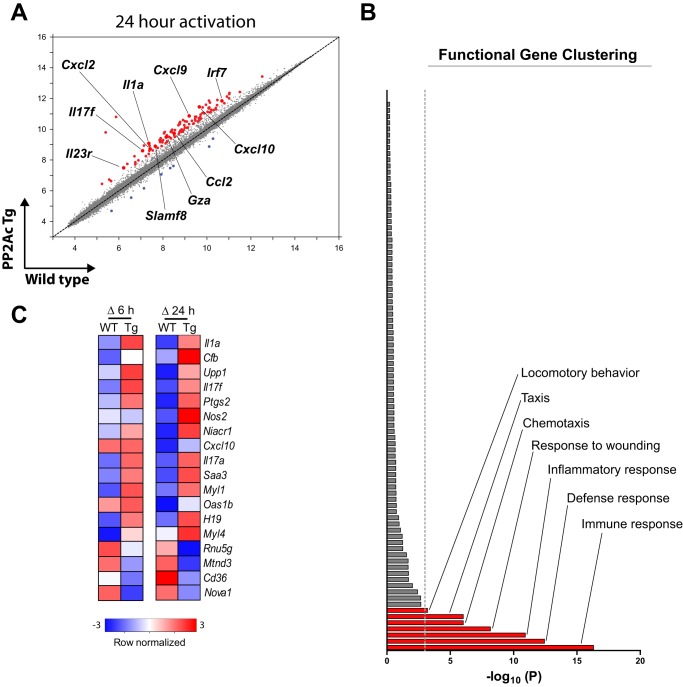
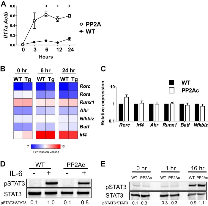
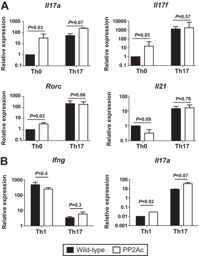
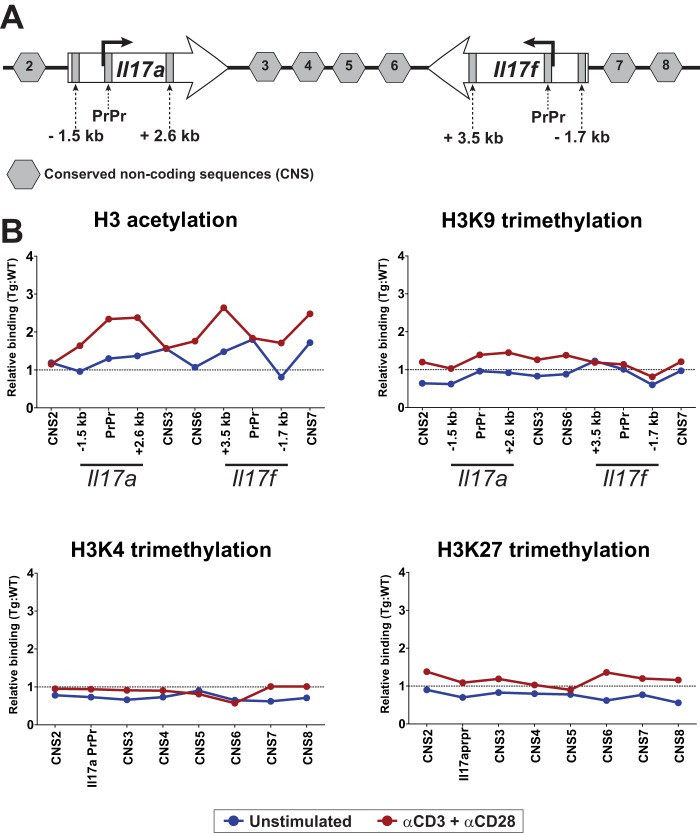
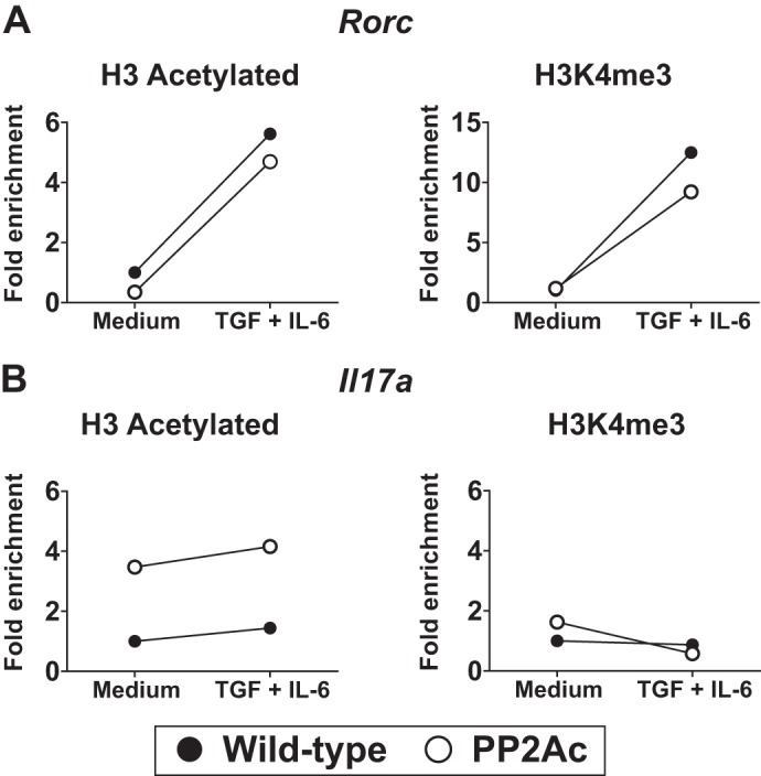
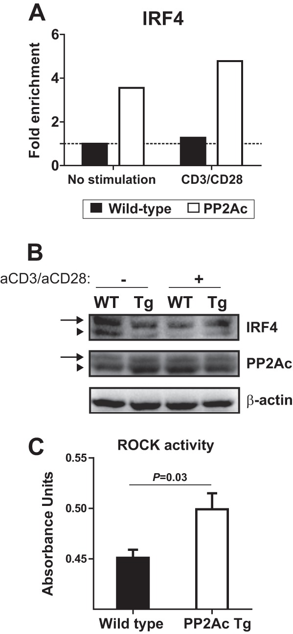
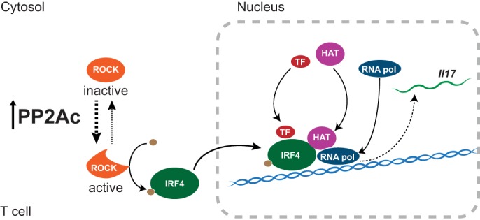
Protein phosphatase 2A (PP2A) is a heterotrimeric serine/threonine phosphatase involved in essential cellular functions. T cells from patients with systemic lupus erythematosus (SLE) express high levels of the catalytic subunit of PP2A (PP2Ac). A mouse overexpressing PP2Ac in T cells develops glomerulonephritis in an IL-17-dependent manner. Here, using microarray analyses, we demonstrate that increased expression of PP2Ac grants T cells the capacity to produce an array of proinflammatory effector molecules. Because IL-17 is important in the expression of glomerulonephritis, we studied the mechanism through which PP2Ac dysregulation facilitates its production. We report that PP2Ac is involved in the regulation of the Il17 locus by enhancing histone 3 acetylation through a mechanism that involves activation of interferon regulatory factor 4. Increased histone 3 acetylation of the Il17 locus is shared between T cells of PP2Ac transgenic mice and patients with SLE. We propose that, by promoting the inflammatory capacity of T cells, PP2Ac dysregulation contributes to the pathogenesis of SLE.

Keywords: Autoimmunity; Chromatin Histone Modification; Inflammation; Interleukin; PP2A; T Cell.


Figures

FIGURE 1.
FIGURE 1.
PP2Ac dysregulation induces a proinflammatory transcriptional profile in CD4 T cells. A, comparison of microarray expression values in naïve CD4 T cells from PP2Ac transgenic and wild-type mice 24 h after activation with anti-CD3 and anti-CD28. Red dots indicate genes up-regulated ≥ 2-fold in transgenic mice. Blue dots indicate genes down-regulated ≥ 2-fold. Some genes involved in the inflammatory response are highlighted. B, functional gene clustering analysis showing that the set of genes up-regulated in the PP2Ac transgenic mice is enriched in genes involved in cell movement and inflammation. The dotted gray line indicates statistical significance after p value correction (p ≤ 0.001). C, microarray expression values were normalized to unstimulated cells, and the magnitude of their increase upon 6 and 24 h of T cell activation (Δ 6 h and Δ 24 h, respectively) was compared between transgenic (Tg) and wild-type mice. Transcripts whose activation-induced regulation differed ≥ 2-fold in transgenic and wild-type CD4 T cells were included in the heat maps.
FIGURE 2.
FIGURE 2.
PP2Ac does not induce Th17-associated transcription factors. A, naïve CD4 T cells from WT or PP2Ac transgenic mice were stimulated for the indicated time periods. Il17a mRNA was normalized to Actb levels (mean ± S.E.). *, p ≤ 0.01. B, microarray expression values of the indicated genes are shown. C, naïve CD4 T cells were stimulated overnight. Expression of the indicated genes was normalized to Actb. Shown is the fold expression relative to WT cells (mean ± S.E.). D, naïve CD4 T cells were stimulated during 48 h, expanded with IL-2 during 5 days, rested 48 h, washed and incubated with IL-6 for 3 h. Cells were lysed in radioimmune precipitation assay buffer in the presence of protease and phosphatase inhibitors. E, naïve CD4 T cells were stimulated with anti-CD3 and anti-CD28 and lysed at the indicated time points. Data are representative of at least four experiments, each with ≥ 3 mice/group (A and C), or ≥ 2 experiments, each performed with cells pooled from ≥ 3 mice (D and E).
FIGURE 3.
FIGURE 3.
The PP2Ac effect is independent of T cell effector differentiation. A, naïve CD4 T cells were stimulated in neutral (Th0) or Th17-inducing conditions (TGF-β1 and IL-6). At day 5, expression of the indicated genes was analyzed by real-time PCR. Results were normalized to Actb. Shown is the fold expression relative to WT Th0 cells (mean ± S.E.). B, naïve CD4 T cells were stimulated in Th1- (IL-12) or Th17-polarizing conditions. After 5 days, expression of Ifng and Il17a was analyzed by real-time PCR. Results are expressed as fold expression relative to Actb. Data are representative of four (A) or three (B) experiments, each with three mice per group.
FIGURE 4.
FIGURE 4.
PP2Ac increases chromatin accessibility at the Il17 locus. A, the mouse Il17 locus is depicted. The conserved non-coding sequences as well as the sites within the Il17a and Il17f genes that were amplified during the ChIP experiments are indicated. B, naïve CD4 T cells were stimulated and fixed, and ChIP was performed for acetylated H3, H3K9me3, H3K4me3, and H3K27me3. Shown is relative binding (PP2Ac:WT ratio) in resting (blue) or stimulated (red) cells. Data were normalized to the corresponding input DNA and are representative of three or four independent experiments, each with ≥ 3 mice/group. PrPr, proximal promoter.
FIGURE 5.
FIGURE 5.
Increased PP2Ac levels induce H3 acetylation in the Il17 locus in a constitutive manner. A and B, naïve CD4 T cells were stimulated in serum-free medium in the absence or presence of TGF-β and IL-6. After 18 h, the promoter regions of the Rorc and Il17a genes were analyzed by ChIP assays using anti-H3 acetylated or anti-H3K4me3 antibodies. Results were normalized against the input and are expressed as fold change over the WT stimulated in culture medium alone.
FIGURE 6.
FIGURE 6.
PP2Ac promotes IRF4 activation and recruitment to the Il17a locus. A, ChIP experiments were performed to analyze IRF4 occupancy of the Il17a promoter region in naïve CD4 T cells before and after stimulation with anti-CD3 and anti-CD28. Results were normalized against the input and are expressed as fold change over the unstimulated WT cells. B, naïve CD4 T cells were isolated from PP2Ac Tg and WT mice and were lysed before or after activation with anti-CD3 and anti-CD28 (1 h). Lysates were separated in a SuperSep Phos-tag acrylamide gel that retards the migration of phosphorylated proteins. Proteins were transferred to a PVDF membrane that was blotted with anti-IRF4, anti-PP2Ac, and anti-β-actin. Arrows indicate the phosphorylated proteins. Arrowheads indicate unphosphorylated proteins. C, cell lysates of stimulated naïve CD4 T cells were probed for Rho kinase activity.
FIGURE 7.
FIGURE 7.
PP2Ac promotes IL-17 production by facilitating IRF4 activity. Increased levels of PP2Ac promote Rho kinase (ROCK) activation. Activated ROCK phosphorylates IRF4, which then localizes to the Il17 locus. There, IRF4 recruits histone acetyltransferases (HAT), as well as other transcription factors (TF) and RNA polymerase (RNA pol) to initiate transcription of Il17.

References

    1. Janssens V., Goris J. (2001) Protein phosphatase 2A: a highly regulated family of serine/threonine phosphatases implicated in cell growth and signalling. Biochem. J. 353, 417–439 - PMC - PubMed
    1. Xu Y., Xing Y., Chen Y., Chao Y., Lin Z., Fan E., Yu J. W., Strack S., Jeffrey P. D., Shi Y. (2006) Structure of the protein phosphatase 2A holoenzyme. Cell 127, 1239–1251 - PubMed
    1. Cho U. S., Xu W. (2007) Crystal structure of a protein phosphatase 2A heterotrimeric holoenzyme. Nature 445, 53–57 - PubMed
    1. Janssens V., Longin S., Goris J. (2008) PP2A holoenzyme assembly. In cauda venenum (the sting is in the tail). Trends Biochem. Sci. 33, 113–121 - PubMed
    1. Lee T. Y., Lai T. Y., Lin S. C., Wu C. W., Ni I. F., Yang Y. S., Hung L. Y., Law B. K., Chiang C. W. (2010) The B56γ3 regulatory subunit of protein phosphatase 2A (PP2A) regulates S phase-specific nuclear accumulation of PP2A and the G1 to S transition. J. Biol. Chem. 285, 21567–21580 - PMC - PubMed

Publication types

Associated data