Unusual branching pattern of axillary artery associated with the high origin of ulnar artery
- PMID: 23919202
- PMCID: PMC3728875
- DOI: 10.4103/2141-9248.113674
Unusual branching pattern of axillary artery associated with the high origin of ulnar artery
Abstract
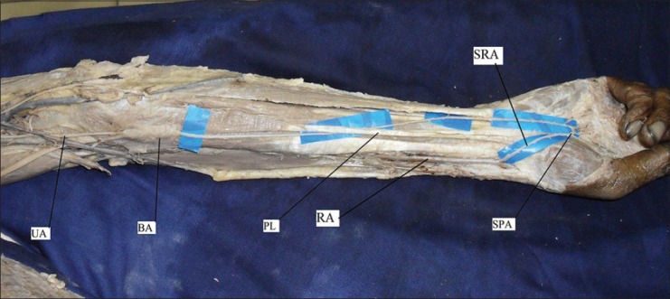
Axillary artery is a continuation of subclavian artery, extending from the outer border of first rib to the lower border of teres major muscle. During routine dissection for the undergraduate medical students, a rare variations was seen in an approximately 55-year-old male cadaver. This case showed a variation in branching pattern of right axillary and subscapular arteries. The subscapular artery originated from 2(nd) part of axillary artery, gave origin to posterior circumflex humeral and lateral thoracic arteries in addition to its normal branches. The ulnar artery originated from the 3(rd) part of the axillary artery, just above the lower border of teres major muscle. The variant ulnar artery passed deep to the median cubital vein, bicipital aponeurosis, and tendon of palmaris longus muscle. Then, it passed superficial to flexor digitorum superficialis muscle and flexor retinaculum to enter the palm. In the palm, it formed the superficial palmar arch. This variant ulnar artery was much smaller in caliber than the radial artery.
Keywords: Axillary artery; Palmar arches; Subscapular artery; Ulnar artery; Variations.
Conflict of interest statement
Figures


References
-
- George BM, Nayak S, Pramod K. Clinically significant neurovascular variations in the axilla and the arm – a case report. Neuroanatomy. 2007;6:36–8.
-
- Williams PL, Warwick R, Dyson M, Bannister LH, editors. 37th ed. London, England: Churchill Livingstone; 1989. Gray's anatomy; pp. 756–64.
-
- Bhat KM, Potu BK, Gowda S. High origin of ulnar artery in south Indian male cadaver: A case report. Rom J Morphol Embryol. 2008;49:573–5. - PubMed
-
- Vollala VR, Jetti R, Soni S. High origin of an ulnar artery – development and surgical significance. Chang Gung Med J. 2011;34:39–42. - PubMed
-
- Gaur S, Katariya SK, Vaishnani H, Wani IN, Bondre KV, Shah GV. A cadaveric study of branching pattern of the axillary artery. Int J Biol Med Res. 2012;3:1388–91.
Publication types
LinkOut - more resources
Full Text Sources
Other Literature Sources
Miscellaneous