. 2014 Feb;13(1):39-48.
doi: 10.1111/acel.12147. Epub 2013 Sep 19.

Mitochondrial energetics is impaired in vivo in aged skeletal muscle

Affiliations

Mitochondrial energetics is impaired in vivo in aged skeletal muscle

Gilles Gouspillou et al. Aging Cell. 2014 Feb.

Abstract

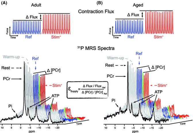
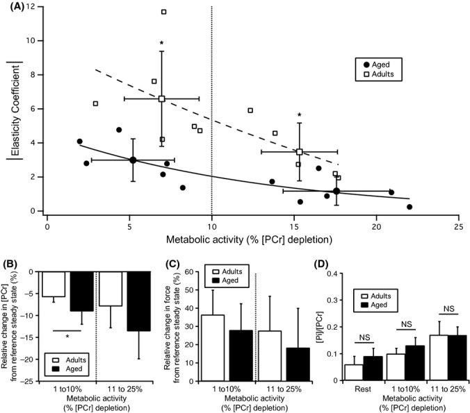
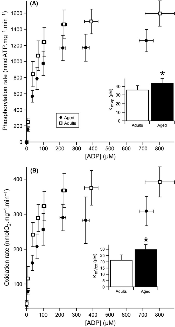
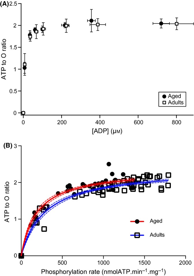
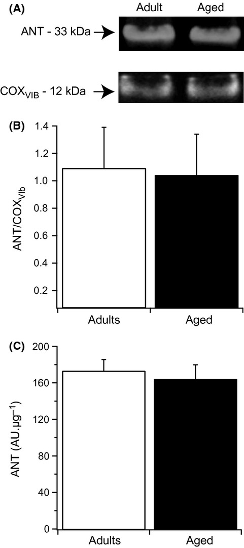
With aging, most skeletal muscles undergo a progressive loss of mass and strength, a process termed sarcopenia. Aging-related defects in mitochondrial energetics have been proposed to be causally involved in sarcopenia. However, changes in muscle mitochondrial oxidative phosphorylation with aging remain a highly controversial issue, creating a pressing need for integrative approaches to determine whether mitochondrial bioenergetics are impaired in aged skeletal muscle. To address this issue, mitochondrial bioenergetics was first investigated in vivo in the gastrocnemius muscle of adult (6 months) and aged (21 months) male Wistar rats by combining a modular control analysis approach with (31) P magnetic resonance spectroscopy measurements of energetic metabolites. Using this innovative approach, we revealed that the in vivo responsiveness ('elasticity') of mitochondrial oxidative phosphorylation to contraction-induced increase in ATP demand is significantly reduced in aged skeletal muscle, a reduction especially pronounced under low contractile activities. In line with this in vivo aging-related defect in mitochondrial energetics, we found that the mitochondrial affinity for ADP is significantly decreased in mitochondria isolated from aged skeletal muscle. Collectively, the results of this study demonstrate that mitochondrial bioenergetics are effectively altered in vivo in aged skeletal muscle and provide a novel cellular basis for this phenomenon.

Keywords: adenosine nucleotide translocator; affinity for ADP; energetics; in vivo; mitochondria; skeletal muscle aging.


Figures

Figure 1
Figure 1
Typical phosphocreatine and contraction records carried out to calculate the elasticity of the energy-supply module in adult (A) and aged (B) animals. Muscle contraction and 31P MRS spectra were recorded simultaneously during each experiment. Magnetic resonance spectra were analyzed to assess steady values of energetic intermediates. First, an MRS spectrum was recorded at rest (black spectrum) to determine the basal level of energetic intermediates. The intensity of stimulation was then progressively increased (warm-up phase, gray spectra), to place the gastrocnemius muscle at an intensity of contraction corresponding to a reference steady state (Ref; spectra & contraction activity in blue). The determination of the energy-supply module elasticity was then performed by increasing the intensity of stimulation (Stim+; spectra & contraction activity in red). The green spectrum corresponds to a transition spectrum. The magnitude of the contraction signal was averaged over the corresponding period of time. Relative changes in contraction and [PCr] from the Ref to Stim+ steady states provided all the required data for the calculation of the energy-supply module elasticity. εSupply, elasticity of the energy-supply module; Pi, inorganic phosphate; ATP, adenosine triphosphate; PCr, phosphocreatine.
Figure 2
Figure 2
Effect of aging on the elasticity of the energy-supply module and on the PCr and Pi content in the gastrocnemius muscle. (A) The elasticity of the energy-supply module was determined under contraction intensities that elicited metabolic activities ranging from 2 to 25% depletion of [PCr] determined at rest. Each value presented in this figure corresponds to the elasticity of the energy-supply module determined in a single animal. Changes in elasticity as a function of metabolic activity were approximated by a mono-exponential function. (B) Relative change in [PCr] induced by the increase in stimulation intensity from reference steady state. (C) Relative change in force induced by the increase in stimulation intensity from reference steady state. (D) The [Pi]/[PCr] ratio determined at rest and under the different metabolic activity ranges corresponding to the reference steady states we studied was similar between adult and aged rats. Values are expressed as mean ± SD (n = 11 for adult and n = 12 for aged rats). *P < 0.05 vs. adult group.
Figure 3
Figure 3
Changes in intramuscular pH during experimental determination of the energy-supply module elasticity. The chemical shift of Pi relative to PCr was used to assess the evolution of intramuscular pH during the experimental determination of the energy-supply elasticity. The different areas defined in this fig. correspond to the successive steps required for the experimental determination of the energy-supply module elasticity. Values are presented as mean ± SD (n = 11 for adult and n = 12 for aged rats).
Figure 4
Figure 4
Dependence of oxidation and phosphorylation rates on ADP concentration in adult (A) and aged (B) gastrocnemius muscle mitochondria. Oxidation and phosphorylation rates were recorded simultaneously in mitochondria isolated from adult (n = 7) and aged (n = 5) muscles oxidizing glutamate + malate + succinate as substrates. Data were fitted using the Michaelis–Menten equation. Km for ADP was determined from both oxidation (KmVox) and phosphorylation (KmVp) rates. Data are presented as mean ± SD. *P < 0.05 vs. adult group.
Figure 5
Figure 5
Changes in the ATP to O ratio as a function of ADP concentration and phosphorylation rate in adult and aged mitochondria. ATP to O ratio was determined by calculating the ratio of phosphorylation to oxidation rates. ATP to O ratios obtained from young and aged mitochondria were plotted as a function of (A) ADP concentration or (B) phosphorylation rates. Data for adult (n = 7) and aged (n = 5) rats are presented as mean ± SD in panel (A). Thick red (aged) and dashed blue (adults) curves in panel B were obtained by fitting data using the Michaelis–Menten equation. Thin dashes red (aged) and blue (adults) curved in panel B represent the 95% confidence interval of each curve fit.
Figure 6
Figure 6
Effect of aging on mitochondrial ANT content. (A) Representative Western blot of ANT and cytochrome c oxidase obtained by the study of mitochondria isolated from adult and aged gastrocnemius muscle. (B) ANT content normalized cytochrome c oxidase content. (C) ANT content normalized to the mitochondrial protein quantity used for Western blot (10 μg). Data presented in (B) and (C) are expressed as mean ± SD (n = 4 per group).

References

    1. Arsac LM, Beuste C, Miraux S, Deschodt-Arsac V, Thiaudiere E, Franconi JM, Diolez PH. In vivo modular control analysis of energy metabolism in contracting skeletal muscle. Biochem. J. 2008;414:391–397. - PubMed
    1. Bastien C, Sanchez J. Phosphagens and glycogen content in skeletal muscle after treadmill training in young and old rats. Eur. J. Appl. Physiol. Occup. Physiol. 1984;52:291–295. - PubMed
    1. Brand MD. Top down metabolic control analysis. J. Theor. Biol. 1996;182:351–360. - PubMed
    1. Brand MD, Curtis RK. Simplifying metabolic complexity. Biochem. Soc. Trans. 2002;30:25–30. - PubMed
    1. Bratic I, Trifunovic A. Mitochondrial energy metabolism and ageing. Biochim. Biophys. Acta. 2010;1797:961–967. - PubMed