. 2014 Feb;13(1):49-59.
doi: 10.1111/acel.12148. Epub 2013 Sep 18.

Calpain cleavage and inactivation of the sodium calcium exchanger-3 occur downstream of Aβ in Alzheimer's disease

Affiliations

Calpain cleavage and inactivation of the sodium calcium exchanger-3 occur downstream of Aβ in Alzheimer's disease

Joe Atherton et al. Aging Cell. 2014 Feb.

Abstract

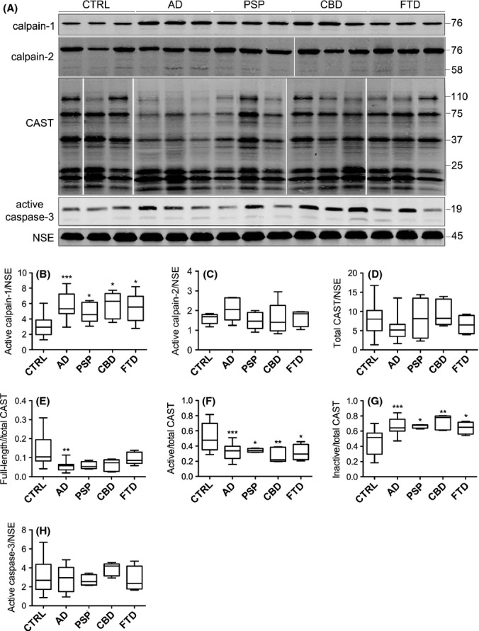
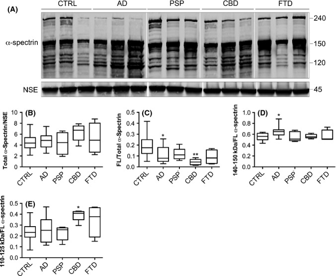
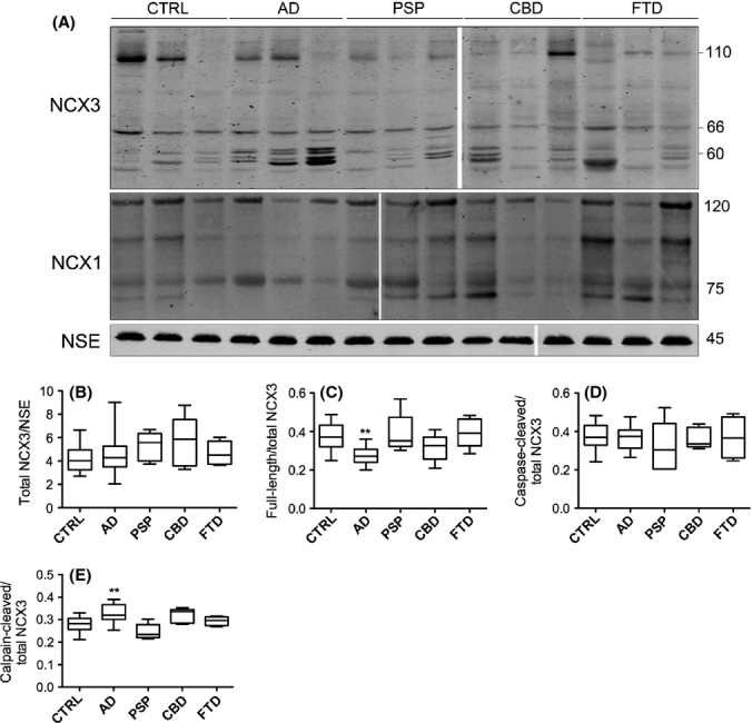
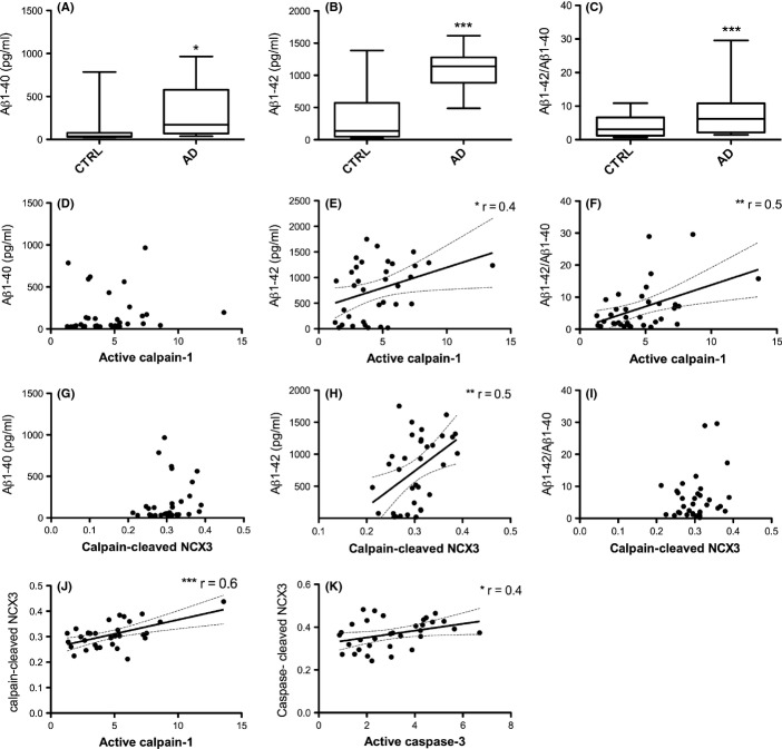
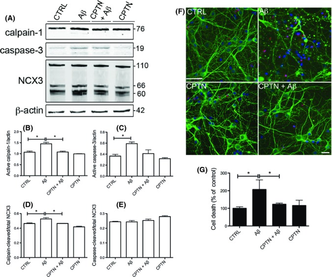
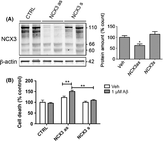
Alzheimer's disease (AD) is a neurodegenerative disorder characterized by pathological deposits of β-amyloid (Aβ) in senile plaques, intracellular neurofibrillary tangles (NFTs) comprising hyperphosphorylated aggregated tau, synaptic dysfunction and neuronal death. Substantial evidence indicates that disrupted neuronal calcium homeostasis is an early event in AD that could mediate synaptic dysfunction and neuronal toxicity. Sodium calcium exchangers (NCXs) play important roles in regulating intracellular calcium, and accumulating data suggests that reduced NCX function, following aberrant proteolytic cleavage of these exchangers, may contribute to neurodegeneration. Here, we show that elevated calpain, but not caspase-3, activity is a prominent feature of AD brain. In addition, we observe increased calpain-mediated cleavage of NCX3, but not a related family member NCX1, in AD brain relative to unaffected tissue and that from other neurodegenerative conditions. Moreover, the extent of NCX3 proteolysis correlated significantly with amounts of Aβ1-42. We also show that exposure of primary cortical neurons to oligomeric Aβ1-42 results in calpain-dependent cleavage of NCX3, and we demonstrate that loss of NCX3 function is associated with Aβ toxicity. Our findings suggest that Aβ mediates calpain cleavage of NCX3 in AD brain and therefore that reduced NCX3 activity could contribute to the sustained increases in intraneuronal calcium concentrations that are associated with synaptic and neuronal dysfunction in AD.

Keywords: Alzheimer's disease; beta-amyloid; calcium; calpain; sodium calcium exchanger; tau.


Figures

Figure 1
Figure 1
Calpain-1 active subunit amounts are elevated in neurodegenerative disease brain. (A) Representative immunoblots of cortical lysates from postmortem brain. Blots were probed with antibodies that detect active fragments of calpain-1 at 76 kDa, calpain-2 holoprotein and cleaved fragments at 76 and 58 kDa, respectively, and active caspase-3 at 17 and 19 kDa. An antibody against calpastatin (CAST) was used that detects active CAST holoprotein at 110 kDa, active fragments of > 25 kDa and inactive fragments of < 25 kDa. Blots were also probed with an antibody- to neuron-specific enolase (NSE), 45 kDa. Box plots show amounts of (B) active calpain-1, (C) active calpain-2 and (D) total CAST, all as a proportion of NSE. (E) Full length (FL) CAST, (F) active CAST and (G) inactive CAST, all as a proportion of total CAST, and (H) active caspase-3 following standardization to NSE content in each sample. CTRL: control (n = 20), AD: Alzheimer’s disease (n = 20), PSP: progressive supranuclear palsy (n = 5), CBD: corticobasal degeneration (n = 5), FTD: frontotemporal dementia with tau mutations (n = 4). *P < 0.05, **P < 0.005, ***P < 0.001.
Figure 2
Figure 2
Calpain- and caspase-mediated proteolysis of α-spectrin in neurodegenerative disease brain. (A) Representative immunoblot of cortical lysates from postmortem brain. Blots were probed with an antibody that detects α-spectrin holoprotein at approximately 240 kDa, calpain- and caspase-cleaved fragments at approximately 140–150 kDa and caspase-cleaved fragments at approximately 110–125 kDa. Blots were also probed with an antibody to neuron-specific enolase (NSE), 45 kDa. Box plots show quantification of (B) total α-spectrin following standardization to NSE amounts. (C) α-spectrin holoprotein, (D) 140- to 150-kDa α-spectrin fragments and (E) 110- to 125-kDa α-spectrin fragments, all quantified as a proportion of total α-spectrin. Mean ± standard error is shown. CTRL: control (n = 20), AD: Alzheimer’s disease (n = 20), PSP: progressive supranuclear palsy (n = 5), CBD: corticobasal degeneration (n = 5), FTD: frontotemporal dementia with tau mutations (n = 4). *P < 0.05, **P < 0.005.
Figure 3
Figure 3
Calpain-mediated cleavage of NCX3, but not NCX1, is increased in AD brain. (A) Representative immunoblots of cortical lysates from postmortem brain. Blots were probed with an antibody that detects NCX3 holoprotein at approximately 110 kDa, a caspase-3-cleaved fragment at approximately 66 kDa, calpain-cleaved fragments at approximately 60 kDa and an antibody against NCX1, which detects full-length NCX1 at 120 kDa and proteolytic fragments at approximately 70–80 kDa. Blots were also probed with an antibody to neuron-specific enolase (NSE), 45 kDa. Plots show quantification of (B) total NCX3 amounts when standardized to NSE, and (C) full-length NCX3, (D) caspase-3-cleaved NCX3 and (E) calpain-cleaved NCX3, all as a proportion of total NCX3. CTRL: control (n = 20), AD: Alzheimer’s disease (n = 20), PSP: progressive supranuclear palsy (n = 5), CBD: corticobasal degeneration (n = 5), FTD: frontotemporal dementia with tau mutations (n = 4). **P < 0.005.
Figure 4
Figure 4
NCX3 cleavage correlates with calpain-1 activity and Aβ burden. Aβ ELISAs were used to measure Aβ amounts in frontal cortex samples from AD and control (CTRL) brain. Box plots show (A) Aβ1–40, and (B) Aβ1–42amounts in pg mL−1, (C) shows the ratio of Aβ1–42 to Aβ1–40. Scatter plots show the correlation between amounts of active calpain-1 and (D) Aβ1–40, (E) Aβ1–42 and (F) Aβ1–42/Aβ1–40, and between calpain-cleaved NCX3 fragments and (G) Aβ1–40, (H) Aβ1–42, (I) Aβ1–42/Aβ1–40, and (J) active calpain-1, and (K) caspase-cleaved NCX-3 fragments with active caspase-3. Parametric Pearson correlation analysis was used to generate r values since all data were normally distributed, and statistical statistical significance is shown for each analysis. CTRL: control (n = 16), AD: Alzheimer’s disease (n = 16).
Figure 5
Figure 5
Aβ-induced calpain-mediated NCX3 cleavage is associated with neurotoxicity in primary cortical cultures. Rat primary cortical cultures were exposed to 10 μm Aβ1–42 (Aβ) for 48 h with and without pretreatment with 5 μm calpeptin (CPTN) for 1 h. (A) Representative Western blots probed with antibodies against active calpain-1 (76 kDa) and that pick up full-length (110 kDa), caspase-cleaved (66 kDa) and calpain-cleaved (60 kDa) NCX3 are shown. Blots were also probed with β-actin (42 kDa) as a loading control. Molecular weight markers are indicated (kDa). Bar charts show amounts of (B) active calpain-1 and (C) active caspase-3 as a proportion of β-actin in each sample. The amounts of (D) calpain-cleaved and (E) caspase-cleaved NCX3, both as a proportion of total NCX3, are also shown. (F) Representative images from cortical cultures incubated with live/dead cell dye (red), fixed and immunostained with an antibody against βIII-tubulin (green). Hoechst 33352 was used to stain nuclei (blue). Scale bar: 25 μm. (G) Lactate dehydrogenase (LDH) release from control and treated primary cortical cultures. Values represent LDH release from cells into the medium as a percentage of total LDH, presented as % control. Data are mean ± SEM. Experiments were performed in triplicate (n = 3). *P < 0.05.
Figure 6
Figure 6
Antisense knockdown of NCX3 sensitizes cortical neurons to Aβ. Rat primary cortical cultures were treated with sense (s) or antisense (as) oligonucleotides against rat NCX3 for 24 h and then treated with a subtoxic concentration (1 μm) of Aβ1–42 (Aβ) for a further 16 h. (A) Representative Western blots probed with antibodies that detect full-length (110 kDa) and cleaved (66, 60 kDa) NCX3. Blots were also probed with β-actin (42 kDa) as a loading control. Molecular weight markers are indicated. Bar charts show (A) total NCX3 expression following standardization to the β-actin loading control and (B) cell death as measured by live/dead cell assay in cells treated with 1 μm Aβ as a proportion of death in control cultures and antisense oligonucleotides 1 μm Aβ standardized to cells treated with sense NCX3 oligonucleotides only. Data are mean ± SEM (n = 3). *P < 0.05, **P < 0.005.

References

    1. Arispe N, Pollard HB, Rojas E. Giant multilevel cation channels formed by Alzheimer disease amyloid beta-protein [A beta P-(1-40)] in bilayer membranes. Proc. Natl Acad. Sci. USA. 1993;90:10573–10577. - PMC - PubMed
    1. Ayala-Grosso C, Tam J, Roy S, Xanthoudakis S, Da Costa D, Nicholson DW, Robertson GS. Caspase-3 cleaved spectrin colocalizes with neurofilament- immunoreactive neurons in Alzheimer’s disease. Neuroscience. 2006;141:863–874. - PubMed
    1. Bano D, Young KW, Guerin CJ, Lefeuvre R, Rothwell NJ, Naldini L, Rizzuto R, Carafoli E, Nicotera P. Cleavage of the plasma membrane Na+/Ca2+ exchanger in excitotoxicity. Cell. 2005;120:275–285. - PubMed
    1. Bano D, Munarriz E, Chen HL, Ziviani E, Lippi G, Young KW, Nicotera P. The plasma membrane Na+/Ca2+ exchanger is cleaved by distinct protease families in neuronal cell death. Ann. N. Y. Acad. Sci. 2007;1099:451–455. - PubMed
    1. Brustovetsky T, Bolshakov A, Brustovetsky N. Calpain activation and Na+/Ca2+ exchanger degradation occur downstream of calcium deregulation in hippocampal neurons exposed to excitotoxic glutamate. J. Neurosci. Res. 2010;88:1317–1328. - PMC - PubMed

Publication types

MeSH terms