

doi: 10.1161/STROKEAHA.113.001972.
Epub 2013 Aug 6.
Recommendations on angiographic revascularization grading standards for acute ischemic stroke: a consensus statement
Affiliations
- PMID: 23920012
- PMCID: PMC4160883
- DOI: 10.1161/STROKEAHA.113.001972
Item in Clipboard
Recommendations on angiographic revascularization grading standards for acute ischemic stroke: a consensus statement
Stroke.
2013 Sep.
No abstract available
Keywords: AOL; IA therapy; TICI; TIMI; embolectomy; endovascular therapy; grading; recanalization; revascularization; score; scoring; stent-retrievers; stroke; thrombectomy.
Figures
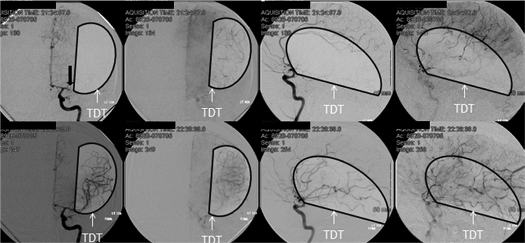
Example of thrombolysis in cerebral infarction (TICI) 0 and TICI 2b. Top, Anteroposterior (first 2 boxes) and lateral (last 2 boxes) in an early arterial and late capillary phases depicting TICI 0 at baseline. Bottom, Same phases depicting TICI 2b after intra-arterial therapy. Black arrow indicating the target arterial lesion (TAL): middle cerebral artery/M1 horizontal segment occlusion (TAL) distal to the lenticulostriate (LS). Black half circles approximate the target downstream territory (TDT; the presumed area supplied by the TAL).
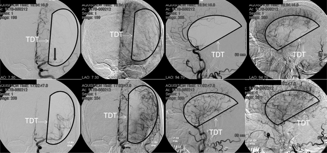
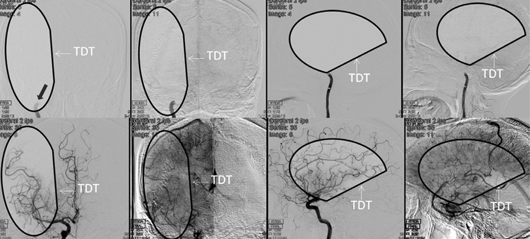
Example of thrombolysis in cerebral infarction (TICI) 1 and TICI 3. Top, Anteroposterior (left) and lateral (right) in an early arterial and late capillary phases depicting TICI 1 at baseline. Bottom, Same phases depicting TICI 3 after intraarterial therapy. Black arrow indicating the target arterial lesion (TAL): middle cerebral artery/M1 horizontal segment occlusion (TAL) distal to the lenticulostriate (LS). Black half circles approximate the target downstream territory (TDT; the presumed area supplied by the TAL). Ischemic arteriovenous shunting is noted with opacification of straight sinus (right bottom corner).
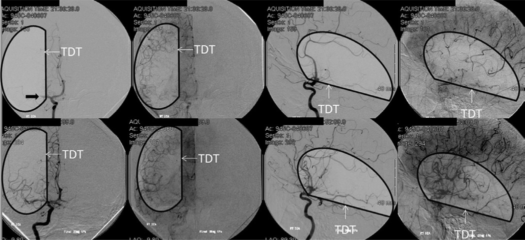
Example of thrombolysis in cerebral infarction (TICI) 0 and TICI 2a (compared with TICI 2b and 3 above). Top, Anteroposterior (first 2 boxes) and lateral (last 2 boxes) in an early arterial and late capillary phases depicting TICI 0 at baseline. Bottom, Same phases depicting TICI 2a after intra-arterial therapy. Black arrow indicating the target arterial lesion (TAL): middle cerebral artery/M1 horizontal segment occlusion (TAL) distal to the lenticulostriate (LS). Black half circles approximate the target downstream territory (TDT; the presumed area supplied by the TAL).
Example of thrombolysis in cerebral infarction (TICI) 0 and TICI 2a (compare with TICI 3 and 2b above). Top, Anteroposterior (first 2 boxes) and lateral (last 2 boxes) in an early arterial and late capillary phases depicting TICI 0 at baseline. Bottom, Same phases depicting TICI 2a after intra-arterial therapy. Black arrow indicating the target arterial lesion (TAL): middle cerebral artery/M1 horizontal segment occlusion (TAL) distal to the lenticulostriate (LS). Black half circles approximate the target downstream territory (TDT; the presumed area supplied by the TAL).
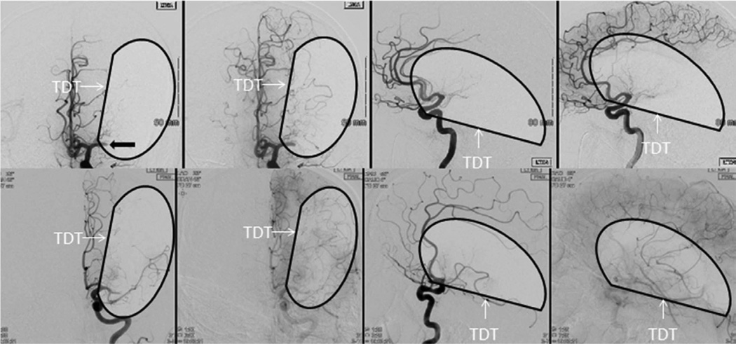
Example of downstream territory (TDT) for an ICA occlusion with pre–intra-arterial therapy (IAT) data from CT angiogram showing cross-filling from the contralateral anterior cerebral artery. Top, Anteroposterior (first 2 boxes) and lateral (last 2 boxes) in an early arterial and late capillary phases depicting thrombolysis in cerebral infarction (TICI) 0 at baseline. Bottom, Same phases depicting TICI 3 after IAT. Black arrow indicating the target arterial lesion (TAL): Distal ICA proximal to the ophthalmic artery. Black half circles approximate the target downstream territory (TDT; the presumed area supplied by the TAL). Early ischemic arteriovenous shunting is noted in the right lower corner.
References
-
- Saver JL, Jahan R, Levy EI, Jovin TG, Baxter B, Nogueira RG, et al. SWIFT Trialists. Solitaire flow restoration device versus the Merci Retriever in patients with acute ischaemic stroke (SWIFT): a randomised, parallel-group, non-inferiority trial. Lancet. 2012;380:1241–1249. - PubMed
Publication types
MeSH terms
Grants and funding
LinkOut - more resources
Full Text Sources
Other Literature Sources
Medical
