. 2013 Nov;110(3):222-30.
doi: 10.1016/j.ymgme.2013.06.020. Epub 2013 Jul 6.

Lethal phenotype in conditional late-onset arginase 1 deficiency in the mouse

Affiliations

Lethal phenotype in conditional late-onset arginase 1 deficiency in the mouse

Jennifer Kasten et al. Mol Genet Metab. 2013 Nov.

Abstract

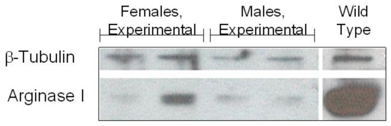
Human arginase deficiency is characterized by hyperargininemia and infrequent episodes of hyperammonemia, which lead to neurological impairment with spasticity, loss of ambulation, seizures, and severe mental and growth retardation; uncommonly, patients suffer early death from this disorder. In a murine targeted knockout model, onset of the phenotypic abnormality is heralded by weight loss at around day 15, and death occurs typically by postnatal day 17 with hyperargininemia and markedly elevated ammonia. This discrepancy between the more attenuated juvenile-onset human disease and the lethal neonatal murine model has remained suboptimal for studying and developing therapy for the more common presentation of arginase deficiency. These investigations aimed to address this issue by creating an adult conditional knockout mouse to determine whether later onset of arginase deficiency also resulted in lethality. Animal survival and ammonia levels, body weight, circulating amino acids, and tissue arginase levels were examined as outcome parameters after widespread Cre-recombinase activation in a conditional knockout model of arginase 1 deficiency. One hundred percent of adult female and 70% of adult male mice died an average of 21.0 and 21.6 days, respectively, after the initiation of tamoxifen administration. Animals demonstrated elevated circulating ammonia and arginine at the onset of phenotypic abnormalities. In addition, brain and liver amino acids demonstrated abnormalities. These studies demonstrate that (a) the absence of arginase in adult animals results in a disease profile (leading to death) similar to that of the targeted knockout and (b) the phenotypic abnormalities seen in the juvenile-onset model are not exclusive to the age of the animal but instead to the biochemistry of the disorder. This adult model will be useful for developing gene- and cell-based therapies for this disorder that will not be limited by the small animal size of neonatal therapy and for developing a better understanding of the characteristics of hyperargininemia.

Keywords: AAV; ANOVA; ARG; Animal model; Arginase deficiency; Conditional knockout; HPF; Hyperargininemia; OAT; PCR; RT-PCR; TBS; Tris-buffered saline; adeno-associated virus; analysis of variance; arginase; high-power field; ornithine amino transferase; polymerase chain reaction; reverse transcription-polymerase chain reaction.


Figures

Fig. 1
Fig. 1
Weight of mice after administration of tamoxifen. Weight of mice at euthanasia or death after tamoxifen was administered. The graph shows the terminal weights of floxed arginase 1 (Arg1) mice compared with heterozygous lox-arginase mice (both hemizygous for Cre-recombinase); 100% is the baseline weight. Error bars represent standard deviation.
Fig. 2
Fig. 2
Survival of mice with tamoxifen administration. Survival in days between the experimental (floxed arginase, hemizygous Cre-recombinase) and control (heterozygous arginase, hemizygous Cre-recombinase) mice after tamoxifen administration. All female mice (n = 10) died 19 to 23 days after tamoxifen administration, whereas 70% (n = 7) of male mice died between days 20 and 26 (p=0.0002). The three surviving male mice retained hepatic arginase expression. f, female; m, male.
Fig. 3
Fig. 3
Ammonia levels with arginase loss. Serum ammonia levels were measured weekly after tamoxifen administration was initiated. The groups presented are male and female floxed (experimental) mice and male and female control mice. Values are presented in micromoles per liter.
Fig. 4
Fig. 4
Serum arginine levels with arginase loss. Serum arginine levels were measured after tamoxifen administration was initiated. The groups presented are male and female floxed (experimental) mice and male and female control mice. Values are presented in micromoles per liter. The final time point for the floxed mice was when the mice were euthanized as in terminal crisis.
Fig. 5
Fig. 5
Arginase expression in hepatocytes by immunohistochemistry. A. Representative sections of immunohistochemically detected endogenous hepatic arginase expression. Images show expression of arginase in liver of (A) baseline animal (no tamoxifen administered), (B) control male (tamoxifen administered), (C) control female (tamoxifen administered), (D) experimental male (tamoxifen administered) with incomplete arginase deletion (‘intermediate’ male), (E) experimental male (tamoxifen administered), and (F) experimental female (tamoxifen administered) mice. B. Count of arginase expressing hepatocytes. Arginase-positive hepatocytes were compared between baseline animals and experimental females (n = 10) and experimental males (n = 7) after tamoxifen administration when animals were euthanized. Error bars represent standard deviation.
Fig. 5
Fig. 5
Arginase expression in hepatocytes by immunohistochemistry. A. Representative sections of immunohistochemically detected endogenous hepatic arginase expression. Images show expression of arginase in liver of (A) baseline animal (no tamoxifen administered), (B) control male (tamoxifen administered), (C) control female (tamoxifen administered), (D) experimental male (tamoxifen administered) with incomplete arginase deletion (‘intermediate’ male), (E) experimental male (tamoxifen administered), and (F) experimental female (tamoxifen administered) mice. B. Count of arginase expressing hepatocytes. Arginase-positive hepatocytes were compared between baseline animals and experimental females (n = 10) and experimental males (n = 7) after tamoxifen administration when animals were euthanized. Error bars represent standard deviation.
Fig. 6
Fig. 6
Hepatic arginase levels in mice administered tamoxifen by arginase assay. Liver tissue was collected from baseline (n = 6), control (n = 10), and floxed (male n = 7, female n = 10) mice at euthanasia. Floxed mice administered tamoxifen exhibited a significant loss of hepatic arginase activity. Error bars represent standard deviation. f, female; m, male.
Fig. 7
Fig. 7
Hepatic arginase expression in mice administered tamoxifen by Western blot. Total protein was isolated from mice administered tamoxifen at euthanasia. Western blot analysis showed that arginase expression in floxed arginase 1 (ARG1) mice was greatly reduced when assayed at euthanasia. GAPDH was used as a control for loading.
Fig. 8
Fig. 8
Hepatic arginase mRNA in mice administered tamoxifen by quantitative reverse transcription-polymerase chain reaction (RT-PCR). A. Relative arginase expression in tamoxifen-treated mice was compared with baseline. Liver tissue was collected from baseline (n = 6) (no tamoxifen), control (n = 10), and experimental animals that received tamoxifen (females, n = 10; males, n = 7; male intermediate, n = 3). Arginase 1 (ARG1) expression by RT-PCR was compared and showed significantly lower levels of ARG1 expression in the liver. Error bars represent standard deviation. f, female; m, male. B. RT-PCR analysis of ARG1 gene expression. Lanes 1 and 2 represent control animals treated, whereas lanes 3 and 4 represent experimental animals; all treatment was with tamoxifen. The GAPDH gene was used as a housekeeping gene standard.

References

    1. Carvalho DR, Brand GD, Brum JM, Takata RI, Speck-Martins CE, Pratesi R. Analysis of novel ARG1 mutations causing hyperargininemia and correlation with arginase I activity in erythrocytes. Gene. 2012;509:124–130. - PubMed
    1. Edwards RL, Moseley K, Watanabe Y, Wong LJ, Ottina J, Yano S. Long-term neurodevelopmental effects of early detection and treatment in a 6-year-old patient with argininaemia diagnosed by newborn screening. J. Inherit. Metab. Dis. 2009;32(Suppl 1):197–200. - PubMed
    1. Grody WW, Klein D, Dodson AE, Kern RM, Wissman PB, Goodman BK, Bassand P, Marescau B, Kang S-S, Leonard JV, Cederbaum SD. Molecular genetic study of human arginase deficiency. Am. J. Hum. Genet. 1992;50:1281–1290. - PMC - PubMed
    1. Jain-Ghai S, Nagamani SC, Blaser S, Siriwardena K, Feigenbaum A. Arginase I deficiency: severe infantile presentation with hyperammonemia: more common than reported? Mol. Genet. Metab. 2011;104:107–111. - PMC - PubMed
    1. Lee BH, Jin HY, Kim GH, Choi JH, Yoo HW. Argininemia presenting with progressive spastic diplegia. Pediatr. Neurol. 2011;44:218–220. - PubMed

Publication types

MeSH terms

LinkOut - more resources