. 2013 Jul;123(7):2832-49.
doi: 10.1172/JCI65859. Epub 2013 Jun 10.

Reciprocal regulation by TLR4 and TGF-β in tumor-initiating stem-like cells

Affiliations

Reciprocal regulation by TLR4 and TGF-β in tumor-initiating stem-like cells

Chia-Lin Chen et al. J Clin Invest. 2013 Jul.

Retraction in

Abstract

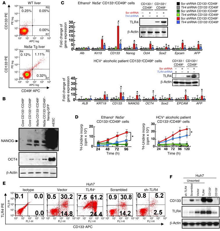
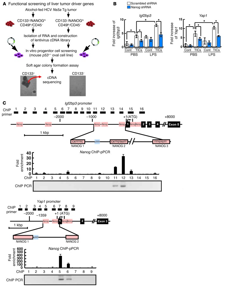
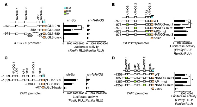
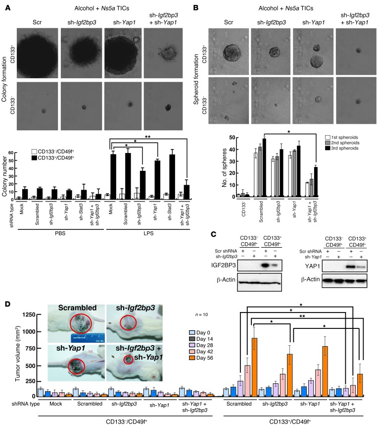
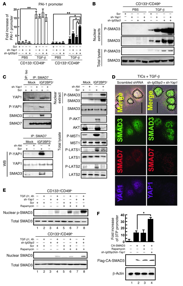
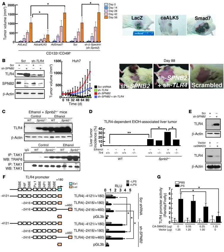
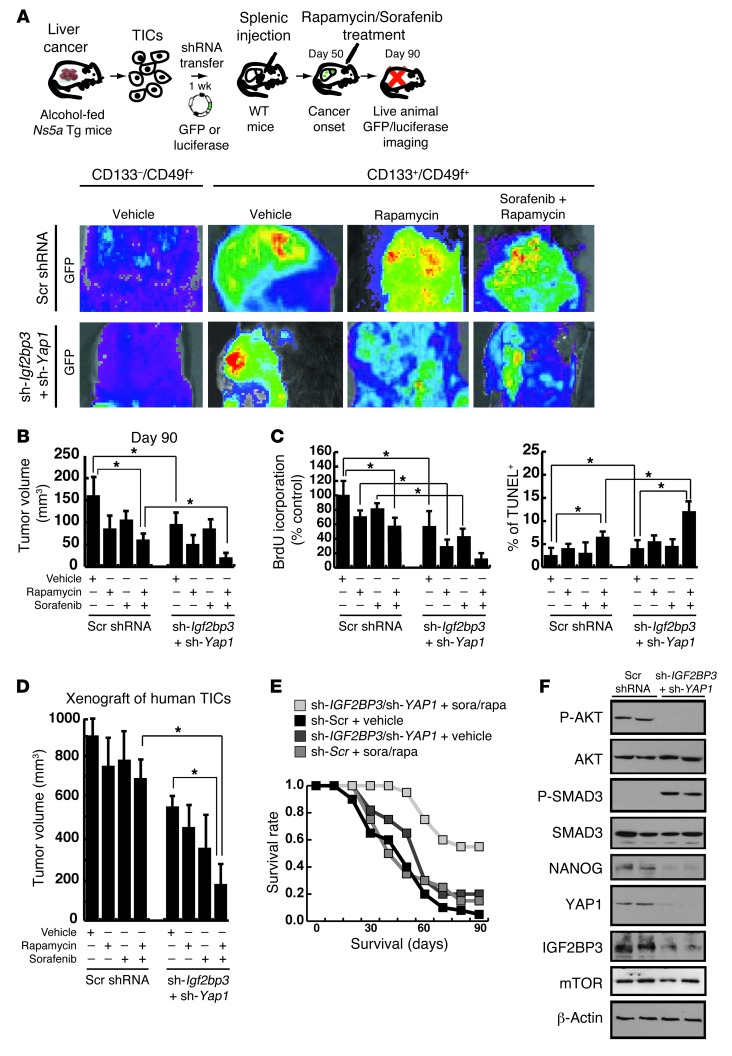
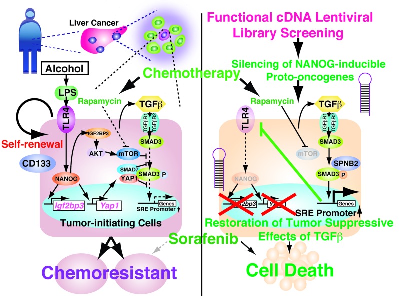
Tumor-initiating stem-like cells (TICs) are resistant to chemotherapy and associated with hepatocellular carcinoma (HCC) caused by HCV and/or alcohol-related chronic liver injury. Using HCV Tg mouse models and patients with HCC, we isolated CD133(+) TICs and identified the pluripotency marker NANOG as a direct target of TLR4, which drives the tumor-initiating activity of TICs. These TLR4/NANOG-dependent TICs were defective in the TGF-β tumor suppressor pathway. Functional oncogene screening of a TIC cDNA library identified Yap1 and Igf2bp3 as NANOG-dependent genes that inactivate TGF-β signaling. Mechanistically, we determined that YAP1 mediates cytoplasmic retention of phosphorylated SMAD3 and suppresses SMAD3 phosphorylation/activation by the IGF2BP3/AKT/mTOR pathway. Silencing of both YAP1 and IGF2BP3 restored TGF-β signaling, inhibited pluripotency genes and tumorigenesis, and abrogated chemoresistance of TICs. Mice with defective TGF-β signaling (Spnb2(+/-) mice) exhibited enhanced liver TLR4 expression and developed HCC in a TLR4-dependent manner. Taken together, these results suggest that the activated TLR4/NANOG oncogenic pathway is linked to suppression of cytostatic TGF-β signaling and could potentially serve as a therapeutic target for HCV-related HCC.


Figures

Figure 1
Figure 1. TLR4/NANOG–dependent TICs from liver tumors in alcohol-fed Ns5a Tg mice and patients.
(A) FACS separation of CD133+/CD49f+ cells from liver tumors of a HCV Ns5a Tg mouse. (B) Immunoblot analysis confirms induction of NANOG and OCT4 in TICs from both Core and Ns5a mice. Mouse embryonic stem cells (mESC) serve as a positive control. (C) TICs isolated from liver tumors of Ns5a Tg mice or alcoholic HCV patients express Nanog, Oct4, Sox2, and Krt19 at higher levels than CD133/CD49f+ or CD133/CD49f cells, as determined by qPCR. Immunoblots reveal increased TLR4 protein levels in TICs, which are effectively silenced by transduction of lentiviral shRNA (insets). This silencing abrogates stemness gene upregulation. *P < 0.05, #P < 0.01, compared with scrambled shRNA. (D) 3H-uridine incorporation assay in culture demonstrates enhanced, TLR4-dependent cell proliferation of TICs from Ns5a Tg model (left) and patients (right). *P < 0.05, compared with scrambled shRNA. (E) TLR4 expression correlates with the stemness marker CD133. Human HCC cell line Huh7 cells were transduced by a retroviral vector expressing TLR4 (TLR4+) or shRNA for TLR4 (sh-TLR4) and examined for surface expression of CD133 by FACS, as compared with those transduced with the control vector (Vector) or scrambled shRNA (Scrambled). (F) An immunoblot confirms induced CD133 expression in Huh7 cells transduced with Tlr4 (TLR4+) and a loss of this expression by transduction of shRNA against Tlr4 (sh-TLR4). A CD133+ population of Huh7 cells (CD133+) express abundant TLR4, and CD133 Huh7 cells (CD133) lack this expression.
Figure 2
Figure 2. TLR4/NANOG–dependent tumor-initiation property of TICs.
(A) GFP-transduced TICs, but not other populations from the Ns5a Tg mouse and patient HCC, give rise to growing tumors, as determined by whole-body imaging following subcutaneous transplantation in NOG mice. This growth is attenuated by Tlr4 shRNA transduction prior to transplantation (*P < 0.05). Final tumor weight is also reduced with Tlr4 shRNA. (B) Tumor growth by TICs from Ns5a Tg mouse or patient HCC is attenuated by transduction of Nanog shRNA. Final tumor weight is also reduced. Immunoblotting of cell lysates collected 10 days after the transplantation confirms expression of NANOG in the TICs and knockdown of this protein with specific shRNA. *P < 0.05.
Figure 3
Figure 3. Identification of TLR4/NANOG–dependent oncogenes, Igf2bp3 and Yap1, from functional screening of a lentiviral cDNA library.
(A) Strategy of in vitro identification of liver oncogenes, with oval cells infected with the TIC library but not with the control cell library forming colonies. (B) Increased expression of Igf2bp3 and Yap1 in the TICs as confirmed by qPCR is further upregulated by LPS treatment. Nanog shRNA significantly reduces these inductions. (C) Schematic diagrams of Igf2bp3 and Yap1 promoters depicting the locations of NANOG consensus binding sequences (orange boxes) and the areas analyzed by ChIP (1–16 black boxes for Igf2bp3 and 1–9 black boxes for Yap1). NANOG ChIP-qPCR data for TICs from the Ns5a Tg model are shown below, with NANOG enrichment in the regions 11 and 12 within NANOG 1 and 2 sites for the Igf2bp3 promoter and in the region 5 encompassing the NANOG 1 site of the Yap1 promoter.
Figure 4
Figure 4. Identification of TLR4/NANOG–dependent oncogenes, Igf2bp3 and Yap1, from functional screening of a lentiviral cDNA library.
(A) Igf2bp3 promoter analysis with deletion constructs demonstrates the importance of the proximal segment (–978/–359 nt) in LPS-induced promoter activity in TLR4-transduced Huh7 cells. NANOG dependence of this promoter activity is also demonstrated by the abrogating effects of NANOG shRNA. (B) Mutations of the NANOG consensus sites within –978/+55 nt (NANOG binding sites 1 and 2 but not 3) reduce LPS/TLR4–induced IGF2BP3 promoter activity. (C) YAP1 promoter analysis with deletion constructs demonstrates the importance of the proximal segment (–1359/–336 nt) in LPS-induced promoter activity in TLR4-transduced Huh7 cells. NANOG shRNA similarly abrogates the promoter activity. (D) Mutation of one of the NANOG consensus sites (NANOG binding site 2) reduces LPS/TLR4–induced YAP1 promoter activity. *P < 0.05.
Figure 5
Figure 5. Validation of Yap1 and Igf2bp3 as TIC oncogenes.
(A) The number of colonies formed by TICs in soft agar is moderately reduced by lentiviral transduction of shRNA against Yap1 or Igf2bp3 but largely abrogated when knocking down both. Stat3 silencing has no effects. *P < 0.05, **P < 0.01. (B) Self-renewal ability, as assessed by spheroid formation assay, is reduced in sh-Yap1/sh-Igf2bp3–transduced TICs. Serial spheroid-forming capacity is also reduced by dual transduction of sh-Yap1 and sh-Igf2bp3. *P < 0.05. (C) Immunoblot analysis demonstrates induced expression of IGF2BP3 and YAP1 in TICs and effective knockdown by respective shRNA. (D) Transduction of Ifg2bp3 shRNA, but not that of Yap1 shRNA, modestly reduces subcutaneous tumor growth by the TICs from the Ns5a Tg mice in NOG mice, while transduction of both shRNA clearly abrogates the growth at the later 3 time points. Representative photos of NOG mice carrying TIC-derived tumors are shown to depict an appreciable reduction of tumor size by Igf2bp3 and Yap1 silencing. *P < 0.05, **P < 0.01 versus scrambled shRNA group.
Figure 6
Figure 6. TICs are defective in TGF-β signaling due to SMAD3 pathway interference by YAP1 and IGF2BP3.
(A) TGF-β–stimulated PAI-1 promoter activity is lower in TICs than in control cells. This defect is modestly improved with Yap1 or Igf2bp3 shRNA alone and synergistically corrected by both. *P < 0.05, **P < 0.01. (B) Nuclear levels of p-SMAD3 or SMAD3 in TGF-β–treated TICs are increased by Yap1 or Igf2bp3 silencing with respective shRNA and conspicuously by silencing of both genes. (C) Interactions of SMAD7 with YAP1, p-YAP1, and SMAD3 are increased by Igf2bp3 transduction in PIL4 cells expressing YAP1 (PIL4-YAP1 cells), and these interactions are attenuated with Yap1 shRNA (top left). In the same PIL4-YAP1 cells with Igf2bp3 transduction, increased SMAD7 interactions with p-YAP1 and SMAD3 are abrogated by Akt silencing. Conversely, reduced nuclear p-SMAD3 level is increased. Phosphorylation of MST1 and LATS1/2 is increased by overexpression of IGF2BP3 but reduced by Akt silencing. (D) Fluorescent microscopy depicting increased nuclear staining of SMAD3 in TGF-β–treated TICs by silencing Igf2bp3 and Yap1. Dotted circles indicate the outline of nuclei. Original magnification, ×200. (E) Rapa (200 nM) further increases TGF-β–induced nuclear p-SMAD3 level, which is already augmented by silencing Yap1 or Igf2bp3. (F) Igf2bp3/Yap1 silencing but not rapa augments 3TP-Luc promoter activity induced by CA-SMAD3. *P < 0.05.
Figure 7
Figure 7. Inhibition of the TGF-β pathway promotes TLR4-mediated oncogenesis.
(A) Subcutaneous tumor growth by TICs from Ns5a Tg mouse tumors in NOG mice is attenuated by a gain-of-function approach for TGF-β signaling using adenoviral transduction of the constitutively active TβRI (caALK5), while it is promoted by overexpression of Smad7 or shRNA against Spnb2. (B) SPNB2 knockdown induces TLR4 and tumor-initiating property of Huh7 cells, which do not cause spontaneous xenograft growth. This growth by SPNB2 knockdown is largely prevented by knockdown of TLR4 with shRNA. Representative pictures of NOG mice bearing tumors at day 88 are shown. (C) Heterozygosity of Spnb2 induces TLR4 expression and downstream signaling (TAK1/TRAF6 association), and these changes are accentuated by alcohol feeding for 12 months. (D) The spontaneous liver tumor incidence in the Spnb2+/– mice, but not in Spnb2+/–Tlr4 mice, is increased by alcohol feeding. (E) Knockdown of SPNB2 with shRNA induces TLR4, while overexpression of caSMAD reduces TLR4 in Huh7 cells. (F) Knockdown of SPNB2 in Huh7 cells equally promotes LPS-induced activity of the TLR4 promoter containing 3 proximal SMAD-responsive elements (SRE). (G) Expression of CA-SMAD3 inhibits LPS-mediated TLR4 promoter (–4121/+180 nt) activity in Huh7 cells. *P < 0.05, **P < 0.01.
Figure 8
Figure 8. Induction of TLR4/NANOG/YAP1/IGF2BP3 pathway components in human HCC.
(A) TLR4, NANOG, IGF2BP3, and YAP1 protein levels are increased in HCC specimens from patients with HCV infection without or with alcoholism, as compared with cirrhotic or healthy livers. (B) Immunofluorescent microscopy demonstrates higher expression of NANOG, TLR4, YAP1, and IGF2BP3, which are often colocalized in patient HCC specimens, as compared with normal liver. Original magnification, ×200. (C) Immunofluorescent microscopy demonstrates increased expression of NANOG, YAP1, and IGF2BP3, which are often colocalized in HCC specimens from patients with nonalcoholic steatohepatitis (NASH), as compared with normal liver. p-SMAD3 staining is conversely reduced in HCC. Original magnification, ×200. (D) The log OR (and 95% CI) of patient death within 5 years from initial diagnosis for HCC patients is increased for those with positive IGF2BP3 or/and YAP1 immunoreactivity, as compared with those with negative IGF2BP3 and YAP1 immunoreactivity (red line).
Figure 9
Figure 9. Silencing of Igf2bp3 and Yap1 sensitizes TICs to drug-induced cell death.
(A) Silencing of Igf2bp3 and Yap1 in TICs from HCV Ns5a Tg mice promotes growth inhibition of tumor upon chemotherapeutic drug treatment (rapa and sorafenib) in immune-competent C57BL/6 mice, as monitored by GFP imaging after splenic injection and engraftment enrichment procedures with retrorsine and CCl4. (B) The tumor volume measurement performed at day 90 after transplantation shows a maximal growth retardation achieved by the combination of the drugs and Igf2bp3/Yap1 silencing (*P < 0.05). (C) TIC proliferation, as determined by BrdU incorporation in vivo at day 90, is most inhibited by the combination of the chemotherapeutic drugs and silencing of Igf2bp3 and Yap1. Concomitantly, the incidence of apoptotic cell death increases conspicuously by the combined treatment. *P < 0.05. (D) Xenograft tumor growth by TICs from patient HCC in NOG mice is most conspicuously suppressed by rapa and sorafanib treatment only in the presence of IGF2BP3 and/or YAP1 silencing (*P < 0.05). (E) Improved survival of mice injected with human TICs transduced with shRNA targeting YAP1 and IGF2BP3 and subjected to injection of the chemotherapeutic drugs, as compared with other groups with single therapies. (F) Immunoblot analysis demonstrates that silencing of both YAP1 and IGF2BP3 induces p-SMAD3 levels, while reducing NANOG expression in xenograft tissue of mice treated with rapa and sorafenib.
Figure 10
Figure 10. A schematic representation of the proposed link between oncogenic TLR4/NANOG signaling and a defective TGF-β tumor suppressor pathway in generating TICs.
Ectopic upregulation of TLR4 and its activation by LPS induce the pluripotency factor NANOG, other stem cell genes, and self-renewal of TICs. NANOG induces IGF2BP3 and YAP1, which in turn inhibit TGF-β signaling at the level of SMAD3 phosphoactivation and p-SMAD3 nuclear translocation. The former effect is dependent on the IGF2BP3/AKT/mTOR pathway, while the latter is caused by p-YAP1/SMAD7/SMAD3 interactions, which are enhanced by IGF2BP3/AKT–mediated YAP1 phosphorylation. Restoration of the TGF-β tumor suppressor pathway by silencing IGF2BP3 and YAP1 downregulates TLR4 via SMAD3-mediated transcriptional repression and inhibits TLR4/NANOG–mediated TIC self-renewal and oncogenic activity while chemosensitizing TICs. A defective canonical TGF-β pathway, as in Spnb+/– mice, upregulates TLR4 by transcriptional derepression and activates the TLR4/NANOG oncogenic pathway for liver tumorigenesis.

References

    1. Lobo NA, Shimono Y, Qian D, Clarke MF. The biology of cancer stem cells. Annu Rev Cell Dev Biol. 2007;23:675–699. doi: 10.1146/annurev.cellbio.22.010305.104154. - DOI - PubMed
    1. Jemal A, Bray F, Center MM, Ferlay J, Ward E, Forman D. Global cancer statistics. CA Cancer J Clin. 2011;61(2):69–90. doi: 10.3322/caac.20107. - DOI - PubMed
    1. Hanahan D, Weinberg RA. Hallmarks of cancer: the next generation. Cell. 2011;144(5):646–674. doi: 10.1016/j.cell.2011.02.013. - DOI - PubMed
    1. Grivennikov SI, Greten FR, Karin M. Immunity, inflammation, and cancer. Cell. 2010;140(6):883–899. doi: 10.1016/j.cell.2010.01.025. - DOI - PMC - PubMed
    1. Bauer AK, et al. Toll-like receptor 4 in butylated hydroxytoluene-induced mouse pulmonary inflammation and tumorigenesis. J Natl Cancer Inst. 2005;97(23):1778–1781. doi: 10.1093/jnci/dji403. - DOI - PubMed

Publication types

MeSH terms

Substances

Associated data