. 2013 Sep 20;288(38):27692-27701.
doi: 10.1074/jbc.M113.479675. Epub 2013 Aug 6.

ATP and AMP mutually influence their interaction with the ATP-binding cassette (ABC) adenylate kinase cystic fibrosis transmembrane conductance regulator (CFTR) at separate binding sites

Affiliations

ATP and AMP mutually influence their interaction with the ATP-binding cassette (ABC) adenylate kinase cystic fibrosis transmembrane conductance regulator (CFTR) at separate binding sites

Christoph O Randak et al. J Biol Chem. .

Abstract

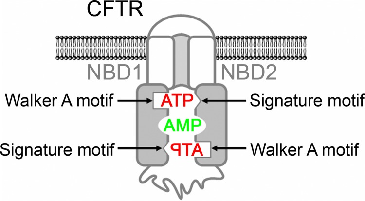
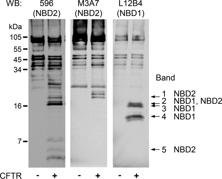
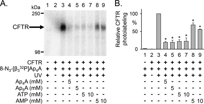
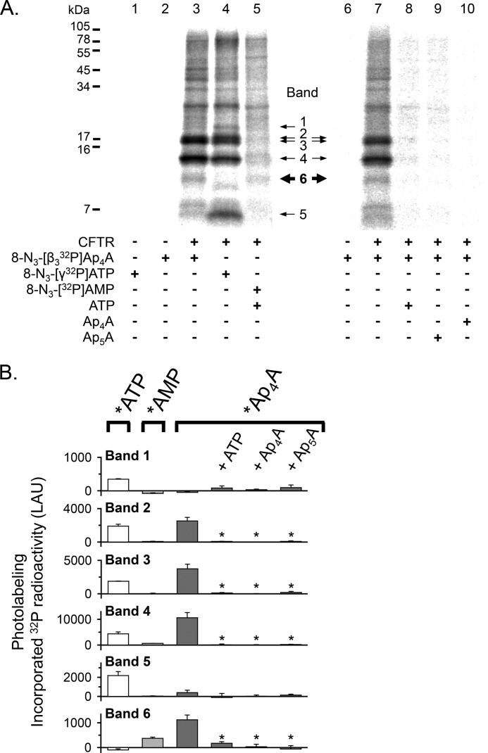
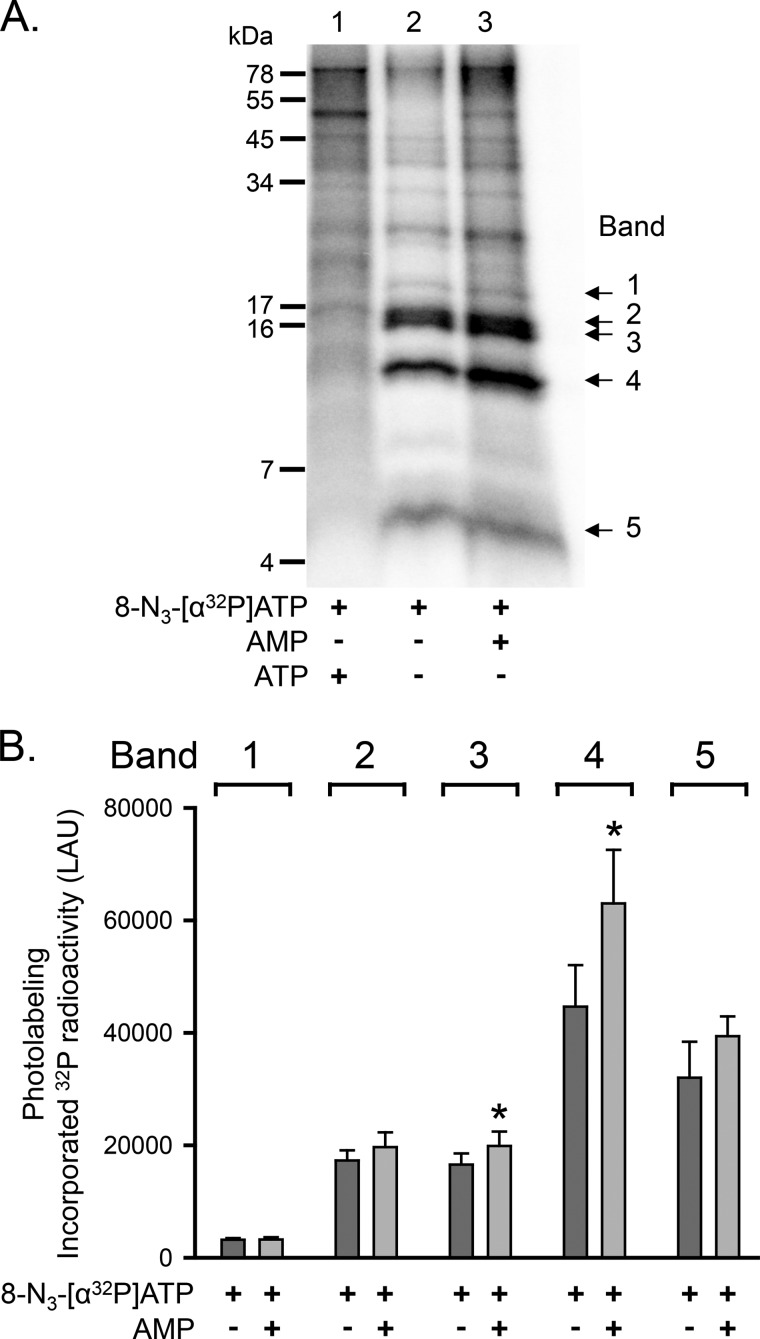
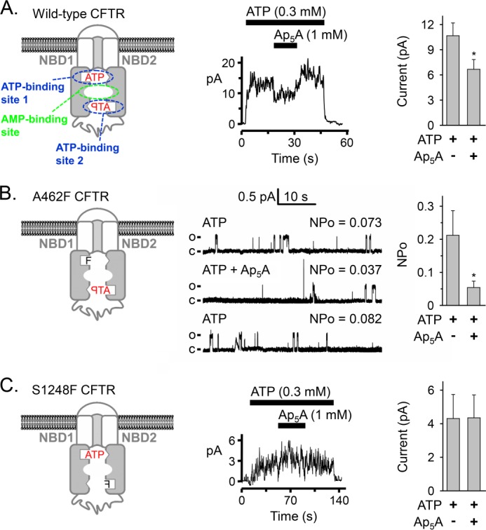
Cystic fibrosis transmembrane conductance regulator (CFTR) is an anion channel in the ATP-binding cassette (ABC) transporter protein family. In the presence of ATP and physiologically relevant concentrations of AMP, CFTR exhibits adenylate kinase activity (ATP + AMP &lrarr2; 2 ADP). Previous studies suggested that the interaction of nucleotide triphosphate with CFTR at ATP-binding site 2 is required for this activity. Two other ABC proteins, Rad50 and a structural maintenance of chromosome protein, also have adenylate kinase activity. All three ABC adenylate kinases bind and hydrolyze ATP in the absence of other nucleotides. However, little is known about how an ABC adenylate kinase interacts with ATP and AMP when both are present. Based on data from non-ABC adenylate kinases, we hypothesized that ATP and AMP mutually influence their interaction with CFTR at separate binding sites. We further hypothesized that only one of the two CFTR ATP-binding sites is involved in the adenylate kinase reaction. We found that 8-azidoadenosine 5'-triphosphate (8-N3-ATP) and 8-azidoadenosine 5'-monophosphate (8-N3-AMP) photolabeled separate sites in CFTR. Labeling of the AMP-binding site with 8-N3-AMP required the presence of ATP. Conversely, AMP enhanced photolabeling with 8-N3-ATP at ATP-binding site 2. The adenylate kinase active center probe P(1),P(5)-di(adenosine-5') pentaphosphate interacted simultaneously with an AMP-binding site and ATP-binding site 2. These results show that ATP and AMP interact with separate binding sites but mutually influence their interaction with the ABC adenylate kinase CFTR. They further indicate that the active center of the adenylate kinase comprises ATP-binding site 2.

Keywords: ABC Transporter; AMP; ATP; CFTR; Cystic fibrosis; Genetic Diseases.


Figures

FIGURE 1.
FIGURE 1.
Model of CFTR with separate binding sites for ATP and AMP. ATP is sandwiched between the Walker A motif of one NBD and the ABC signature motif of the other NBD. The residues interacting with AMP are not known.
FIGURE 2.
FIGURE 2.
Photolabeling of CFTR with 8-N3-[32P]AMP. A, Western blot (WB) probed with CFTR antibody 13-4. Letters mark highly glycosylated (C) and core-glycosylated (B) CFTR. CFTR was immunoprecipitated from 30 μg of HeLa cell membrane protein as described under “Experimental Procedures.” No CFTR was detected in membranes from HeLa cells not infected with the recombinant vaccinia virus encoding CFTR (lane 1). B, autoradiograph (left) and Western blot (probed with CFTR antibodies 13-1 and M3A7) (right) of the same gel. Membranes were mixed on ice with 25 μm 8-N3-[32P]AMP in the absence and presence of 8.3 mm non-radioactive ATP as indicated below the lanes of the autoradiograph. The samples were immediately irradiated with UV light for 30 s. The sample of lane 1 was not UV-irradiated. Comparing autoradiograph and Western blot corroborated that the labeled band was CFTR. C, autoradiographs from three different experiments labeling CFTR with 8-N3-[32P]AMP. Experimental conditions are indicated below the lanes. The concentration of non-radioactive ATP, AMP, Ap4A, and Ap5A was 8.3 mm. D, summary data. To compare the results from different autoradiographs, data were normalized to CFTR radioactivity under control conditions indicated below bars 1, 4, and 11. Dark gray bars mark labeling conditions in the presence of ATP. *, p = 0.012 (bar 5) and p < 0.001 (bars 6 and 8) compared with control (bars 4 and 11) (one-way repeated measures analysis of variance followed by Holm-Sidak's method of multiple comparisons versus control group, n = 2–10). ‡, p ≤ 0.015 compared with bars 6 and 8 (one-way repeated measures analysis of variance followed by Holm-Sidak's method of multiple comparisons versus control group, n = 2–10). Error bars, S.E.
FIGURE 3.
FIGURE 3.
Photolabeling of CFTR with 8-N3-[α-32P]ATP. A, autoradiographs from two different experiments. Membranes were incubated on ice for 10 min with 50 μm 8-N3-[α-32P]ATP in the absence and presence of 5 mm non-radioactive ATP, AMP, Ap4A, or Ap5A as indicated below the lanes of the autoradiographs. The samples were subsequently irradiated with UV light for 90 s. The sample of lane 5 was not UV-irradiated. CFTR was solubilized, immunoprecipitated, and fractionated on 8% (lanes 1–4) and 6% (lanes 5–8) SDS-polyacrylamide gels as described under “Experimental Procedures.” B, summary data. Experiments were performed as in A. Radioactivity incorporated into CFTR was normalized to radioactivity for the conditions indicated below bars 2 and 6. *, p ≤ 0.001 compared with control (bars 2 and 6) (one-way repeated measures ANOVA followed by Holm-Sidak's method of multiple comparisons versus control group, n = 6–7). Error bars, S.E.
FIGURE 4.
FIGURE 4.
Ap5A and Ap4A do not interact with all CFTR ATP-binding sites. A, left, autoradiograph. Photolabeling of CFTR with 50 μm 8-N3-[α-32P]ATP was performed as described in the legend to Fig. 3. 5 mm non-radioactive ATP, Ap4A, or Ap5A were present as indicated below the lanes of the autoradiograph. After photolabeling, CFTR was solubilized, immunoprecipitated, and subjected to partial proteolysis with the proteinase Arg-C. The digestion products were fractionated on a 16% Tricine gel. To compare with undigested photolabeled and immunoprecipitated CFTR, see Fig. 3. Middle, summary of Western blot analysis results (see Fig. 5). Right, quantitative data in linear arbitrary units (LAU) for radioactivity incorporated into the different CFTR fragments (bands 1–5) after labeling with 8-N3-[α32P]ATP. *, p ≤ 0.024 compared with background control of no CFTR present (Mann-Whitney rank sum test, n = 3–8). B, quantitative data for photolabeling bands 1–5 with 50 μm 8-N3-[α-32P]ATP in the presence or absence of 5 mm non-radioactive ATP, Ap4A, or Ap5A. Amount of radioactivity incorporated into each band was normalized to radioactivity for control conditions (i.e. absence of non-radioactive ATP, Ap4A, or Ap5A). *, p ≤ 0.001 (one-way repeated measures ANOVA followed by Holm-Sidak's method of multiple comparisons versus control group, n = 2–5). Error bars, S.E.
FIGURE 5.
FIGURE 5.
Identification of CFTR NBD fragments produced by limited Arg-C digestion by Western blot analysis. CFTR was solubilized, immunoprecipitated, and subjected to partial proteolysis with the proteinase Arg-C as described in the legend to Fig. 4. After fractionating on 16% Tricine gel, Western blots (WB) were probed with monoclonal antibodies to NBD1 (L12B4) and NBD2 (M3A7 and 596). These antibodies recognize epitopes within the following CFTR amino acid sequences: L12B4, 385–410; 596, 1204–1211; M3A7, 1373–1382 (42). Bands 1–5 were identified by comparing autoradiographs and Western blots of the same gels.
FIGURE 6.
FIGURE 6.
Photolabeling of CFTR with 8-N3-[β3-32P]Ap4A. A, autoradiograph. Membranes were mixed on ice with 25 μm 8-N3-[β3-32P]Ap4A in the absence and presence of non-radioactive ATP, AMP, Ap4A, or Ap5A, as indicated below the lanes of the autoradiograph. The samples were immediately irradiated with UV light for 30 s. B, summary data. Amount of radioactivity incorporated into CFTR was normalized to CFTR radioactivity under the conditions indicated below bar 3. *, p < 0.001 compared with bar 3 (one-way repeated measures ANOVA followed by Holm-Sidak's method of multiple comparisons versus control group, n = 2–4). Error bars, S.E.
FIGURE 7.
FIGURE 7.
Photolabeling of CFTR with 8-N3-[β3-32P]Ap4A, 8-N3-[γ-32P]ATP, and 8-N3-[32P]AMP. A, autoradiographs from two different experiments. Photolabeling of CFTR was performed as described in the legend to Fig. 6. Non-radioactive ATP, Ap4A, or Ap5A was present in a concentration of 8.3 mm, as indicated below the lanes of the autoradiographs. In lanes 1–5, the three different radionucleotides, 8-N3-[β3-32P]Ap4A (10.3 Ci/mmol), 8-N3-[γ-32P]ATP (4.1 Ci/mmol), and 8-N3-[32P]AMP (5.8 Ci/mmol), were added at equal amounts of radioactivity (12.4 μCi). After photolabeling, CFTR was solubilized, immunoprecipitated, and subjected to partial proteolysis with the proteinase Arg-C. The digestion products were fractionated on a 16% Tricine gel. B, quantitative data in linear arbitrary units (LAU) for radioactivity incorporated into the different CFTR fragments after labeling with *Ap4A (dark gray bars), *ATP (white bars), and *AMP (light gray bars). *, p ≤ 0.005 compared with 8-N3-[β3-32P]Ap4A labeling under control conditions (i.e. absence of non-radioactive ATP, Ap4A, or Ap5A (dark gray bars)) (one-way repeated measures ANOVA followed by Holm-Sidak's method of multiple comparisons versus control group, n = 3–5). Error bars, S.E.
FIGURE 8.
FIGURE 8.
Increased photolabeling of CFTR with 8-N3-[α-32P]ATP at one ATP-binding site in the presence of AMP. A, autoradiograph. Experiments were performed as described in the legend to Fig. 4. The concentration of non-radioactive ATP and AMP was 5 mm. B, quantitative data in linear arbitrary units (LAU) for radioactivity incorporated into bands 1–5 after labeling with 8-N3-[α-32P]ATP. The absence (dark gray bars) or presence of AMP (light gray bars) is indicated below each bar. *, p ≤ 0.016 (paired t test, n = 4). Error bars, S.E.
FIGURE 9.
FIGURE 9.
Effect of phenylalanine mutations in the phosphate-binding loop (Walker A motif) of either ATP-binding site on Ap5A inhibition of CFTR current. A, left, model of CFTR. The phosphate-binding loops are depicted as open rectangles, and the ABC signature motifs are shown as open triangles. The binding site for AMP is not known. Middle, current recording (100 ms averages) from an excised inside-out membrane patch containing multiple CFTR channels. ATP and Ap5A were present during the times and at the concentrations indicated by bars. ATP was added together with PKA catalytic subunit. Holding voltage was −50 mV. Right, CFTR Cl current before and after adding 1 mm Ap5A. Experiments were performed as shown in the middle panel with 0.3 mm ATP and PKA present. *, p < 0.001 (Wilcoxon signed rank test, n = 13 paired experiments of current measurements before and after adding Ap5A obtained from five membrane patches). B, left, model of A462F CFTR. Middle, current recording from one excised inside-out membrane patch containing at least two A462F CFTR channels perfused on cytosolic surface with ATP and Ap5A as indicated. PKA catalytic subunit was present throughout the recording. Holding voltage was −50 mV. Each lane shows 48 s of recording and mean NPo. The first lane shows the trace in the presence of 0.3 mm ATP, the second lane shows the recording after 1 mm Ap5A was added, and the third lane shows the recording after Ap5A was removed again. For illustration purposes, traces were digitally low pass-filtered at 50 Hz. c, channel closed state; o, single channel open state. Right, NPo of A462F CFTR with 0.3 mm ATP and PKA present in the bath solution before and after adding 1 mm Ap5A. *, p = 0.002 (Wilcoxon signed rank test, n = 10 membrane patches). C, left, model of S1248F CFTR. This mutant contained an N-terminal 6-histidine tag between CFTR amino acids 2 and 3. Middle, current recording (100 ms averages) from an excised inside-out membrane patch containing multiple S1248F CFTR channels. ATP and Ap5A were present during the times and at the concentrations indicated by bars. ATP was added together with PKA catalytic subunit. Holding voltage was −80 mV. Right, S1248F CFTR Cl current before and after adding 1 mm Ap5A. Experiments were performed as shown in the middle panel with 0.3 mm ATP and PKA present. No significant differences were detected (p = 0.463, Wilcoxon signed rank test, n = 14 paired experiments of current measurements before and after adding Ap5A obtained from four membrane patches). Error bars, S.E.
FIGURE 10.
FIGURE 10.
Three-dimensional model of the CFTR NBD1-NBD2 heterodimer. The model was constructed as described under “Experimental Procedures.” Left, stick model of the overall heterodimer structure with NBD1 in red and NBD2 in blue. Two ATP molecules (in a space-filling representation) are bound between the Walker A motif of one NBD and the ABC signature motif of the other NBD. A central space between the two NBDs is evident, into which one adenosine moiety of Ap5A could extend. Residues lining this space from both NBDs are depicted in green. Right, close-up view of the central space region between the two NBDs. The residues in green lining the cavity might interact with AMP. Histidine 1348 might prevent Ap5A from interacting with ATP-binding site 1. See supplemental Movie S1 to facilitate visualizing the tree-dimensional positioning of these residues.

Similar articles

Cited by

References

    1. Hanrahan J. W., Gentzsch M., Riordan J. R. (2003) The cystic fibrosis transmembrane conductance regulator (ABCC7). in ABC Proteins from Bacteria to Man (Holland I. B., Cole S. P. C., Kuchler K., Higgins C. F., eds) pp. 589–618, Academic Press, Amsterdam
    1. Hwang T. C., Kirk K. L. (2013) The CFTR ion channel. Gating, regulation, and anion permeation. Cold Spring Harb. Perspect. Med. 3, a009498. - PMC - PubMed
    1. Davidson A. L., Dassa E., Orelle C., Chen J. (2008) Structure, function, and evolution of bacterial ATP-binding cassette systems. Microbiol. Mol. Biol. Rev. 72, 317–364 - PMC - PubMed
    1. Walker J. E., Saraste M., Runswick M. J., Gay N. J. (1982) Distantly related sequences in the α- and β-subunits of ATP synthase, myosin, kinases and other ATP-requiring enzymes and a common nucleotide binding fold. EMBO J. 1, 945–951 - PMC - PubMed
    1. Shyamala V., Baichwal V., Beall E., Ames G. F. (1991) Structure-function analysis of the histidine permease and comparison with cystic fibrosis mutations. J. Biol. Chem. 266, 18714–18719 - PubMed

Publication types

MeSH terms

Substances