Review
. 2013 Oct;9(10):2263-76.
doi: 10.4161/hv.25181. Epub 2013 Jun 4.

A novel, disruptive vaccination technology: self-adjuvanted RNActive(®) vaccines

Affiliations
Review

A novel, disruptive vaccination technology: self-adjuvanted RNActive(®) vaccines

Karl-Josef Kallen et al. Hum Vaccin Immunother. 2013 Oct.

Abstract

Nucleotide based vaccines represent an enticing, novel approach to vaccination. We have developed a novel immunization technology, RNActive(®) vaccines, that have two important characteristics: mRNA molecules are used whose protein expression capacity has been enhanced by 4 to 5 orders of magnitude by modifications of the nucleotide sequence with the naturally occurring nucleotides A (adenosine), G (guanosine), C (cytosine), U (uridine) that do not affect the primary amino acid sequence. Second, they are complexed with protamine and thus activate the immune system by involvement of toll-like receptor (TLR) 7. Essentially, this bestows self-adjuvant activity on RNActive(®) vaccines. RNActive(®) vaccines induce strong, balanced immune responses comprising humoral and cellular responses, effector and memory responses as well as activation of important subpopulations of immune cells, such as Th1 and Th2 cells. Pre-germinal center and germinal center B cells were detected in human patients upon vaccination. RNActive(®) vaccines successfully protect against lethal challenges with a variety of different influenza strains in preclinical models. Anti-tumor activity was observed preclinically under therapeutic as well as prophylactic conditions. Initial clinical experiences suggest that the preclinical immunogenicity of RNActive(®) could be successfully translated to humans.

Keywords: RNA; vaccines.


Figures

None
Figure 1. Schematic representation of mRNA and expression levels reached by modifactions thereof. (A) The principle structure of an mRNA-molecule consists of a cap-region, followed by an (optional) 5′-untranslated region, the open reading frame, an (optional) 3′-untranslated region and the poly-A-tail. Sequence modifications of each depicted subunit of an mRNA molecule that only comprise the naturally occurring nucleotides A, G, C, U and that do not affect the primary amino acid sequence encoded by the open reading frame constitute the basis of modified mRNA-molecules used in RNActive® vaccines. Details in ref. (B) Different generations of PpLuc-coding mRNAs produced over the last years were electroporated into HeLa cells (generation 1 to 4) and compared for their in vitro expression of luciferase. The luciferase level was determined at 6, 24, and 48 h or 72 h post transfection. The expression power of generation 4 and 5 was compared in human dermal fibroblasts after lipofection. The dynamic range of the assay was not sufficient to compare all mRNA molecules in one experiment.
None
Figure 2. Protein expression in vivo is strongly prolonged using CureVac’s proprietary mRNA technology and lasts for many days. Firefly luciferase-encoding mRNA, optimized for translation and stability, was injected intradermally into a BALB/c mouse (4 injection sites). At various time points after mRNA injection, luciferase expression was visualized in the living animal by optical imaging. (A) Visualization of luciferase expression at selected time points, showing maximal protein levels 24 to 48 h after mRNA injection. (B) Quantitative expression of luciferase over time until 9 d after mRNA injection. Results are shown on a linear scale (left-hand panel) or on a semi-logarithmic scale (right-hand panel). The figure is adapted with permission from ref. , details therein.
None
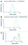
Figure 3. Size analysis of RNActive® vaccines as shown by a vaccine encoding Ppluc that was produced using the RNActive® technology. The size distribution of particles composing the vaccine solution was analyzed using a Malvern Zetasizer Scattering Instrument. Closely similar data were generated with several different mRNAs (A) The naked mRNA has a size of around 50 nm. (B) mRNA is complexed with protamine at a mass ratio of mRNA:protamine of 2:1. The resulting particles are distinctly larger than the ones consisting of naked mRNA. (C) The size distribution analysis of a complete RNActive® vaccine shows that the particle sizes of the two individual components are maintained in the RNActive® vaccines. Further analysis with fluorescent correlation spectroscopy demonstrated that the protamine/ mRNA complexes are very tight, so that naked, free mRNA does not exchange with the protamine-complexed mRNA. Figure was adapted with permission from ref. , details therein.
None
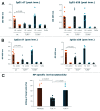
Figure 7. Effect of pattern recognition receptors on RNActive vaccination. (A and B) Male TLR7−/− or TLR9−/− BALB/c mice (purchased from Bioindustry Division. Oriental Yeast Co., Tokyo, Japan) were vaccinated intradermally on day 0 and day 7 with 20 µg of RNActive vaccines encoding HA (PR8) or ovalbumin as control. Serum samples were taken 7 d (A) and 28 d (B) after the final vaccination and IgG1 and IgG2a antibodies against HA determined with ELISAs (methods in Petsch et al.25). Data points represent single mice. (C) One month later, the same mice were vaccinated with 20 µg of RNActive encoding nucleoprotein (NP) from PR8 with the same vaccination schedule. Splenocytes from vaccinated mice were stimulated with a peptide (amino acids 147–155) from NP 7 d after the last vaccination and the cytolytic activity determined with an in vivo killing assay (described in ref. 20).
None
Figure 4. RNActive® vaccines induce effective B cell and T cell responses. Mice were vaccinated intradermally with an RNActive® vaccine encoding ovalbumin. (A) The presence of ovalbumin-specific antibodies was measured in serially diluted sera of vaccinated and control mice taken 11 d after the last vaccination and analyzed using ELISA. Data points represent antibody endpoint titers calculated for individual mice. (B) Ex vivo ELISpot analysis of the secretion of IFNγ in splenocytes from vaccinated and control mice. Cells were isolated on day 6 after the last vaccination and stimulated either with antigenic or with control peptide. The graph shows single data points for individual mice. (C) In vivo cytotoxicity against target cells loaded with the ovalbumin derived SIINFEKL peptide on day 5 after the last injection. The graph shows single data points for individual mice. (D) Ex vivo ELISpot analysis of the secretion of IFNγ in sorted CD4+ T cells from mice vaccinated with an RNActive® vaccine encoding prostate specific membrane antigen (PSMA) and control mice. Cells were isolated on day 6 after the last vaccination and stimulated either with PSMA-derived or control peptide library. (E) Frequencies of IFNγ+ CD44+ CD62L- CCR7- memory T cells in sorted CD8+ T cells from vaccinated and control mice. Cells were isolated on day 55 after last vaccination and stimulated ex vivo either with antigenic or with control peptide library and anti-CD28 antibody for 6 h. After intracellular staining of IFNγ secretion, cells were stained for surface markers of memory T cells. Adapted with permission from refs. and .
None
Figure 5. RNActive® vaccines in prophylactic and therapeutic tumor models. (A) C57BL/6 mice were immunized intradermally with an RNActive® vaccine encoding ovalbumin on day 1 and week 3. Six days after the last vaccination, C57BL/6 mice were challenged subcutaneously with 1 × 10⁶ syngeneic E.G7-OVA tumor cells. Tumor growth was monitored by measuring the tumor size in three dimensions using calipers. (B) Outgrown tumors were excised and ovalbumin expression was quantified via RT-PCR relative to murine GAPDH from total RNA isolates. (C) C57BL/6 mice (n = 9) were challenged subcutaneously with 0.3 × 10⁶ syngeneic E.G7- OVA tumor cells on day 0. Tumors were palpable on day 3. Mice were treated according to the schedule specified in the graph with either the RNActive® ovalbumin or with control vaccine (32 µg/vaccination) or with buffer. Therapy was started either on day 4 or on day 7. (D) Mice were treated as indicated in (C), but outgrown tumors were excised to quantify the expression of ovalbumin via RT-PCR. Figure adapted with permission from refs. and .
None
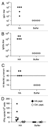
Figure 6. Immunological characterization of an RNActive® vaccine encoding hemagglutinin from influenza virus PR8. BALB/c mice were vaccinated intradermally with an RNActive® vaccine encoding hemagglutinin from influenza virus PR8 on day 1 and day 22. PR8HA-specific antibodies in the serum were quantified 4 weeks after the last immunization by IgG1- (A) and IgG2a-specific ELISA (B), and by hemagglutination inhibition (HI) assays (C). The dashed line in c indicates the conventionally defined protective HI titer of 1:40. (D) ELISPOT of IFNγ production in CD4+ T-cells sorted from vaccinated and buffer control mice on day 28. CD4+ T-cells were stimulated with a pool of five MHC class II–restricted peptides from HA (HA pept.) or as a control from the HIVpol protein (control; ctrl. pept.). Figure adapted with permission from ref. .
None
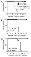
Figure 8. Protective efficacy of mRNA vaccine against lethal virus challenge in BALB/c mice. (A) BALB/c mice (n = 5/group) were injected intradermally with 80 µg of PR8HA mRNA that was either frozen at −20°C before immunization (HA mRNA) or lyophilized and stored for 3 weeks at 37°C before immunization (HA mRNA 37°C). In one group of mice immunized with PR8HA mRNA (that had been frozen), T cells were depleted at days −1 and +3 with respect to challenge infection (HA T depl.). Control mice were injected intradermally with buffer or intramuscularly with 10 µg of inactivated PR8 virus (Inact. PR8). Vaccine or control injections were done on days 0 and 21. On day 56 mice were infected with 10x LD50 of PR8 virus and survival assessed. (B and C). BALB/c mice (n = 5/group for Re6; n = 8/group for Vn04) were injected intradermally with 80 µg of mRNA encoding HA from influenza strain A/Regensburg/D6/2009 (Re6/H1N1v) or A/Vietnam/1194/2004 (Vn04/H5N1), three independent experiments. Control mice were injected with (B) 80 µg of ovalbumin RNActive® vaccines or Ringer′s lactate buffer (C). Immunizations were done at day 0 and booster injections at day 21. Five weeks after booster injection mice were challenged with virus expressing the homologous HA. For Vn04 and PR98 10x LD50 were used as challenge dose. Due to technical limitations a 6.8x LD50 was used for the challenge with Re6. Statistical analysis was done using a log rank analysis (Mantel Cox test): (A) p = 0.0017, (B) p = 0.001, (C) p = < 0.0001. Further information in ref. .
None
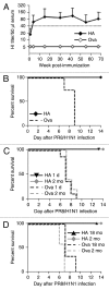
Figure 9. RNActive® vaccines elicit durable immune responses, circumvent immunosenescence in old mice, but are also immunogenic in newborn mice. (A) 8-week-old female BALB/c mice were injected intradermally on days 0 and 7 with 20 µg of PR8HA RNActive® (n = 5) or ovalbumin RNActive® (n = 4). For each condition, two independent experiments were performed. HI titers were monitored over a period of 70 weeks (16 mo) and plotted as mean + s.d. (B) Sixteen months after immunization, mice were challenged with 10 × LD50 of PR8 and survival was monitored. Newborn mice (1 d old, 1d, n = 9/group; three independent experiments) (C), aged (18 mo old, 18 mo, n = 3/group; one experiment) (D) or adult (2 mo old, 2 mo, n = 5/group) BALB/c mice were injected intradermally with 80 µg of PR8HA or ovalbumin RNActive® with an interval of 7 d. Five weeks after the second immunization, mice were challenged with 10 × LD50 of live PR8 virus and survival monitored for 14 d post-infection. Statistical analysis was done using a log rank analysis (Mantel Cox test): (B) p = 0.005, (C) 1 d: p = 0.0007; 2 mo: p = 0.0015, (D) 2 mo: p = 0.031; 18 mo: p = 0.01,
None
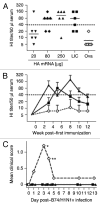
Figure 10. Immune effects of RNActive® vaccines in ferrets and pigs and protective efficacy in pigs. (A, B) Immunogenicity of RNActive® vaccines was assessed in six-month-old male ferrets (n = 6/group) that were immunized intradermally with 20, 80 or 250 µg of Re6HA RNActive® vaccine or 80 µg of Ova RNActive® vaccine, or intramuscularly with 500 µl of the licensed vaccine Celvapan® (LIC) which contains 7.5 µg C7HA. mRNA was injected at weeks 0, 1, and 6. Celvapan® was injected at weeks 0 and 3. (A) HI titers were measured 2 weeks after the booster vaccination (week 3 for RNActive® vaccine, week 5 for Celvapan®). (B) The kinetics of HI titers was recorded over the whole experiment for groups treated with 250 µg Re6HA, 80 µg ovalbumin (ova) or Celvapan (symbols are the same in panels A and B). Data are expressed as mean + s.d. for clarity and represent two independent experiments. (C) 2-mo-old seronegative pigs (n = 5/group) were immunized on days 0 and 21 with 250 µg of each Re6HA, Re6NA, and PR8NP RNActive® vaccine, 500 µl Mutagrip 2011/2012, or buffer. On day 16 post-immunization, animals were infected with 106.5 TCID50 of A/Bayern/74/2009 (B74/H1N1v) virus. Clinical symptoms were measured in a blinded fashion and were recorded over the ensuing 13 d (*: impaired general condition 5/5 buffer-treated animals, **: impaired general condition 4/5 buffer-treated animals). Further details in Petsch et al.25.
None
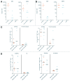
Figure 11. Intradermal vs. intranodal administration of RNActive® vaccines. Mice were vaccinated with 16 or 32 µg of RNActive® vaccine encoding ovalbumin or HA from PR8 as control either intradermally or intranodally on days 0, 3, 6, and 9. A maximum volume of 10 µl could be injected into lymph nodes, for which reason the 32 µg dose was administered to two different lymph nodes. (A and B) On day 15, serum samples were taken and IgG1 and IgG2a antibody titers against ovalbumin were determined (method described in Fotin-Mleczek et al.20). (A) 16 µg dose, (B) 32 µg dose. (C and D) Splenocytes were also isolated from vaccinated mice and the frequency of IFNγ+ or TNFα+ CD8+ T cells determined by intracellular cytokine staining after stimulation with the SIINFEKL peptide from ovalbumin or HA derived epitopes as control., (C) 16 µg dose, (D) 32 µg dose

References

    1. Rappuoli R, Mandl CW, Black S, De Gregorio E. Vaccines for the twenty-first century society. Nat Rev Immunol. 2011;11:865–72. - PMC - PubMed
    1. Wolff JA, Malone RW, Williams P, Chong W, Acsadi G, Jani A, et al. Direct gene transfer into mouse muscle in vivo. Science. 1990;247:1465–8. doi: 10.1126/science.1690918. - DOI - PubMed
    1. Martinon F, Krishnan S, Lenzen G, Magné R, Gomard E, Guillet JG, et al. Induction of virus-specific cytotoxic T lymphocytes in vivo by liposome-entrapped mRNA. Eur J Immunol. 1993;23:1719–22. doi: 10.1002/eji.1830230749. - DOI - PubMed
    1. Conry RM, LoBuglio AF, Wright M, Sumerel L, Pike MJ, Johanning F, et al. Characterization of a messenger RNA polynucleotide vaccine vector. Cancer Res. 1995;55:1397–400. - PubMed
    1. Hoerr I, Obst R, Rammensee HG, Jung G. In vivo application of RNA leads to induction of specific cytotoxic T lymphocytes and antibodies. Eur J Immunol. 2000;30:1–7. doi: 10.1002/1521-4141(200001)30:1<1::AID-IMMU1>3.0.CO;2-#. - DOI - PubMed

MeSH terms