. 2013 Aug 28;32(17):2392-406.
doi: 10.1038/emboj.2013.176. Epub 2013 Aug 6.

WHSC1 links transcription elongation to HIRA-mediated histone H3.3 deposition

Affiliations

WHSC1 links transcription elongation to HIRA-mediated histone H3.3 deposition

Naoyuki Sarai et al. EMBO J. .

Abstract

Actively transcribed genes are enriched with the histone variant H3.3. Although H3.3 deposition has been linked to transcription, mechanisms controlling this process remain elusive. We investigated the role of the histone methyltransferase Wolf-Hirschhorn syndrome candidate 1 (WHSC1) (NSD2/MMSET) in H3.3 deposition into interferon (IFN) response genes. IFN treatment triggered robust H3.3 incorporation into activated genes, which continued even after cessation of transcription. Likewise, UV radiation caused H3.3 deposition in UV-activated genes. However, in Whsc1(-/-) cells IFN- or UV-triggered H3.3 deposition was absent, along with a marked reduction in IFN- or UV-induced transcription. We found that WHSC1 interacted with the bromodomain protein 4 (BRD4) and the positive transcription elongation factor b (P-TEFb) and facilitated transcriptional elongation. WHSC1 also associated with HIRA, the H3.3-specific histone chaperone, independent of BRD4 and P-TEFb. WHSC1 and HIRA co-occupied IFN-stimulated genes and supported prolonged H3.3 incorporation, leaving a lasting transcriptional mark. Our results reveal a previously unrecognized role of WHSC1, which links transcriptional elongation and H3.3 deposition into activated genes through two molecularly distinct pathways.


Conflict of interest statement

The authors declared that they have no conflict of interests.

Figures

Figure 1
Figure 1
WHSC1 is required for induction of ISG transcription and H3.3 incorporation. (A) Intranuclear localization of H3.3-YFP was visualized by immunostaining of WT and Whsc1−/− MEFs with anti-GFP antibody (green), counterstained with Hoechst 33342 for DNA (red). (B) Schematic map of ISGs and Gtf2b. The arrow indicates the transcribed region. Exons are marked by grey boxes. Positions of primers used in ChIP analysis are shown underneath and colour-coded. (C) IFN induced H3.3 incorporation. H3.3-YFP expressing WT and Whsc1−/− cells were treated with IFN for 1, 3, 6, 12, 24, and 48 h, and ChIP assays were performed using anti-GFP antibody at the indicated sites of ISGs. Gtf2b was tested as a control. Values represent the average of duplicate determinations ±s.d. (D) Induction of ISG mRNA in the above cells was detected by qRT–PCR, normalized by Gapdh and expressed as fold induction. Values represent the average of two determinations ±s.d.
Figure 2
Figure 2
WHSC1 reintroduction rescues ISG transcription and H3.3 incorporation in Whsc1−/− cells. (A) Whsc1−/− cells expressing H3.3-YFP were transduced with WT WHSC1 or the catalytically defective mutant (H1143G) vectors. Cells were treated with IFN for indicated times, and Whsc1 mRNA was detected by qRT–PCR. Values represent the average of two determinations ±s.d. Untransduced Whsc1−/− cells were tested as a negative control. (B) WHSC1 protein expression in above cells was detected by immunoblotting with anti-WHSC1 antibody. β-actin was tested as a loading control. (C) Above cells were tested for IFN-induced H3.3 incorporation by ChIP assays as in Figure 1C. Values represent the average of duplicate determinations ±s.d. (D) Above cells were tested for ISG mRNA expression by qRT-PCR. Values represent the average of two determinations ±s.d. Source data for this figure is available on the online supplementary information page.
Figure 3
Figure 3
WHSC1 is recruited to ISGs upon IFN stimulation. (AC) Recruitment of WHSC1 (A), HIRA (B), and H3K36me3 (C) on indicated position of Ifit1 was tested in WT and Whsc1−/− cells treated with IFN for indicated times by ChIP analysis using respective antibodies. In (A), binding of WHSC1 was plotted in an expanded X axis to better depict early recruitment kinetics. Values represent the average of duplicate determinations ±s.d. (D) NIH3T3 cells were transduced with WT H3.3-YFP or mutant H3.3K36R-YFP, treated with IFN for indicated times, and their deposition into Ifit1 was detected by ChIP using anti-GFP antibody. Values represent the average of duplicate determinations ±s.d.
Figure 4
Figure 4
Defective ISG elongation in Whsc1−/− cells. WT and Whsc1−/− cells were treated with IFN for indicated times, and the recruitment of Pol II-2P (A), Pol II-HP (B), Pol II (C), Pol II-5P (D), CDK9 (E), and BRD4 (F) to indicated position of Ifit1 was tested by ChIP using respective antibodies. Values represent the average of duplicate determinations ±s.d.
Figure 5
Figure 5
WHSC1 interacts with BRD4 and HIRA independently. (A) Whole cell extracts from Whsc1−/− cells transiently expressing FLAG-WHSC1 were immunoprecipitated with anti-FLAG antibody, and tested for co-precipitation of indicated proteins by immunoblotting. Normal IgG was used as a control. Input represents 10% of total protein. (B) Extracts from NIH3T3 cells expressing BRD4-YFP were immunoprecipitated with anti-GFP antibody, and tested for co-precipitation of indicated proteins by immunoblotting. (C) Whole cell extracts from WT cells transiently expressing HIRA-YFP were immunoprecipitated with anti-GFP antibody, and tested for co-precipitation of indicated proteins by immunoblotting. (D) Whole cell extracts from WT cells treated with IFN for indicated times were immunoprecipitated with anti-WHSC1 antibody, and tested for co-precipitation of BRD4 and HIRA. (E) Graphic representation of the experiments shown in (D). The amounts of precipitated proteins were quantified and normalized by input protein. Values are the average of two assays ±s.d. (F) A diagram of FLAG-tagged WHSC1 deletion mutants. The red dots indicate deleted regions. (G) Whole cell extracts from Whsc1−/− cells transiently expressing above deletion constructs were immunoblotted with anti-WHSC1 antibody (left) or immunoprecipitated with anti-FLAG antibody, followed by immunoblotting with anti-WHSC1 antibody (right). (H) Proteins immunoprecipitated from above cells with anti-FLAG antibody were immunoblotted for BRD4, HIRA, H3K36me3, or total H3. Source data for this figure is available on the online supplementary information page.
Figure 6
Figure 6
WHSC1 co-occupies the ISG chromatin with BRD4 and HIRA independently. WT cells were treated with IFN for indicated times, and sequential ChIP assays were performed first with anti-WHSC1 antibody (A), then with anti-BRD4 (B) or anti-HIRA (C) antibody. Values represent the average of duplicate determinations ±s.d.
Figure 7
Figure 7
BRD4 is required for H3.3 deposition and ISG induction. (A) WT cells expressing Brd4 shRNA or control shRNA were treated with IFN for indicated times, and mRNA levels of Brd4, Ifit1, and Whsc1 were measured by qRT-PCR. Values represent the average of two determinations ±s.d. (BD) ChIP analysis was performed to detect recruitment of BRD4 (B), WHSC1 (C), and H3.3-YFP (D) into Ifit1 using respective antibodies. Values represent the average of duplicate determinations ±s.d.
Figure 8
Figure 8
WHSC1 directs ISG elongation and H3.3 deposition through different molecular processes. (A, B) 10 μM of (+)-JQ1 or the inactive stereoisomer (−)-JQ1 (Ctrl) was added to the cells 1 h prior to IFN treatment for indicated times. ChIP assays were performed for recruitment of BRD4 (A) and WHSC1 (B) into Ifit1 using respective antibodies. Values represent the average of duplicate determinations ±s.d. (C) ChIP analysis was performed for WT cells expressing H3.3-YFP after IFN and JQ1 treatment using anti-GFP antibody. Values represent the average of duplicate determinations ±s.d. (D, E) 100 nM of Flavopiridol or vehicle (Ctrl) was added to the cells simultaneously with IFN treatment for indicated times. ChIP assays were performed for recruitment of CDK9 (D) and WHSC1 (E) into Ifit1 using respective antibodies. Values represent the average of duplicate determinations ±s.d. (F) ChIP analysis was performed for WT cells expressing H3.3-YFP after IFN and Flavopiridol treatment using anti-GFP antibody. (G, H) WT cells were treated with IFN in the presence of JQ1 (G) or Flavopiridol (H), and mRNA levels of Ifit1 and Whsc1 were measured by qRT-PCR. Values represent the average of two determinations ±s.d.
Figure 9
Figure 9
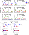
WHSC1 is required for UV-induced transcription and H3.3 incorporation. (A) WT and Whsc1−/− cells expressing H3.3-YFP were irradiated with UV-B (4 mJ/cm2) and induction of c-Fos and c-Jun mRNAs was detected at indicated times by qRT–PCR. Values represent the average of two determinations ±s.d. Gtf2b mRNA was tested as a control. (B) H3.3 incorporation into the indicated regions within the c-Fos (top), c-Jun (middle), and Gtf2b (bottom) genes was tested by ChIP analysis using anti-GFP antibody. Values represent the average of duplicate determinations ±s.d. (C) A model for WHSC1-dependent ISG elongation and H3.3 deposition. Upon IFN stimulation, BRD4 is recruited to ISGs, which in turn recruits WHSC1 and P-TEFb at the TSS (top). P-TEFb phosphorylates Pol II at serine 2 in the C-terminal domain to start elongation. WHSC1 recruits HIRA and these proteins travel together along with Pol II-2 P across the ISG coding region, launching elongation-coupled H3.3 deposition (middle). WHSC1 and HIRA continue to stay on the ISG coding region even after completion of ISG transcription to support sustained, post-elongation phase of H3.3 deposition (bottom).

Similar articles

Cited by

References

    1. Bartkowiak B, Liu P, Phatnani HP, Fuda NJ, Cooper JJ, Price DH, Adelman K, Lis JT, Greenleaf AL (2010) CDK12 is a transcription elongation-associated CTD kinase, the metazoan ortholog of yeast Ctk1. Genes Dev 24: 2303–2316 - PMC - PubMed
    1. Bergemann AD, Cole F, Hirschhorn K (2005) The etiology of Wolf-Hirschhorn syndrome. Trends Genet 21: 188–195 - PubMed
    1. Carrozza MJ, Li B, Florens L, Suganuma T, Swanson SK, Lee KK, Shia WJ, Anderson S, Yates J, Washburn MP, Workman JL (2005) Histone H3 methylation by Set2 directs deacetylation of coding regions by Rpd3S to suppress spurious intragenic transcription. Cell 123: 581–592 - PubMed
    1. Chao SH, Fujinaga K, Marion JE, Taube R, Sausville EA, Senderowicz AM, Peterlin BM, Price DH (2000) Flavopiridol inhibits P-TEFb and blocks HIV-1 replication. J Biol Chem 275: 28345–28348 - PubMed
    1. Chen S, Dai Y, Pei XY, Myers J, Wang L, Kramer LB, Garnett M, Schwartz DM, Su F, Simmons GL, Richey JD, Larsen DG, Dent P, Orlowski RZ, Grant S (2012) CDK inhibitors upregulate BH3-only proteins to sensitize human myeloma cells to BH3 mimetic therapies. Cancer Res 72: 4225–4237 - PMC - PubMed

Publication types

MeSH terms