. 2013 Oct;41(19):8926-42.
doi: 10.1093/nar/gkt706. Epub 2013 Aug 5.

A Common Docking Domain in Progesterone Receptor-B links DUSP6 and CK2 signaling to proliferative transcriptional programs in breast cancer cells

Affiliations

A Common Docking Domain in Progesterone Receptor-B links DUSP6 and CK2 signaling to proliferative transcriptional programs in breast cancer cells

Christy R Hagan et al. Nucleic Acids Res. 2013 Oct.

Abstract

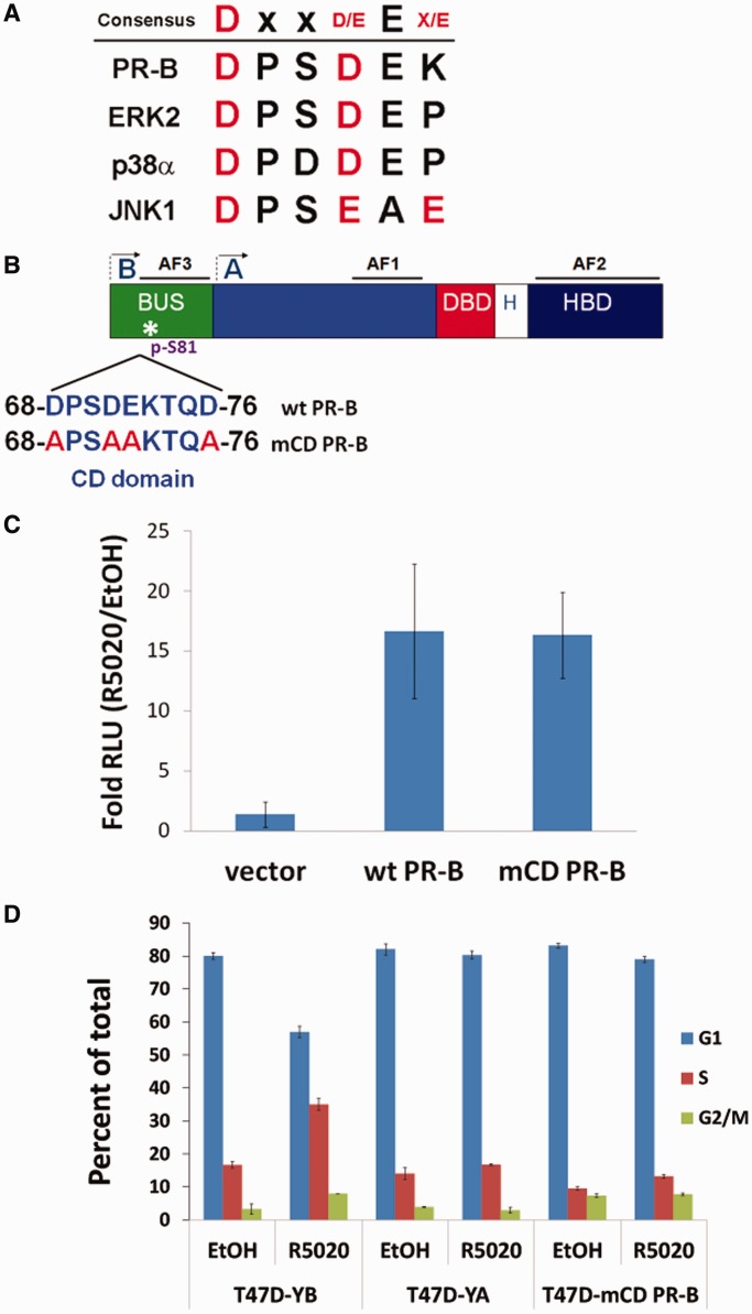
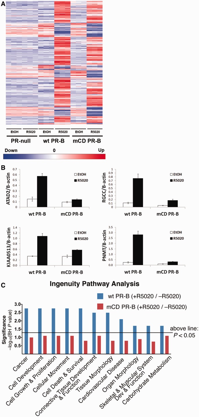
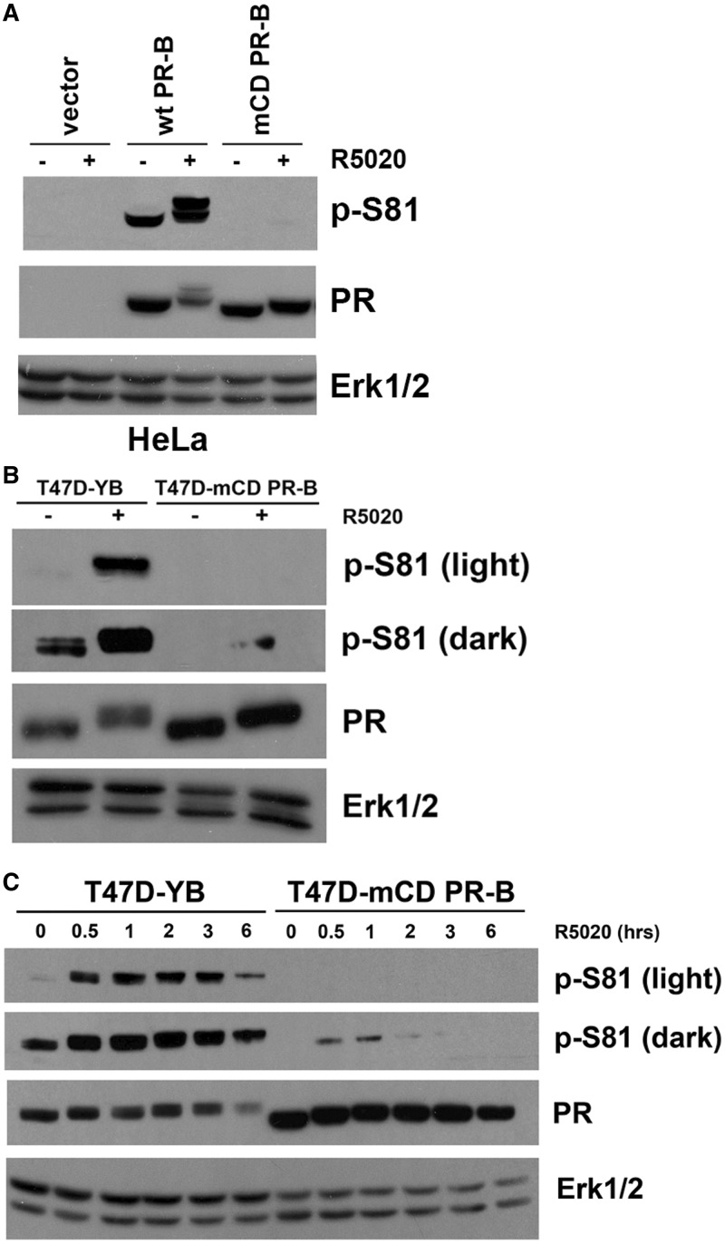
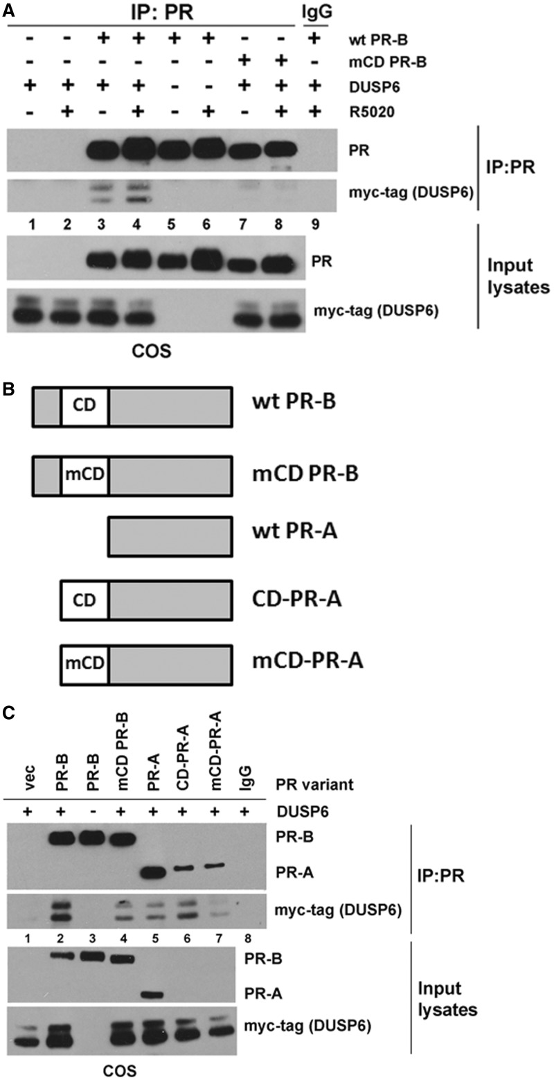
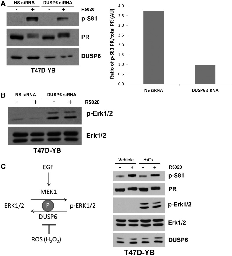
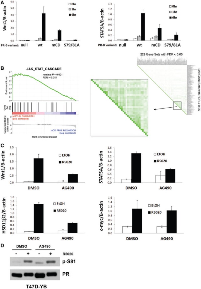
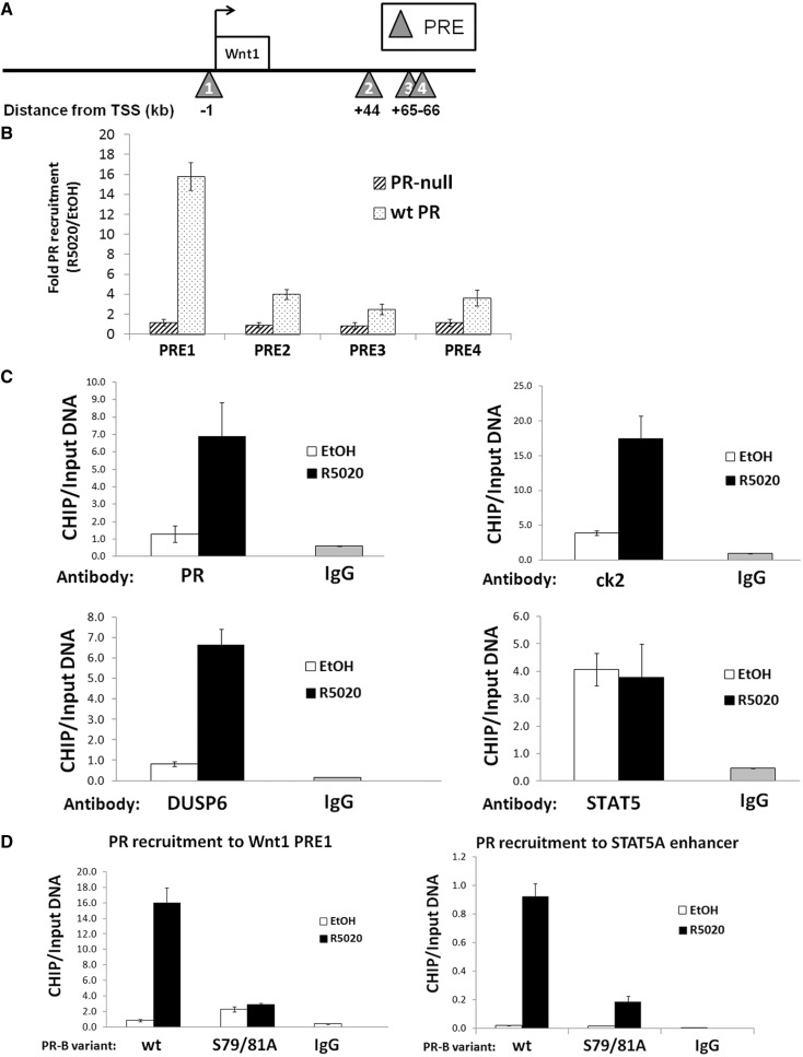
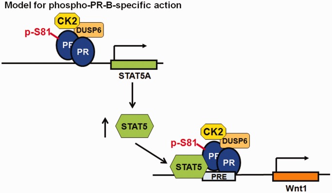
Progesterone receptors (PR) are transcription factors relevant to breast cancer biology. Herein, we describe an N-terminal common docking (CD) domain in PR-B, a motif first described in mitogen-activated protein kinases. Binding studies revealed PR-B interacts with dual-specificity phosphatase 6 (DUSP6) via the CD domain. Mutation of the PR-B CD domain (mCD) attenuated cell cycle progression and expression of PR-B target genes (including STAT5A and Wnt1); mCD PR-B failed to undergo phosphorylation on Ser81, a ck2-dependent site required for expression of these genes. PR-B Ser81 phosphorylation was dependent on binding with DUSP6 and required for recruitment of a transcriptional complex consisting of PR-B, DUSP6 and ck2 to an enhancer region upstream of the Wnt1 promoter. STAT5 was present at this site in the absence or presence of progestin. Furthermore, phospho-Ser81 PR-B was recruited to the STAT5A gene upon progestin treatment, suggestive of a feed-forward mechanism. Inhibition of JAK/STAT-signaling blocked progestin-induced STAT5A and Wnt1 expression. Our studies show that DUSP6 serves as a scaffold for ck2-dependent PR-B Ser81 phosphorylation and subsequent PR-B-specific gene selection in coordination with STAT5. Coregulation of select target genes by PR-B and STAT5 is likely a global mechanism required for growth promoting programs relevant to mammary stem cell biology and cancer.


Figures

Figure 1.
Figure 1.
CD domain in PR-B is required for progestin-induced S-phase entry. (A) Table comparing the CD domain core amino acid sequences in PR-B and selected MAPKs. (B) Schematic representing functional domains in wt PR-B. The full-length receptor has an N-terminal region unique to PR-B, termed the BUS. Both PR-B and the shorter form, PR-A, contain two activating function (AF 1 and 2) domains, a DNA-binding domain (DBD), hinge region (H) and a hormone-binding domain (HBD). PR-B contains an additional AF3, located in the BUS. Arrows mark the translational start sites for PR-B and PR-A. The CD domain is located between amino acids 68–76, immediately upstream of a ck2-dependent phosphorylation site, Ser81. Negatively charged amino acids were mutated to alanines (in red) to create the mCD PR-B mutant. (C) Reporter gene assays measuring transcriptional activity of PR constructs. HeLa cells were transiently transfected with plasmids expressing wt PR-B, mCD PR-B or vector, as well as a firefly PRE-luciferase reporter construct and Renilla expression control. Luciferase assays were performed as described in the ‘Materials and Methods’ section. Fold relative luciferase units (PRE-luciferase over Renilla luciferase controls) of R5020/EtOH-treated cells are plotted. Error bars represent ± SD of three independent experiments. (D) PR-B CD domain required for progestin-induced S-phase entry. T47D cells stably expressing wt PR-B (T47D-YB), PR-A (T47D-YA) or mCD PR-B (T47D-mCD PR-B) were starved for 18 h in serum-free media, followed by treatment with 10 nM R5020 or vehicle for 18 h. Single cells were analyzed by flow cytometry.
Figure 2.
Figure 2.
Gene expression analysis in mCD PR-B cells. (A) Heat map highlighting transcriptional differences between cells stably expressing wt PR-B, mCD PR-B or PR-null. The indicated T47D cells were treated with R5020 or vehicle. Genes differentially expressed >1.5 fold (BH-adjusted P < 0.01) are displayed for each treatment group. The experiment was performed in triplicate. (B) Validation of CD-regulated genes. T47D-Y cells stably expressing either wt or mCD PR-B were starved for 18 h in serum-free media, followed by treatment with 10 nM R5020 or EtOH for 6 h. mRNA levels for selected genes were analyzed by qPCR. Error bars represent ± SD. (C) IPA comparing >1.5-fold upregulated genes from wt PR-B and mCD PR-B expressing cells. Line represents P = 0.05 significance.
Figure 3.
Figure 3.
PR-B phosphorylation is altered by mutation to the CD of PR-B. (A) PR-B Ser81 phosphorylation is reduced in cells expressing mCD PR-B. HeLa cells were transiently transfected with wt PR-B, mCD PR-B or vector only. Twenty-four hours after transfection, cells were starved for 18 h in serum-free media and then treated with 10 nM R5020 or EtOH for 60 min. Lysates were analyzed via western blotting using p-S81, PR-B and Erk1/2 antibodies. (B) T47D cells stably expressing wt PR-B or mCD PR-B were starved and treated as in (A). Lysates were analyzed as in (A). A light and dark exposure of the western blotting film is shown for blotting with the p-Ser81 antibody. (C) Time course of Ser81 phosphorylation in cells expressing wt and mCD PR-B. T47D cells stably expressing wt PR-B or mCD PR-B were starved for 18 h in serum-free media, followed by treatment with 10 nM R5020 for 0–6 h. Lysates were analyzed via western blotting as in (A).
Figure 4.
Figure 4.
PR-B interacts with DUSP6 through the CD domain. (A) CD-domain dependent interaction between PR-B and DUSP6. COS cells were transiently transfected with wt PR-B, mCD PR-B or vector alone, as well as myc-tagged DUSP6 (or vector). Twenty-four hours after transfection, cells were starved for 18 h in serum-free media and then treated with 10 nM R5020 or EtOH for 60 min. Lysates were IP'd with PR antibody or mouse IgG (control). IP lysates or input lysates (25 µg not subjected to immunoprecipitation) were analyzed via western blotting using PR-B or myc-tag (to detect myc-tagged DUSP6) antibodies. Myc-tagged DUSP6 resolves as a doublet by western blotting due to alternate translational start sites. (B) Schematic representing wt and mutant PR-B constructs. The CD domain in wt PR-B (top) was mutated to create mCD PR-B (as described above). PR-A lacks a BUS region where the CD domain is located. A synthetic wt or mutant CD domain was fused to the N-terminus of wt PR-A to create CD-PR-A or mCD-PR-A, respectively. (C) CD domain-mediated interaction between PR-B and DUSP6. COS cells were transiently transfected with the indicated PR constructs or vector alone, as well as myc-tagged DUSP6 (or vector). CoIP and western blotting were performed as described in (A).
Figure 5.
Figure 5.
PR-B’s interaction with DUSP6 is required for PR-B Ser81 phosphorylation. (A) DUSP6 is necessary for ligand-induced PR-B Ser81 phosphorylation. Left: T47D cells stably expressing wt PR-B (T47D-YB) were transfected with 50 nM nonsilencing (NS) or DUSP6 siRNA. Seventy-two hours after transfection, cells were treated with 10 nM R5020 or EtOH for 60 min. Lysates were analyzed via western blotting using p-S81, PR-B and DUSP6 antibodies. Right: Densitometry was used to determine the ratio of Ser81 phosphorylated PR-B in R5020-treated cells to total PR-B in cells transfected with NS or DUSP6 siRNA. (B) Knockdown of DUSP6 increased MAPK activation. T47D-YB cells were transfected and treated as in (A), and lysates were analyzed via western blotting using p-Erk1/2 and Erk1/2 antibodies. (C) DUSP6 phosphatase activity is not needed to mediate ligand-induced PR-B Ser81 phosphorylation. Left: Schematic showing how ROS production (increased through treatment with H2O2) decreases DUSP6 phosphatase activity, which subsequently leads to an increase in Erk1/2 activity and phosphorylation. Right: T47D-YB cells were pretreated with 1 mM H2O2 for 20 min, followed by 10 nM R5020 (or EtOH) for 30 min. Lysates were analyzed via western blotting using p-S81, PR, p-Erk1/2, Erk1/2 and DUSP6 antibodies.
Figure 6.
Figure 6.
Endogenous phospho-Ser81 PR-B-target genes are JAK/STAT-dependent. (A) Select genes regulated by PR-B Ser81 phosphorylation. T47D-Y cells stably expressing either wt PR-B, mCD PR-B, S79/81A PR-B or unmodified (PR-null) cells were starved for 18 h in serum-free media, followed by treatment with 10 nM R5020 or EtOH for 0–6 h. mRNA levels were analyzed by qPCR. Error bars represent ±SD. (B) GSEA comparing gene expression data sets obtained using the Illumina Microarray described in Figure 3. Left: wt PR-B R5020/EtOH compared with mCD PR-B R5020/EtOH is shown as compared with the c5 Gene Ontology database, JAK_STAT_Cascade gene set. Right: Leading Edge analysis within GSEA (c2 Curated Gene Set) included 229 gene sets significantly enriched (FDR < 0.05) in wt PR-B cells, as compared with mCD PR-B. A subset of 38 gene sets (boxed region) contained substantial overlap (green shading) and were significantly involved in interferon signaling pathways. See Supplementary Table S1 for gene set names and statistics. (C) Ser81-dependent PR-B target genes are JAK/STAT-dependent. T47D-Y cells stably expressing wt PR-B (T47D-YB) were starved for 18 h in serum-free media. Cells were then pretreated (60 min) with 50 μM AG490, followed by 10 nM R5020 for 6 h. mRNA levels were analyzed by qPCR. Error bars represent ±SD. (D) PR-B Ser81 phosphorylation is unaffected by JAK/STAT-inhibition. T47D-YB cells were starved for 18 h in serum-free media. Cells were then pretreated (60 min) with 50 uM AG490, followed by 10 nM R5020 for 30 min. Lysates were analyzed via western blotting using p-S81 and total PR antibodies.
Figure 7.
Figure 7.
PR-B regulates Wnt1 transcription through binding to Wnt1 enhancer regions. (A) Schematic of the Wnt1 promoter/enhancer regions. PRE sites (triangles) are located upstream and downstream of the Wnt1 transcription start site (TSS; denoted with an arrow). Distance of the regulatory regions from the TSS is listed in kilobases (kb). (B) PR-B is recruited to Wnt1 PREs. T47D-Y cells stably expressing wt PR-B or unmodified cells (PR-null) were serum-starved for 18 h. Cells were then treated with 10 nM R5020 or EtOH for 60 min. Fixed lysates were subjected to ChIP with antibodies against PR-B or species-specific IgG (control), and qPCR was performed on the isolated DNA using primers designed to amplify the respective regulatory region (PREs 1–4). Recruitment of PR-B to PREs is shown as fold change between R5020- or EtOH-treated cells (R5020/EtOH). Error bars represent ±SD of triplicate experiments. (C) PR-B Ser81 phosphorylation-scaffolding complex is recruited to Wnt1 enhancer. T47D-Y cells stably expressing wt PR-B were serum-starved for 18 h. Cells were then treated with 10 nM R5020 or EtOH for 60 min. Fixed lysates were subjected to ChIP with antibodies against PR-B, ck2, DUSP6, STAT5 or species-specific IgG (controls), and qPCR was performed on the isolated DNA using primers designed to amplify Wnt1 PRE1. ChIP experiments were performed in triplicate. A representative experiment is shown here; error bars represent ±SD of technical replicates. (D) PR-B Ser81 phosphorylation is necessary for PR-B recruitment to Ser81-regulated genes. T47D-Y cells stably expressing wt PR-B or S79/81A PR-B were serum-starved for 18 h. Cells were then treated with 10 nM R5020 or EtOH for 60 min. Fixed lysates were subjected to ChIP with antibodies against PR or species-specific IgG (controls), and qPCR was performed on the isolated DNA using primers designed to amplify Wnt1 PRE1 or a STAT5A-enhancer site. ChIP experiments were performed in triplicate. A representative experiment is shown here; error bars represent ±SD of technical replicates.
Figure 8.
Figure 8.
Model of phospho-PR-B–specific action. PR-B CD domain-dependent recruitment of DUSP6 and ck2 is required for PR-B phosphorylation on Ser81. This complex is required for PR-B–dependent expression of STAT5A, whose protein product in turn then complexes with Ser81-phosphorylated PR-B on a specific-subset of PR-target genes, such as Wnt1. JAK/STAT-dependent regulation of phospho-Ser81 PR-B target genes regulates critical genes involved in mammary gland development, mammary stem cell maintenance/expansion and early events in breast cancer progression.

Similar articles

Cited by

References

    1. Kraus WL, Montano MM, Katzenellenbogen BS. Cloning of the rat progesterone receptor gene 5′-region and identification of two functionally distinct promoters. Mol. Endocrinol. 1993;7:1603–1616. - PubMed
    1. Kastner P, Krust A, Turcotte B, Stropp U, Tora L, Gronemeyer H, Chambon P. Two distinct estrogen-regulated promoters generate transcripts encoding the two functionally different human progesterone receptor forms A and B. EMBO J. 1990;9:1603–1614. - PMC - PubMed
    1. Kastner P, Krust A, Turcotte B, Stropp U, Tora L, Gronemeyer H, Chambon P. Two distinct estrogen-regulated promoters generate transcripts encoding the two functionally different human progesterone receptor forms A and B. EMBO J. 1990;9:1603–1614. - PMC - PubMed
    1. Hill KK, Roemer SC, Churchill ME, Edwards DP. Structural and functional analysis of domains of the progesterone receptor. Mol. Cell. Endocrinol. 2012;348:418–429. - PMC - PubMed
    1. Sartorius CA, Melville MY, Hovland AR, Tung L, Takimoto GS, Horwitz KB. A third transactivation function (AF3) of human progesterone receptors located in the unique N-terminal segment of the B-isoform. Mol. Endocrinol. 1994;8:1347–1360. - PubMed

Publication types

MeSH terms