

Neuron biomechanics probed by atomic force microscopy
- PMID: 23921683
- PMCID: PMC3759903
- DOI: 10.3390/ijms140816124
Neuron biomechanics probed by atomic force microscopy
Abstract
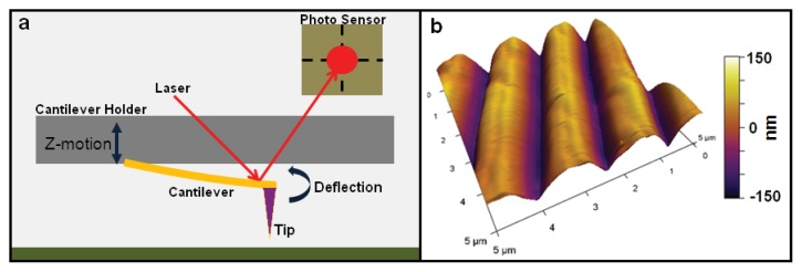
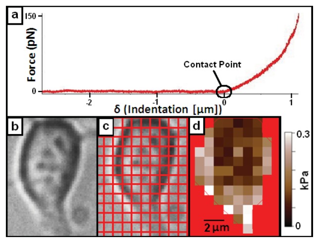
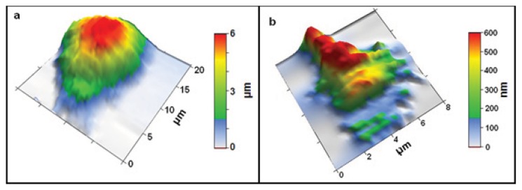
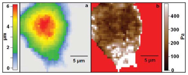
Mechanical interactions play a key role in many processes associated with neuronal growth and development. Over the last few years there has been significant progress in our understanding of the role played by the substrate stiffness in neuronal growth, of the cell-substrate adhesion forces, of the generation of traction forces during axonal elongation, and of the relationships between the neuron soma elastic properties and its health. The particular capabilities of the Atomic Force Microscope (AFM), such as high spatial resolution, high degree of control over the magnitude and orientation of the applied forces, minimal sample damage, and the ability to image and interact with cells in physiologically relevant conditions make this technique particularly suitable for measuring mechanical properties of living neuronal cells. This article reviews recent advances on using the AFM for studying neuronal biomechanics, provides an overview about the state-of-the-art measurements, and suggests directions for future applications.
Figures
Similar articles
-
Substrate Deformation Predicts Neuronal Growth Cone Advance.Biophys J. 2015 Oct 6;109(7):1358-71. doi: 10.1016/j.bpj.2015.08.013. Biophys J. 2015. PMID: 26445437 Free PMC article.
-
Combined Traction Force-Atomic Force Microscopy Measurements of Neuronal Cells.Biomimetics (Basel). 2022 Oct 8;7(4):157. doi: 10.3390/biomimetics7040157. Biomimetics (Basel). 2022. PMID: 36278714 Free PMC article.
-
Variations of Elastic Modulus and Cell Volume with Temperature for Cortical Neurons.Langmuir. 2019 Aug 20;35(33):10965-10976. doi: 10.1021/acs.langmuir.9b01651. Epub 2019 Aug 9. Langmuir. 2019. PMID: 31380651 Free PMC article.
-
Nanomechanics of Cells and Biomaterials Studied by Atomic Force Microscopy.Adv Healthc Mater. 2015 Nov 18;4(16):2456-74. doi: 10.1002/adhm.201500229. Epub 2015 Jul 22. Adv Healthc Mater. 2015. PMID: 26200464 Review.
-
Atomic force microscopy and its contribution to understanding the development of the nervous system.Curr Opin Genet Dev. 2011 Oct;21(5):530-7. doi: 10.1016/j.gde.2011.07.001. Epub 2011 Aug 16. Curr Opin Genet Dev. 2011. PMID: 21840706 Review.
Cited by
-
Quantifying the Local Mechanical Properties of Cells in a Fibrous Three-Dimensional Microenvironment.Biophys J. 2019 Sep 3;117(5):817-828. doi: 10.1016/j.bpj.2019.07.042. Epub 2019 Jul 31. Biophys J. 2019. PMID: 31421835 Free PMC article.
-
Modeling of the axon membrane skeleton structure and implications for its mechanical properties.PLoS Comput Biol. 2017 Feb 27;13(2):e1005407. doi: 10.1371/journal.pcbi.1005407. eCollection 2017 Feb. PLoS Comput Biol. 2017. PMID: 28241082 Free PMC article.
-
Atomic Force Microscopy Protocol for Measurement of Membrane Plasticity and Extracellular Interactions in Single Neurons in Epilepsy.Front Aging Neurosci. 2016 May 4;8:88. doi: 10.3389/fnagi.2016.00088. eCollection 2016. Front Aging Neurosci. 2016. PMID: 27199735 Free PMC article.
-
An Integrated Cytoskeletal Model of Neurite Outgrowth.Front Cell Neurosci. 2018 Nov 26;12:447. doi: 10.3389/fncel.2018.00447. eCollection 2018. Front Cell Neurosci. 2018. PMID: 30534055 Free PMC article. Review.
-
Heavy ion and X-ray irradiation alter the cytoskeleton and cytomechanics of cortical neurons.Neural Regen Res. 2014 Jun 1;9(11):1129-37. doi: 10.4103/1673-5374.135315. Neural Regen Res. 2014. PMID: 25206772 Free PMC article.
References
-
- Franze K., Guck J. The biophysics of neuronal growth. Rep. Prog. Phys. 2010;73 doi: 10.1088/0034-4885/73/9/094601. - DOI
-
- Franze K., Janmey P.A., Guck J. Mechanics in neuronal development and repair. Annu. Rev. Biomed. Eng. 2013;15:227–251. - PubMed
-
- Sokolov I. Atomic force microscopy in cancer cell research. In: Nalwa H.S., Webster T.J., editors. Cancer Nanotechnology: Nanomaterials for Cancer Diagnostics and Therapy. Vol. 1. American Scientific Publishers; Valencia, CA, USA: 2007. pp. 1–17.
-
- García R., Pérez R. Dynamic atomic force microscopy methods. Surf. Sci. Rep. 2002;47:197–301.
Publication types
MeSH terms
LinkOut - more resources
Full Text Sources
Other Literature Sources
Miscellaneous
