. 2013 Nov;33(11):1658-65.
doi: 10.1038/jcbfm.2013.139. Epub 2013 Aug 7.

The role of PARL and HtrA2 in striatal neuronal injury after transient global cerebral ischemia

Affiliations

The role of PARL and HtrA2 in striatal neuronal injury after transient global cerebral ischemia

Hideyuki Yoshioka et al. J Cereb Blood Flow Metab. 2013 Nov.

Abstract

The presenilin-associated rhomboid-like (PARL) protein and high temperature requirement factor A2 (HtrA2) are key regulators of mitochondrial integrity and play pivotal roles in apoptosis. However, their roles after cerebral ischemia have not been thoroughly elucidated. To clarify these roles, mice were subjected to transient global cerebral ischemia, and striatal neuronal injury was assessed. Western blot and coimmunoprecipitation analyses revealed that PARL and processed HtrA2 localized to mitochondria, and that PARL was bound to HtrA2 in sham animals. Expression of PARL and processed HtrA2 in mitochondria significantly decreased 6 to 72 hours after ischemia, and the binding of PARL to HtrA2 disappeared after ischemia. In contrast, expression of processed HtrA2 increased 24 hours after ischemia in the cytosol, where HtrA2 was bound to X chromosome-linked inhibitor-of-apoptosis protein (XIAP). Administration of PARL small interfering RNA inhibited HtrA2 processing and worsened ischemic neuronal injury. Our results show that downregulation of PARL after ischemia is a key step in ischemic neuronal injury, and that it decreases HtrA2 processing and increases neuronal vulnerability. In addition, processed HtrA2 released into the cytosol after ischemia contributes to neuronal injury via inhibition of XIAP.


Figures

Figure 1
Figure 1
Western blot analysis of presenilin-associated rhomboid-like (PARL) protein in the striatum. (A) Western blot analysis of PARL from the cytosolic (Cyt) and mitochondrial (Mt) fractions showed mitochondrial localization of PARL under normal conditions. The immunoreactivity of PARL was evident with a molecular mass of 36 kDa. (B) Western blot analysis of PARL in the striatum after transient global cerebral ischemia (tGCI). Expression of PARL was not observed in the cytosolic fraction even after tGCI. In the mitochondrial fraction, expression of PARL decreased significantly 6 to 72 hours after tGCI (n=6, *P<0.05). As internal controls, β-actin and cytochrome oxidase subunit IV (COX-IV) were used for the cytosolic and mitochondrial fractions, respectively. OD, optical density; S, sham.
Figure 2
Figure 2
Representative photomicrographs of presenilin-associated rhomboid-like (PARL) protein immunohistochemistry. (A) Time course of PARL immunohistochemistry in the striatum after transient global cerebral ischemia (tGCI). Expression of PARL was decreased after tGCI. Scale bar=100 μm. (B) Double immunofluorescence of PARL (green) and neuronal nuclei (NeuN; a neuronal marker, red) in the striatum of sham animals. In the NeuN-positive striatal neurons, PARL was expressed. Scale bar=50 μm. (C) Double immunofluorescence of cytochrome oxidase subunit IV (COX-IV) (a mitochondrial marker; green) and PARL (red) in the striatum of sham animals. In the striatum, PARL colocalized with COX-IV. Scale bar=50 μm. (D) Double immunofluorescence of PARL (red) and terminal deoxynucleotidyl transferase-mediated uridine 5′-triphosphate-biotin nick end labeling (TUNEL) staining (green) in the striatum 24 hours after tGCI. In TUNEL-positive cells, PARL was not expressed. Scale bar=50 μm. DAPI, 4′,6-diamidino-2-phenylindole.
Figure 3
Figure 3
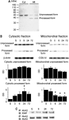
Western blot analysis of high temperature requirement factor A2 (HtrA2) in the striatum. (A) Western blot analysis of HtrA2 under normal conditions showed unprocessed forms at 50 kDa and processed forms at 38 kDa. In the cytosolic fraction (Cyt), only the unprocessed forms were observed. In contrast, both unprocessed and processed forms were observed in the mitochondrial fraction (Mt). (B) Western blot analysis of HtrA2 in the striatum after transient global cerebral ischemia (tGCI). In the cytosolic fraction, expression of processed forms increased significantly 24 hours after tGCI (n=6, *P<0.05), although unprocessed forms did not show a significant change. In the mitochondrial fraction, expression of processed HtrA2 showed a gradual decrease in a time-dependent manner after tGCI. A quantitative analysis showed that processed HtrA2 in mitochondria was decreased significantly 6 to 72 hours after tGCI (n=6, *P<0.05). As internal controls, β-actin and cytochrome oxidase subunit IV (COX-IV) were used for the cytosolic and mitochondrial fractions, respectively. OD, optical density; S, sham. (C) Coimmunoprecipitation analysis for immunoreactivity of X chromosome-linked inhibitor-of-apoptosis protein (XIAP) precipitated by HtrA2 in the striatum after tGCI. Immunoreactivity of XIAP showed a gradual increase after tGCI. To show equal precipitation, HtrA2 was used. IB, immunoblotting; IP, immunoprecipitation.
Figure 4
Figure 4
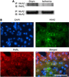
Interaction between presenilin-associated rhomboid-like (PARL) protein and high temperature requirement factor A2 (HtrA2). (A) Coimmunoprecipitation analysis for PARL immunoreactivity precipitated by HtrA2 in the striatum after transient global cerebral ischemia (tGCI). The immunoreactivity of PARL disappeared 24 hours after tGCI. To show equal precipitation, HtrA2 was used. (B) Representative photomicrographs of PARL and HtrA2 double immunostaining in the striatum of sham animals. Presenilin-associated rhomboid-like protein colocalized with HtrA2. Scale bar=50 μm. DAPI, 4′,6-diamidino-2-phenylindole; IB, immunoblotting; IP, immunoprecipitation.
Figure 5
Figure 5
The effects of presenilin-associated rhomboid-like protein-small interfering RNA (PARL-siRNA) on the expression of PARL and high temperature requirement factor A2 (HtrA2). Control and PARL-siRNA (0.5, 1, or 2 μg) were administered into the right striatum. The animals were killed 48 hours after the siRNA injection. Western blot analysis showed that expression of PARL and processed HtrA2 in the mitochondrial fraction (Mt) was significantly decreased in the animals treated with 2 μg PARL-siRNA compared with the nontreated animals (n=4, *P<0.05). In the cytosolic fraction (Cyt), expression of HtrA2 and PARL was not changed with siRNA treatment. As internal controls, β-actin and cytochrome oxidase subunit IV (COX-IV) were used for the cytosolic and mitochondrial fractions, respectively. C, control; NT, nontreated; OD, optical density.
Figure 6
Figure 6
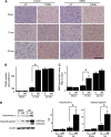
Treatment with 2 μg presenilin-associated rhomboid-like protein-small interfering RNA (PARL-siRNA) worsened ischemic injury in the striatum after transient global cerebral ischemia (tGCI). Small interfering RNA was injected into the right striatum 48 hours before surgery. (A) Representative photomicrographs of cresyl violet (CV) and terminal deoxynucleotidyl transferase-mediated uridine 5′-triphosphate-biotin nick end labeling (TUNEL) staining. Although the ischemic damage 3 days after 17 minutes of tGCI was mild in the control siRNA group, severe ischemic damage in the PARL-siRNA group was observed. In the 22-minute tGCI group, severe ischemic change was observed in both the control and PARL-siRNA groups. (B) Cell counting study showed that the number of TUNEL-positive cells in the PARL-siRNA group was significantly higher than in the control-siRNA group 3 days after 17 minutes of tGCI (n=5, *P<0.05). (C) Cell death assay showed that DNA fragmentation 3 days after 17 minutes of tGCI in the PARL-siRNA group was significantly higher than in the control group (n=5, *P<0.05). (D) Western blot analysis of cytochrome c and spectrin using the cytosolic fraction. Treatment with PARL-siRNA significantly increased cytochrome c release into the cytosol 24 hours after 17 minutes of tGCI (n=5, *P<0.05). Cleaved spectrin products (both 150 and 120 kDa) 24 hours after 17 minutes of tGCI in the PARL-siRNA group were significantly higher than in the control group (n=5, *P<0.05). C, control; Isc, ischemia; OD, optical density; siR, PARL-siRNA.
Figure 7
Figure 7
Summary of the roles of high temperature requirement factor A2 (HtrA2) and presenilin-associated rhomboid-like (PARL) protein in ischemic neuronal injury. Processed HtrA2, maintaining mitochondrial integrity under physiologic conditions, decreased after ischemia caused by the downregulation of PARL, which results in mitochondria (Mt) dysfunction and, finally, ischemic neuronal injury. In addition, the residual processed HtrA2 is released into the cytosol, which induces apoptosis via interacting X chromosome-linked inhibitor-of-apoptosis protein (XIAP).

Comment in

Similar articles

Cited by

References

    1. Duchen MR. Mitochondria in health and disease: perspectives on a new mitochondrial biology. Mol Aspects Med. 2004;25:365–451. - PubMed
    1. Orrenius S, Gogvadze V, Zhivotovsky B. Mitochondrial oxidative stress: implications for cell death. Annu Rev Pharmacol Toxicol. 2007;47:143–183. - PubMed
    1. Kroemer G, Reed JC. Mitochondrial control of cell death. Nat Med. 2000;6:513–519. - PubMed
    1. Lin MT, Beal MF. Mitochondrial dysfunction and oxidative stress in neurodegenerative diseases. Nature. 2006;443:787–795. - PubMed
    1. Chan PH. Reactive oxygen radicals in signaling and damage in the ischemic brain. J Cereb Blood Flow Metab. 2001;21:2–14. - PubMed

Publication types

MeSH terms

LinkOut - more resources