. 2013 Dec;21(12):2247-57.
doi: 10.1038/mt.2013.189. Epub 2013 Aug 7.

Silencing PP2A inhibitor by lenti-shRNA interference ameliorates neuropathologies and memory deficits in tg2576 mice

Affiliations

Silencing PP2A inhibitor by lenti-shRNA interference ameliorates neuropathologies and memory deficits in tg2576 mice

Gong-Ping Liu et al. Mol Ther. 2013 Dec.

Abstract

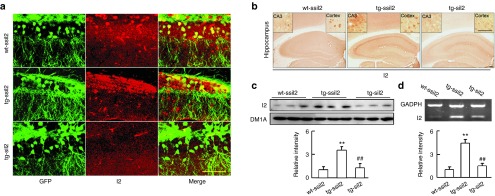
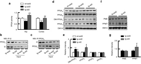
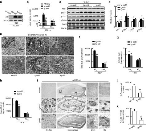
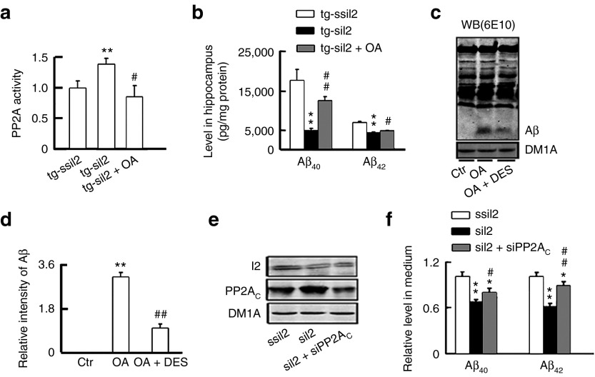
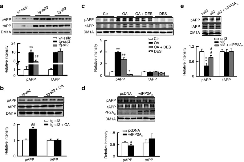
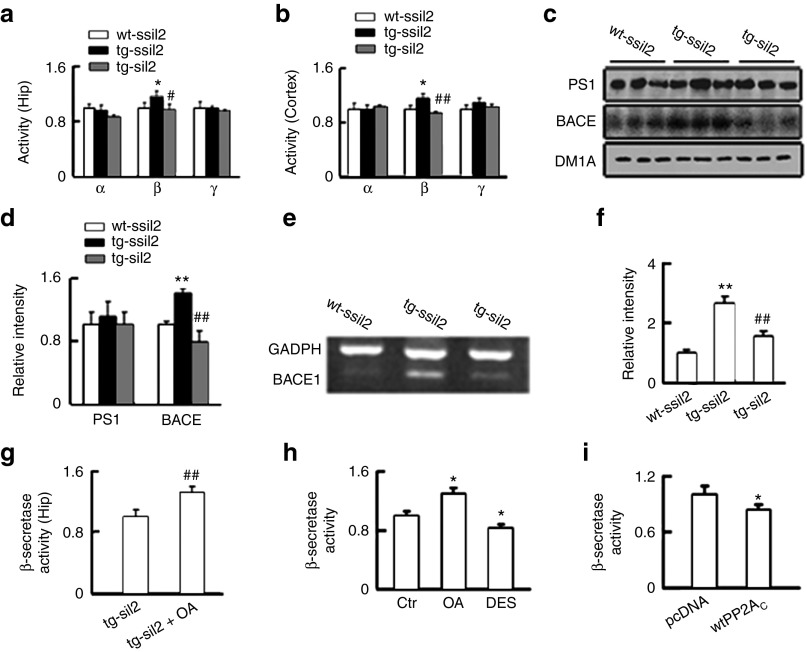
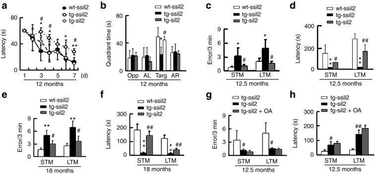
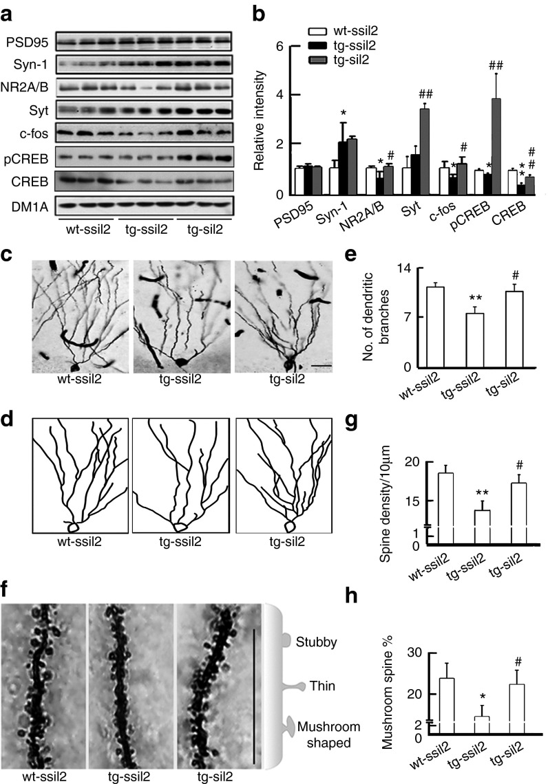
Deficits of protein phosphatase-2A (PP2A) play a crucial role in tau hyperphosphorylation, amyloid overproduction, and synaptic suppression of Alzheimer's disease (AD), in which PP2A is inactivated by the endogenously increased inhibitory protein, namely inhibitor-2 of PP2A (I2(PP2A)). Therefore, in vivo silencing I2(PP2A) may rescue PP2A and mitigate AD neurodegeneration. By infusion of lentivirus-shRNA targeting I2(PP2A) (LV-siI2(PP2A)) into hippocampus and frontal cortex of 11-month-old tg2576 mice, we demonstrated that expression of LV-siI2(PP2A) decreased remarkably the elevated I2(PP2A) in both mRNA and protein levels. Simultaneously, the PP2A activity was restored with the mechanisms involving reduction of the inhibitory binding of I2(PP2A) to PP2A catalytic subunit (PP2AC), repression of the inhibitory Leu309-demethylation and elevation of PP2AC. Silencing I2(PP2A) induced a long-lasting attenuation of amyloidogenesis in tg2576 mice with inhibition of amyloid precursor protein hyperphosphorylation and β-secretase activity, whereas simultaneous inhibition of PP2A abolished the antiamyloidogenic effects of I2(PP2A) silencing. Finally, silencing I2(PP2A) could improve learning and memory of tg2576 mice with preservation of several memory-associated components. Our data reveal that targeting I2(PP2A) can efficiently rescue Aβ toxicities and improve the memory deficits in tg2576 mice, suggesting that I2(PP2A) could be a promising target for potential AD therapies.


Figures

Figure 1
Figure 1
Expression of LV-siI2PP2A downregulates I2PP2A (I2). Virus expressing eGFP-labeled LV-I2PP2A (lentivirus) shRNA (siI2) or the scrambled control (ssiI2) (2 × 109 TU/ml) were injected into the hippocampal CA3 and CA1 and the frontal cortex of tg2576 mice (tg-ssiI2 and tg-siI2) or the wild type littermates (wt-ssiI2) at ~11 months old under a stereotaxic apparatus (2 µl per site). At ~12.5 months, the mice were killed for the following measurements. (a) The representative images show expression of LV-siI2 (green, direct fluorescent image) and the silencing efficacy of I2 (red, with I2 antibody) at hippocampal dentate gyrus (DG) of ~12.5-month mice (n = 3). Scale bars: 100 µm. (b) Level of I2 protein increased in cortex and hippocampus of tg2576 mice compared with the wt littermates, and expression of siI2 reduced the I2 signal measured by immunohistochemistry staining using I2 antibody (the inserts show enlarged images, n = 3). (c) Level of I2 protein in hippocampus of tg2576 mice increased ~3.5-fold of the control level and expression of LV-siI2 reduced I2 protein almost to the normal level measured by Western blotting (n = 3–5). (d) Level of I2 mRNA in hippocampus of tg2576 mice increased ~4.5-fold of the control level and expression of LV-siI2 partially reduced I2 mRNA level measured by RT-PCR (n = 3–5). **P < 0.01 versus wt-ssiI2; ##P < 0.01 versus tg-ssiI2 (mean ± SD).
Figure 2
Figure 2
Silencing I2PP2A activates PP2A by multiple mechanisms. The mice were treated as described in Figure 1. (a) The activity of PP2A in hippocampus and cortex extracts of ~12.5-month tg2576 mice decreased and silencing I2PP2A (I2) by expression of LV-siI2 restored or further stimulated PP2A activity, measured by using a Serine/Threonine Phosphatase Assay Kit (n = 3–5). (b–c) Silencing I2 reduced the inhibitory binding of I2 with PP2AC in hippocampus of tg2576 mice, measured by immunoprecipitation (IP) and Western blot (WB) using anti-I2 or anti-PP2AC antibodies as indicated (n = 3). (d–e) Level of Leu309-demethylated PP2AC (DM-PP2AC) increased in hippocampus of tg2576 mice, and silencing I2 reduced the inhibitory demethylation of PP2AC with a significantly increased total level of PP2AC, whereas the protein levels PP2AA and PP2AB were not changed, measured by Western blotting (n = 3~5). (f–g) Silencing I2 increased protein level of PP2A methyltransferase (PPMT) with no change of PP2A methylesterase (PME) in hippocampus of tg2576 mice measured by Western blotting (n = 3–5). **P < 0.01 versus wt-ssiI2; #P < 0.05; ##P < 0.01 versus tg-ssiI2 (mean ± SD).
Figure 3
Figure 3
Silencing I2PP2A induces a long-lasting attenuation of amyloidogenesis in tg2576 mice. The mice were treated as described in Figure 1. (a) The representative Western blot show an increased Aβ level in hippocampal extracts of ~12.5-month tg2576 mice infused with the scrambled siI2PP2A (tg-ssiI2) compared with the age-matched wild-type littermates (wt-ssiI2), and silencing I2 by LV-siI2PP2A (tg-siI2) reduced Aβ level compared with the tg-ssiI2 controls (n = 3). (b) ELISA data show increased levels of Aβ40 and Aβ42 in hippocampal extracts of tg2576 mice and the reduction of Aβ by silencing I2. The experiments were repeated for at least three times with triplicates. (c–d) An increased tau phosphorylation at multiple AD-related sites in tg2576 mice and the attenuation by I2 knockdown detected by Western blotting (note that tau-1 reacts with the unphosphorylated tau, therefore, an increased immunoreaction indicates reduced tau phosphorylation) (n = 3–5). (e) The representative silver staining images show an enhanced intracellular accumulation of the argyrophilic substances in hippocampus and the cortex of tg2576 mice, and the reduced accumulation by LV-siI2PP2A. Scale bars: 100 µm (n = 3). (f–h) At 18 months, LV-siI2 infused (at ~11 months) tg2576 mice still show lower levels of total and insoluble Aβ in hippocampus than the LV-ssiI2-infused control mice. The ELISA assay was repeated at least three times in triplicates. (i–k) The representative images and the quantitative analyses show increase of plaque load in hippocampus and cortex of ~23-month tg2576 mice immunostained with Aβ antibody, and attenuation of by I2 knockdown measured at the virus injection sites. Scale bars: 100 µm (n = 3). **P < 0.01 versus wt-ssiI2; #P < 0.05; ##P < 0.01 versus tg-ssiI2 (mean ± SD).
Figure 4
Figure 4
Simultaneous inhibition of PP2A abolishes the siI2PP2A-induced attenuation of Aβ accumulation. (a) The mice were first injected as described in Figure 1. After ~1.5 months, okadaic acid (OA) was infused into the hippocampal CA3 and CA1 (0.2 µmol/l, 2 µl per site), and inhibition of PP2A activity by OA was detected in hippocampal extracts by using a Serine/Threonine Phosphatase Assay Kit (n = 3–5 in triplicates). (b) The mice were treated as described in panel a. Levels of Aβ were reduced by expression of LV-siI2 in 12.5-month tg2576 mice, while simultaneous inhibition of PP2A by OA reversed partially the siI2-induced reduction of Aβ levels measured by ELISA assay (n = 3–5 in triplicates). (c–d) N2a/APP cells treated with 5 nmol/l OA for 24 hours increased Aβ levels, whereas PP2A activator C6-ceramide (DES) (7 nmol/l) treatment attenuated the OA-induced Aβ production. The proteins were isolated by native Tris-tricine gradient gel electrophoresis (8–12%) and probed by antibody 6E10. The experiment was repeated for three times. (e) Silencing I2 increased PP2AC in N2a/APP cells, whereas simultaneous expression of siPP2AC for 24 hours downregulated the siI2-induced overexpression of PP2AC measured by Western blotting. The experiment was repeated at least three times. (f) Silencing I2 reduced Aβ levels, whereas simultaneous downregulation of PP2AC by siPP2AC partially reversed the siI2-induced Aβ reduction in the culture medium of N2a/APP cells measured by ELISA. The experiment was repeated at least three times with triplicates. *P < 0.05 versus ssiI2; **P < 0.01 versus tg-ssiI2 or ssiI2 or Ctr; #P < 0.05; ##P < 0.01 versus tg-siI2 or siI2 or OA (mean ± SD).
Figure 5
Figure 5
Activation of PP2A mediates the siI2PP2A-induced APP dephosphorylation at Thr668. (a) The mice were treated as described in Figure 1. The levels of total APP (tAPP) and the Thr668-phosphorylated APP (pAPP) were increased in hippocampal extracts of ~12.5 months tg2576 mice (tg-ssiI2) compared with the age-matched wild-type littermates (wt-ssiI2), both mice were transfected with the scrambled siI2PP2A; expression of siI2 (tg-siI2) decreased the phosphorylation level of APP compared with tg-ssiI2, measured by Western blotting (n = 6). (b) In tg2576 mice, simultaneous inhibition of PP2A by hippocampal (CA3 and CA1) infusion of okadaic acid (2 µl each) reversed the siI2PP2A-suppressed APP phosphorylation (n = 6). (c) In N2a/APP cells, inhibition of PP2A by OA increased APP phosphorylation compared with the control (Ctr), whereas simultaneous activation of PP2A by DES attenuated the OA-induced APP phosphorylation. The experiments were repeated two times in triplicates. (d) In N2a/APP cells, upregulation of PP2A by overexpression of wild type PP2AC (wtPP2AC) reduced the phosphorylation level of APP. The experiments were repeated two times in triplicates. (e) In N2a/APP cells, I2 knockdown by expression of siI2PP2A (siI2) reduced APP phosphorylation, while simultaneous inhibition of PP2A by expression of siPP2AC reversed the siI2-suppressed APP phosphorylation. The experiments were repeated at least for three times. *P < 0.05; **P < 0.01 versus wt-ssiI2 or Ctr or ssiI2; #P < 0.05 versus pcDNA; ##P < 0.01 vs tg-ssiI2 or tg-siI2 or siI2 or OA (mean ± SD).
Figure 6
Figure 6
Activation of PP2A mediates the siI2PP2A-induced inhibition of β-secretase. (a–b) The mice were treated as described in Figure 1, and then the activity of α-, β-, and γ-secretases in hippocampus and cortex extracts of ~12.5 months tg2576 mice and the wild-type littermates (wt) was measured. β-secretase was activated in tg2576 mice compared with the wt, and expression of LV-siI2PP2A (siI2) inhibited β-secretase. The experiments were repeated at least two times in triplicates. (c–d) The protein level of β-site APP-cleaving enzyme 1 (BACE1) was increased in hippocampal extracts of ~12.5 months tg2576 mice measured by Western blotting, and expression of LV-siI2PP2A reduced the protein level of BACE1 (n = 3–5). (e–f) The mRNA level of BACE1 was increased in hippocampal extracts of ~12.5 months tg2576 mice measured by RT-PCR, and expression of LV-siI2PP2A reduced the mRNA level of BACE1 (n = 3–5). (g) Simultaneous inhibition of PP2A by hippocampal infusion of OA (2 µl each at CA3 and CA1) for 48 hours abolished the LV-siI2-induced inhibition of β-secretase in ~12.5 months tg2576 mice after injection of LV-siI2 for 6 weeks (n = 3). (h) In N2a/APP cells, inhibition of PP2A by OA (5 nmol/l) for 24 hours activated β-secretase, whereas activation of PP2A by DES (7 nmol/l) for 24 hours inhibited β-secretase. The experiments were repeated at least three times in triplicates. (i) In N2a/APP cells, overexpression of wild-type PP2AC (wtPP2AC) for 24 hours inhibited β-secretase when compared with pcDNA vector transfected cells. The experiments were repeated at least three times in duplicates. *P < 0.05; **P < 0.01 versus wt-ssiI2 or Ctr or pcDNA; #P < 0.05; ##P < 0.01 versus tg-ssiI2 or tg-siI2 or OA (mean ± SD).
Figure 7
Figure 7
Silencing I2PP2A improves learning and memory independent of PP2A activation in tg2576 mice. (a) Tg2576 mice (tg) at ~12 months had spatial learning deficit compared with the wild-type littermates (wt), and silencing I2PP2A (I2) improved the learning ability measured by the latency to find the hidden platform during 7 days water maze training (n = 15–20 each group). (b) At ~12 months, the tg-mice showed spatial memory deficits and silencing I2 improved the memory ability measured by the time spent in each quadrant after removed platform at day 9 (OPP, opposite quadrant; AL, adjacent left quadrant; Targ, target quadrant; AR, adjacent right quadrant; n = 15–20 each group). (c–d) At ~12.5 months, the tg-mice showed deficits of short-term memory (STM) (measured at 15 minutes after training) and long-term memory (LTM) (measured at 24 hours after training) by step-down avoidance test, and knockdown I2 improved the memory (n = 15–20 for each group). (e–f) At ~18 months, the tg-mice showed deficits of STM and LTM measured by step-down avoidance test, and knockdown I2 improved the memory (n = 8–10 each group). (g–h) Simultaneous inhibition of PP2A by hippocampal (CA3 and CA1) infusion of OA after expression of LV-siI2PP2A for 6 weeks did not significantly affect the siI2-induced improvement of STM and LTM measured at ~12.5 months (n = 6–7 each group). *P < 0.05; **P < 0.01 versus wt-ssiI2; #P < 0.05; ##P < 0.01 versus tg-ssiI2 (mean ± SD).
Figure 8
Figure 8
Silencing I2PP2A preserves several memory-associated components. (a–b) Levels of NR2A/B, c-fos, CREB, and pCREB were reduced in ~12.5 months tg2576 mice, whereas knockdown I2PP2A (I2) restored the levels of the memory-associated proteins analyzed by Western blot (n = 3–5). (c–e) The representative images showing decreased dendrite branch numbers in hippocampal DG region of ~12.5-month tg2576 mice, and preservation of the dendrite complexity by knockdown I2. (c) Dendritic arbors shown by Golgi stain. (d) Reconstruction of the dendritic arbors. (e) Quantitative analysis of dendrite branch numbers. (f–h) The total spine number and the mushroom-shaped spines were decreased in ~12.5-month tg2576 mice, and knockdown I2 preserved spine plasticity analyzed by Golgi stain and NeuronStudio system. Scale bars: 100 µm. *P < 0.05; **P < 0.01 versus wt-ssiI2; #P < 0.05; ##P < 0.01 versus tg-ssiI2 (mean ± SD).

Similar articles

Cited by

References

    1. Braak H, Braak E. Neuropathological stageing of Alzheimer-related changes. Acta Neuropathol. 1991;82:239–259. - PubMed
    1. Wang JZ, Gong CX, Zaidi T, Grundke-Iqbal I, Iqbal K. Dephosphorylation of Alzheimer paired helical filaments by protein phosphatase-2A and -2B. J Biol Chem. 1995;270:4854–4860. - PubMed
    1. Liu F, Grundke-Iqbal I, Iqbal K, Gong CX. Contributions of protein phosphatases PP1, PP2A, PP2B and PP5 to the regulation of tau phosphorylation. Eur J Neurosci. 2005;22:1942–1950. - PubMed
    1. Gong CX, Shaikh S, Wang JZ, Zaidi T, Grundke-Iqbal I, Iqbal K. Phosphatase activity toward abnormally phosphorylated tau: decrease in Alzheimer disease brain. J Neurochem. 1995;65:732–738. - PubMed
    1. Sun L, Liu SY, Zhou XW, Wang XC, Liu R, Wang Q, et al. Inhibition of protein phosphatase 2A- and protein phosphatase 1-induced tau hyperphosphorylation and impairment of spatial memory retention in rats. Neuroscience. 2003;118:1175–1182. - PubMed

Publication types

MeSH terms