. 2013 Aug 6;2(8):e113.
doi: 10.1038/mtna.2013.38.

Toxicology and Biodistribution Studies for MGH2.1, an Oncolytic Virus that Expresses Two Prodrug-activating Genes, in Combination with Prodrugs

Affiliations

Toxicology and Biodistribution Studies for MGH2.1, an Oncolytic Virus that Expresses Two Prodrug-activating Genes, in Combination with Prodrugs

Kazue Kasai et al. Mol Ther Nucleic Acids. .

Abstract

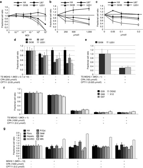
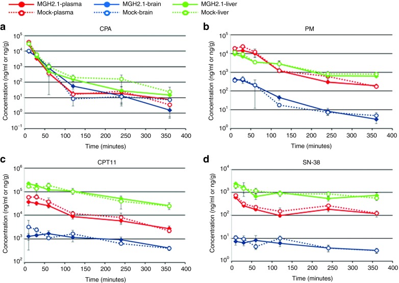
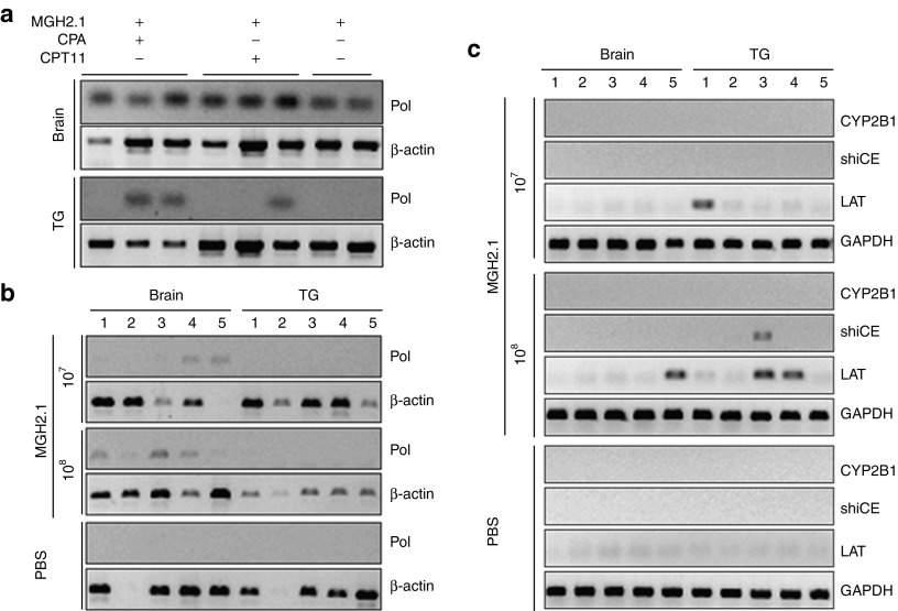
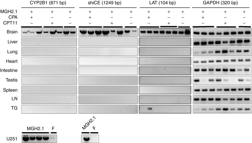
MGH2.1 is a herpes simplex virus type 1 (HSV1) oncolytic virus that expresses two prodrug-activating transgenes: the cyclophosphamide (CPA)-activating cytochrome P4502B1 (CYP2B1) and the CPT11-activating secreted human intestinal carboxylesterase (shiCE). Toxicology and biodistribution of MGH2.1 in the presence/absence of prodrugs was evaluated in mice. MGH2.1 ± prodrugs was cytotoxic to human glioma cells, but not to normal cells. Pharmacokinetically, intracranial MGH2.1 did not significantly alter the metabolism of intraperitoneally (i.p.) administered prodrugs in mouse plasma, brain, or liver. MGH2.1 did not induce an acute inflammatory reaction. MGH2.1 DNA was detected in brains of mice inoculated with 10(8) pfus for up to 60 days. However, only one animal showed evidence of viral gene expression at this time. Expression of virally encoded genes was restricted to brain. Intracranial inoculation of MGH2.1 did not induce lethality at 10(8) pfus in the absence of prodrugs and at 10(6) pfus in the presence of prodrugs. This study provides safety and toxicology data justifying a possible clinical trial of intratumoral injection of MGH2.1 with peripheral administration of CPA and/or CPT11 prodrugs in humans with malignant gliomas.Molecular Therapy-Nucleic Acids (2013) 2, e113; doi:10.1038/mtna.2013.38; published online 6 August 2013.


Figures

Figure 1
Figure 1
In vitro cytotoxicity assays. (a) Dose effect of MGH2.1 alone. HA and human glioma cell lines (Gli36, U87, and U251) were treated with increasing concentrations of MGH2.1. Three hours after infection with MGH2.1 at 37 °C, cells were washed to remove unattached viral particles and fresh medium was added back. Five days later, surviving cells were measured colorimetrically with CytoTox 96 Non-Radioactive Cytotoxicity Assay and confirmed numerically with a Coulter counter. Plotted values represent the mean ratio of surviving cells after treatment with MGH2.1 versus mock compared with mock, from experiments performed in triplicate and are defined as the fractional cell ratio. Standard deviations are indicated by error bars. While MGH2.1 did not show statistically significant cytotoxicity on HA, MGH2.1 at MOI = 1 and 10 caused significant reduction of cell survival of glioma cell lines (P < 0.0001). (b) Dose effect of CPA alone. Cells were incubated in medium containing CPA at 37 °C for 3 hours and then transferred to a 39.8 °C incubator. Four days later, surviving cells were measured. CPA caused significant cytotoxicity on Gli36 at 250 µmol/l (P = 0.0041), 500 µmol/l (P = 0.0050), and 1,000 µmol/l (P < 0.0001); U87 at 250 µmol/l (P = 0.0020), 500 µmol/l (P = 0.0391), and 1,000 µmol/l (P = 0.0084); and U251 at 500 µmol/l (P = 0.0043) and 1,000 µmol/l (P < 0.0001). (c) Dose effect of CPT11 alone. Cells were treated similarly to cells treated in b. CPT11 caused significant cytotoxicity to Gli36 at 0.05 µmol/l (P = 0.0195), 0.1 µmol/l (P = 0.0355), and 0.2 µmol/l (P = 0.0002); U87 at 0.1 µmol/l (P = 0.0324) and 0.2 µmol/l (P = 0.0054); and U251 at 0.05 µmol/l (P = 0036), 0.1 µmol/l (P = 0.0005), and 0.2 µmol/l (P < 0.0001). (d) Effect of TS MGH2.1 in combination with CPA and/or CPT11 in HA (MOI = 10) and glioma cell lines (MOI = 0.1). Cells were incubated in medium containing CPA at 37 °C for 3 hours and then transferred to a 39.8 °C incubator to stop MGH2.1 replication. By stopping viral replication, cytotoxicity mediated by CYP2B1 and shiCE conversion of CPA and CPT11, respectively, into their active anticancer metabolites can be assayed without the confounding variable of cytotoxicity mediated by the replicating oncolytic virus, as detailed in Aghi et al. (1999). Four days later, surviving cells were measured. Compared with no-treatment controls, the combination of MGH2.1 and prodrugs caused significant cytotoxicity to Gli36 (P < 0.001), U87 (P = 0.003), and U251 (P < 0.001), while no effect was observed for HA. (e) Effect of CPA and CPT11 versus TS MGH2.1 + CPA and CPT11 in Gli36 and U251 cells. The MOI of MGH2.1 was increased to 1 and the dose of prodrugs was halved compared with that in d, in order to maximize cell infectivity and minimize effects of prodrugs alone. (f) Effect of TS MGH2.1 and CPA and/or CPT11 on glioma “stem-like” cells, G97, G68, OG02, and X12. (g) Effect of replicative MGH2.1 with or without prodrugs against a panel of normal cells: R. Epi., Hepato., SM, HUVEC, HA, SkM, Fibro., and MN cells. Cells were infected with MGH2.1 for 1 hour at 37 °C and then fresh medium containing 1,000 µmol/l CPA and/or CPT11 was added. Unlike above experiments, incubation was carried out at 37 °C for the full 5 days, before measuring surviving cells. Statistical analyses were conducted by Dunnet's method to evaluate (a) MGH2.1 dose effect, linear models (analysis of variance) with Bonferroni correction to control for type I error to evaluate dose effects of (b) CPA, or (c) CPT11, and Student's t test was used to evaluate combination effects of (eg) MGH2.1 ± CPA ± CPT11. *P < 0.05; #P < 0.01. All comparisons are made between the treatment effect and the mock control. CPA, cyclophosphamide; Fibro., pulmonary fibroblast; HA, human astrocyte; Hepato., hepatocyte; HUVEC, human umbilical venous endothelial cell; MN, mouse neural cells; MOI, multiplicity of infection; R.Epi., renal epithelial cells; SkM, skeletal muscle cells; SM, smooth muscle cells; TS, temperature-shifted.
Figure 2
Figure 2
Prodrug metabolite assay. Concentrations of the prodrug, (a) CPA, and its metabolite, (b) PM, and of the prodrug, (c) CPT11, and its metabolite, (d) SN-38, were measured in plasma, brain, and liver of mice injected with MGH2.1 or mock in the presence of systemic CPA and CPT11. Viable MGH2.1 (1 × 106 pfus) or heat-inactivated mock was inoculated into mouse brains, stereotactically. The next day, 2 mg of CPA and 2 mg of CPT11 was injected intraperitoneally, and blood, brain, and liver were collected at 10 minutes and 30 minutes, 1, 2, 4 and 6 hours after dosing. The concentrations of analytes are shown as ng/ml for the plasma samples and ng/g for brain or liver samples. Each plot represents the average of duplicate samples and standard deviations were indicated by error bars. Student's t test was used for comparison between viable MGH2.1 and mock groups. CPA, cyclophosphamide; PM, phosphoramide mustard.
Figure 3
Figure 3
Detection of viral genomic DNA and LATs. (a) MGH2.1 (5 × 107 pfus) ± CPA (intraperitoneally (i.p.) at 1, 3, 5, and 7 days) or CPT11 (i.p. at 1 day) was intracerebrally injected in mice and viral genomic DNA was isolated from brain and TG 7 days after virus inoculation. (b) MGH2.1 (107 and 108 pfus) was intracerebrally injected in mice and viral genomic DNA was isolated from brain and TG 60 days after virus inoculation. PCR for viral DNA pol was carried out by 35-cycle amplification, and amplified products were separated by agarose gel electrophoresis. Mouse β-actin was used as an internal control for genomic DNA. (c) Expression of transgenes (rat CYP2B1 and human shiCE) and LAT were analyzed by reverse transcription-PCR 60 days after intracerebral inoculation of MGH2.1 or PBS. Mouse GAPDH was used as an internal control for mRNA. CPA, cyclophosphamide; CYP2B1, cytochrome P4502B1; LAT, latency-associated transcript; PBS, phosphate-buffered saline; pol, polymerase; shiCE, secreted human intestinal carboxylesterase; TG, trigeminal ganglia.
Figure 4
Figure 4
Biodistribution of intracerebrally injected MGH2.1. MGH2.1 (5 × 107 pfus) ± CPA (intraperitoneally (i.p.) at 1, 3, 5, and 7 days) or CPT11 (i.p. at 1 day) was intracerebrally injected in mice and viral mRNA was isolated after 7 days. Reverse transcription-PCR for the two encoded transgenes (rat CYP2B1 and human shiCE), for the viral LAT gene and, for mouse GAPDH as a control was carried out in brains, liver, lung, heart, intestines, testis, spleen, lymph nodes, and TG. RNA from U251 infected with MGH2.1 or wild-type F strain was used as control. RNA after reverse-transcription was amplified for 35 cycles, and amplified products were separated by agarose gel electrophoresis. CPA, cyclophosphamide; CYP2B1, cytochrome P4502B1; LAT, latency-associated transcript; LN, lymph node; shiCE, secreted human intestinal carboxylesterase; TG, trigeminal ganglia.

Similar articles

Cited by

References

    1. Stupp R, Mason WP, van den Bent MJ, Weller M, Fisher B, Taphoorn MJ, European Organisation for Research and Treatment of Cancer Brain Tumor and Radiotherapy Groups; National Cancer Institute of Canada Clinical Trials Group et al. Radiotherapy plus concomitant and adjuvant temozolomide for glioblastoma. N Engl J Med. 2005;352:987–996. - PubMed
    1. Kohler BA, Ward E, McCarthy BJ, Schymura MJ, Ries LA, Eheman C, et al. Annual report to the nation on the status of cancer, 1975-2007, featuring tumors of the brain and other nervous system. J Natl Cancer Inst. 2011;103:714–736. - PMC - PubMed
    1. Quick A, Patel D, Hadziahmetovic M, Chakravarti A, Mehta M. Current therapeutic paradigms in glioblastoma. Rev Recent Clin Trials. 2010;5:14–27. - PubMed
    1. Ballman KV, Buckner JC, Brown PD, Giannini C, Flynn PJ, LaPlant BR, et al. The relationship between six-month progression-free survival and 12-month overall survival end points for phase II trials in patients with glioblastoma multiforme. Neuro-oncology. 2007;9:29–38. - PMC - PubMed
    1. Furnari FB, Fenton T, Bachoo RM, Mukasa A, Stommel JM, Stegh A, et al. Malignant astrocytic glioma: genetics, biology, and paths to treatment. Genes Dev. 2007;21:2683–2710. - PubMed