. 2013 Nov;31(11):2408-19.
doi: 10.1002/stem.1502.

Early outgrowth cells release soluble endocrine antifibrotic factors that reduce progressive organ fibrosis

Affiliations

Early outgrowth cells release soluble endocrine antifibrotic factors that reduce progressive organ fibrosis

Darren A Yuen et al. Stem Cells. 2013 Nov.

Abstract

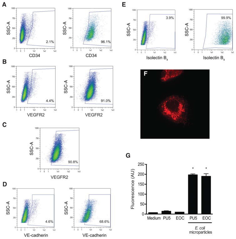
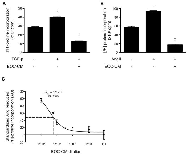
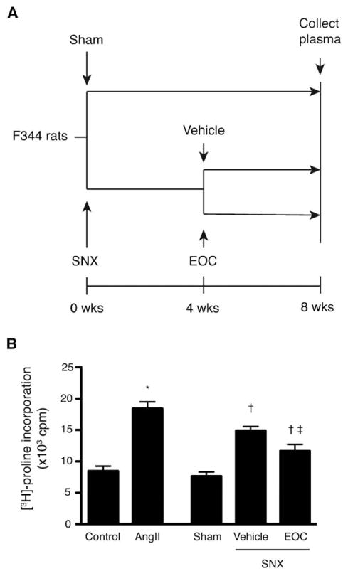
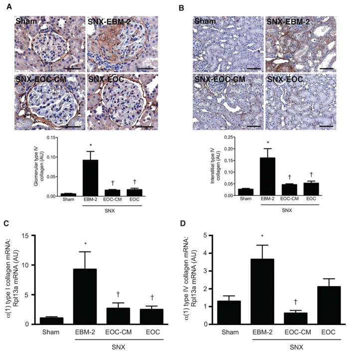
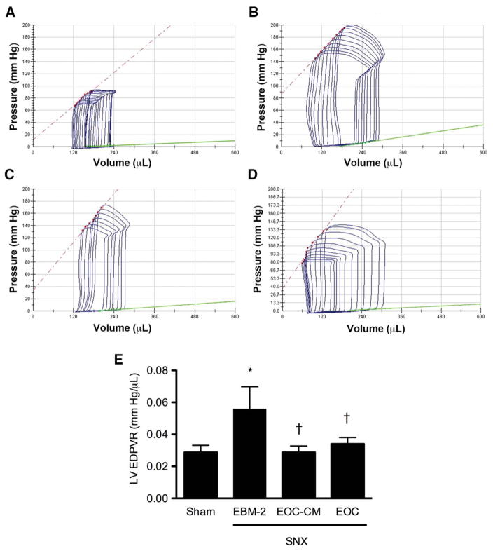
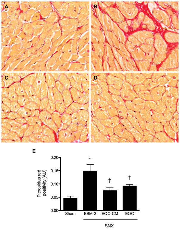
Adult bone marrow-derived cells can improve organ function in chronic disease models, ostensibly by the release of paracrine factors. It has, however, been difficult to reconcile this prevailing paradigm with the lack of cell retention within injured organs and their rapid migration to the reticuloendothelial system. Here, we provide evidence that the salutary antifibrotic effects of bone marrow-derived early outgrowth cells (EOCs) are more consistent with an endocrine mode of action, demonstrating not only the presence of antifibrotic factors in the plasma of EOC-treated rats but also that EOC conditioned medium (EOC-CM) potently attenuates both TGF-β- and angiotensin II-induced fibroblast collagen production in vitro. To examine the therapeutic relevance of these findings in vivo, 5/6 subtotally nephrectomized rats, a model of chronic kidney and heart failure characterized by progressive fibrosis of both organs, were randomized to receive i.v. injections of EOC-CM, unconditioned medium, or 10(6) EOCs. Rats that received unconditioned medium developed severe kidney injury with cardiac diastolic dysfunction. In comparison, EOC-CM-treated rats demonstrated substantially improved renal and cardiac function and structure, mimicking the changes found in EOC-treated animals. Mass spectrometric analysis of EOC-CM identified proteins that regulate cellular functions implicated in fibrosis. These results indicate that EOCs secrete soluble factor(s) with highly potent antifibrotic activity, that when injected intravenously replicate the salutary effects of the cells themselves. Together, these findings suggest that an endocrine mode of action may underlie the effectiveness of cell therapy in certain settings and portend the possibility for systemic delivery of cell-free therapy.

Keywords: Cell therapy; Chronic kidney disease; Congestive heart failure; Early outgrowth cell; Fibrosis; Kidney.


Conflict of interest statement

Disclosure of Potential Conflicts of Interest

The authors indicate no potential conflicts of interest.

Figures

Figure 1
Figure 1
Early outgrowth cells (EOCs) are a heterogeneous bone marrow-derived cell population with endothelial and monocyte/macrophage-like properties. EOCs were characterized with a set of flow cytometric and functional assays. (A–C): Expression of commonly measured EOC surface markers was analyzed by flow cytometry after staining with the following antibodies: (A) Alexa Fluor 647-conjugated anti-CD34 and (B) VioBlue-conjugated anti-VEGFR2. Left panels: Unstained cells. Right panels: Antibody-stained cells. The percentage of positive cells for a given area is listed on each plot. (C): Single positive CD34+ cells were gated as shown in panel (A) and examined for VEGFR2 expression. (D, E): To assess for expression of additional endothelial surface markers, EOCs were stained with (D) PE-conjugated anti-VE-cadherin and (E) fluorescein-conjugated Griffonia (Bandeiraea) simplicifolia lectin I—isolectin B4. Left panels: Unstained cells. Right panels: Stained cells. (F): EOCs were incubated with DiI-conjugated acetylated LDL (red) for 30 minutes and then imaged with an inverted epifluorescence microscope equipped with a digital camera. Original magnification: ×10. Representative image from n = 3 independent experiments. (G): EOCs were incubated with fluorescently labeled E. coli microparticles using a commercially available phagocytosis assay kit. After washing and quenching of uningested fluorescent microparticles, fluorescence was read by a plate reader. PU5-1.8 mouse macrophages were used as a positive control. *, p < .05 versus medium alone. Results are representative of a minimum of n = 6 replicates per condition. Abbreviations: AU, arbitrary units; SSC-A, side scatter; VEGFR, vascular endothelial growth factor receptor 2; VE, vascular endothelium.
Figure 2
Figure 2
Cell-free EOC-CM demonstrates potent antifibrotic activity in vitro. Cultured fibroblasts were preincubated with EOC-CM for 4 hours prior to stimulation with TGF-β 10 ng/mL or angiotensin II 10−7 mol/L. EOC-CM significantly attenuated both (A) TGF-β- and (B) angiotensin II-stimulated [3H]-proline incorporation, a robust marker of collagen production (n = 3 replicates per condition for each panel. Each EOC-CM replicate represents an aliquot from pooled EOC-CM collected from n ≥ 5 animals). (C): EOC-CM was next serially diluted as shown and tested in the same fibroblast collagen production assay, demonstrating significant inhibition of [3H]-proline incorporation even with significant EOC-CM dilution (n = 3–6 replicates per dilution. Each replicate represents an aliquot from pooled EOC-CM collected from n ≥ 5 animals). The measured IC50 was equivalent to a 1:1,780 dilution of EOC-CM. The results in panel (C) are standardized with the effects of serum-free endothelial basal medium-2 (EBM-2) set as 0 and the effects of AngII stimulation set as 100. *, p < .05 versus serum-free EBM-2 medium; , p < .05 versus TGF-β stimulation; , p < .05 versus AngII stimulation. Abbreviations: AngII, angiotensin II. AU, arbitrary units; cpm, counts per minute; EOC-CM, early outgrowth cell-conditioned media; IC50, concentration of EOC-CM leading to a 50% reduction in anti-fibrotic activity when compared with undiluted EOC-CM; TGF-β, transforming growth factor β.
Figure 3
Figure 3
The plasma of rats injected with EOCs contains detectable circulating antifibrotic factors. (A): Fischer 344 rats were subjected to SNX or sham surgery at t = 0. Four weeks after surgery, SNX rats were randomized to receive an i.v. injection of 106 EOCs or vehicle. Eight weeks after surgery, a time point at which EOC injection has been previously shown to inhibit the renal and cardiac fibrosis that develops in untreated SNX animals [11], plasma from each treatment group was collected. (B): Collected plasma was then assayed in the [3H]-proline incorporation assay to test for effects on fibroblast collagen production in the setting of AngII. *, p < .05 versus serum-free endothelial basal medium-2 medium; , p < .05 versus sham rat plasma; , p < .05 versus vehicle-treated SNX rat plasma. n = 3 animals per condition. Abbreviations: AngII, angiotensin II; cpm, counts per minute; EOC, early outgrowth cell; SNX, subtotal nephrectomy.
Figure 4
Figure 4
Both EOC and EOC-CM injections significantly reduce renal fibrosis in the SNX rat 4 weeks post-therapy initiation. (A, B): Kidney sections were prepared 8 weeks after surgery and immunostained with an antibody specific for type IV collagen. (A): Representative glomerular images with quantitative analysis below. Original magnification ×400. Scale bars = 50 μm. (B): Representative cortical interstitial images with quantitative analysis below. Original magnification ×160. Scale bars = 100 μm. (C, D): RNA isolated from snap-frozen kidney tissue was reverse transcribed and analyzed via quantitative real-time polymerase chain reaction for levels of α (1) type I collagen (C) and α (1) type IV collagen mRNA (D). All values are referenced to levels of Rpl13a, a housekeeper transcript, and are expressed relative to sham-operated animal values. n = 4–9 animals per condition. *, p < .05 versus sham-operated animals; , p < .05 versus SNX-EBM-2 animals. Abbreviations: AU, arbitrary units; EBM-2, endothelial basal medium-2; EOC-CM, early outgrowth cell conditioned media; SNX, subtotal nephrectomy.
Figure 5
Figure 5
Both EOC and EOC-CM injections significantly improve left ventricular chamber compliance in the SNX rat 4 weeks post-therapy initiation. 8 weeks after surgery, animals were subjected to invasive cardiac catheterization for cardiac functional analysis. The slope of the green line indicates the left ventricular end-diastolic pressure-volume relationship (LV EDPVR), an index of LV chamber compliance. (A–D): Representative pressure-volume loops. (A): Sham animal. (B): SNX-EBM-2 animal. (C): SNX-EOC-CM animal. (D): SNX-EOC animal. (E): Quantitative analysis of LV EDPVR. *, p < .05 versus sham-operated animals; , p < .05 versus SNX-EBM-2 animals. Abbreviations: EBM-2, endothelial basal medium-2; EOC-CM, early outgrowth cell conditioned media; SNX, subtotal nephrectomy.
Figure 6
Figure 6
Both EOC and EOC-CM injections attenuate cardiac injury in the SNX rat 4 weeks post-therapy initiation. Subendocardial heart sections were prepared 8 weeks after surgery and stained with picrosirius red for assessment of interstitial fibrosis. (A–D): Representative cardiac interstitial images. Original magnification ×160. (A): Sham animal. (B): SNX-EBM-2 animal. (C): SNX-EOC-CM animal. (D): SNX-EOC animal. (E): Quantitative analysis of subendocardial interstitial fibrosis. *, p < .05 versus sham-operated animals; , p < .05 versus SNX-EBM-2 animals. Abbreviations: AU, arbitrary units; EBM-2, endothelial basal medium-2; EOC-CM, early outgrowth cell conditioned media; SNX, subtotal nephrectomy.

References

    1. Asahara T, Murohara T, Sullivan A, et al. Isolation of putative progenitor endothelial cells for angiogenesis. Science. 1997;275:964–967. - PubMed
    1. Kalka C, Masuda H, Takahashi T, et al. Transplantation of ex vivo expanded endothelial progenitor cells for therapeutic neovascularization. Proc Natl Acad Sci USA. 2000;97:3422–3427. - PMC - PubMed
    1. Kocher AA, Schuster MD, Szabolcs MJ, et al. Neovascularization of ischemic myocardium by human bone-marrow-derived angioblasts prevents cardiomyocyte apoptosis, reduces remodeling and improves cardiac function. Nat Med. 2001;7:430–436. - PubMed
    1. Murohara T, Ikeda H, Duan J, et al. Transplanted cord blood-derived endothelial precursor cells augment postnatal neovascularization. J Clin Invest. 2000;105:1527–1536. - PMC - PubMed
    1. Ormiston ML, Deng Y, Stewart DJ, et al. Innate immunity in the therapeutic actions of endothelial progenitor cells in pulmonary hypertension. Am J Respir Cell Mol Biol. 2010;43:546–554. - PubMed

Publication types

MeSH terms