. 2013 Aug 20;110(34):13916-20.
doi: 10.1073/pnas.1311113110. Epub 2013 Aug 6.

Structural basis of the C1q/C1s interaction and its central role in assembly of the C1 complex of complement activation

Affiliations

Structural basis of the C1q/C1s interaction and its central role in assembly of the C1 complex of complement activation

Umakhanth Venkatraman Girija et al. Proc Natl Acad Sci U S A. .

Erratum in

Abstract

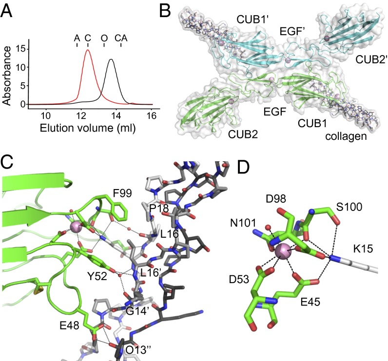
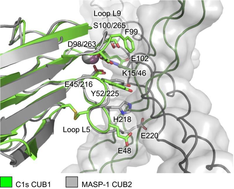
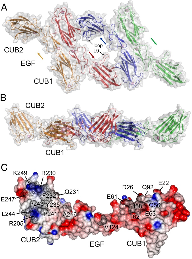
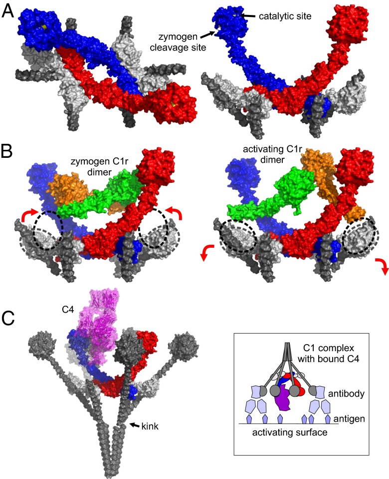
Complement component C1, the complex that initiates the classical pathway of complement activation, is a 790-kDa assembly formed from the target-recognition subcomponent C1q and the modular proteases C1r and C1s. The proteases are elongated tetramers that become more compact when they bind to the collagen-like domains of C1q. Here, we describe a series of structures that reveal how the subcomponents associate to form C1. A complex between C1s and a collagen-like peptide containing the C1r/C1s-binding motif of C1q shows that the collagen binds to a shallow groove via a critical lysine side chain that contacts Ca(2+)-coordinating residues. The data explain the Ca(2+)-dependent binding mechanism, which is conserved in C1r and also in mannan-binding lectin-associated serine proteases, the serine proteases of the lectin pathway activation complexes. In an accompanying structure, C1s forms a compact ring-shaped tetramer featuring a unique head-to-tail interaction at its center that replicates the likely arrangement of C1r/C1s polypeptides in the C1 complex. Additional structures reveal how C1s polypeptides are positioned to enable activation by C1r and interaction with the substrate C4 inside the cage-like assembly formed by the collagenous stems of C1q. Together with previously determined structures of C1r fragments, the results reported here provide a structural basis for understanding the early steps of complement activation via the classical pathway.

Keywords: innate immunity; structural biology.


Conflict of interest statement

The authors declare no conflict of interest.

Figures

Fig. 1.
Fig. 1.
Structure of C1s–C1q collagen complex. (A) Gel filtration of the CUB1-EGF-CUB2 fragment of C1s (35 kDa) in Ca2+ (red) and EDTA (black; each at 2 mM). Samples were separated on a Superdex 200 column equilibrated in 25 mM Tris⋅HCl at pH 7.4, containing 150 mM NaCl. Elution positions of aldolase (A; 158 kDa), conalbumin (C; 75 kDa), ovalbumin (O; 43 kDa), and carbonic anhydrase (CA; 29 kDa) are shown. (B) Structure of the C1s X-shaped dimer (green and blue) bound to the collagen-like peptide of C1q. (C) Close-up of the C1s/C1q collagen interface showing the residues that participate in the interaction. The leading, middle, and trailing strands are in black, gray, and white, respectively. (D) The Ca2+-binding site of the CUB1 domain of C1s showing the six coordinating ligands arranged in a tetragonal bipyramid and the interaction with Lys15 of the C1q collagen-like peptide. Waters are shown as red spheres and Ca2+ are in pink, throughout.
Fig. 2.
Fig. 2.
Comparison of CUB1–collagen and CUB2–collagen complexes of the classical and lectin pathways. Overlay of the C1s–C1q collagen structure (green) with the MASP-1/-3-MBL collagen structure (gray; PDB ID code: 3POB) (15). Key elements of the complexes superimpose including the lysine residue of the collagen, the Ca2+ and Ca2+-coordinating residues in the CUB domains together with a tyrosine residue, which contacts the collagen in each structure. However, the orientation of the collagen helices differs by ∼90°. Glu48 of loop L5 and Glu102 in loop L9 of C1s sandwich the collagen helix creating the shallow binding groove. Glu102 and other residues in loop L9 prevent binding in the alternative (horizontal) orientation through steric clashes. Loop L5 is longer in MASP-1 than in C1s and contributes toward binding via His218, which forms hydrogen bonds to the collagen, and blocks binding in the alternative (vertical) orientation (e.g., through Glu220, which would clash with the collagen chains).
Fig. 3.
Fig. 3.
Structure of the C1s tetramer. Top (A) and side (B) views of the C1s tetramer in which two X-shaped dimers are connected via a unique central interface. Arrows indicate the direction of the C1s chains. (C) Side view of the central C1s–C1s interface showing the electrostatic potential. Residues buried by the interaction are labeled and shaded in gray.
Fig. 4.
Fig. 4.
Structural organization of the C1 complex and the steps leading to activation and substrate recognition. (A) Superposition of the C1s tetramer, the C1s–collagen complex and the catalytic domains of C1s reveals the arrangement of subcomponents at the center of the C1 complex. C1s polypeptides are show in red and blue, the collagen is in gray, and the putative C1r CUB1-EGF-CUB2 fragments are in white. The MASP-1 CUB2-MBL collagen complex (PDB ID code: 3POB) (15) was overlaid onto the CUB2 domains of C1r to position the additional two collagen helices. (B) The catalytic domains of zymogen C1r (PDB ID code: 1GPZ) (18) and active C1r in a putative enzyme-product conformation (PDB ID code: 2QYO) (31) fit between the arms of C1s and can be connected by rotation of the CUB2 domains of C1r. The positions of the C1r CUB2 domains together with the resting position (based on the structure of the C1s tetramer) are indicated by the dotted lines. (C) Superposition of the catalytic domains of C1s with the corresponding domains of MASP-2 bound to C4 (PDB ID code: 4FXG) (24) shows that C4 can fit inside the cage-like assembly formed by the collagenous stems of C1q. The stems have been extended to their full length, and the globular domains of C1q (PDB ID code: 1PK6) (32) are positioned on top. The upper part of the front left stem and the globular domain is partially transparent so that the position of C4 (purple) can be seen. (Inset) Schematic representation of the C1 complex with C4 attached bound to antibody molecules on an activating surface.

References

    1. Ricklin D, Hajishengallis G, Yang K, Lambris JD. Complement: A key system for immune surveillance and homeostasis. Nat Immunol. 2010;11(9):785–797. - PMC - PubMed
    1. Wallis R, Mitchell DA, Schmid R, Schwaeble WJ, Keeble AH. Paths reunited: Initiation of the classical and lectin pathways of complement activation. Immunobiology. 2010;215(1):1–11. - PMC - PubMed
    1. Forneris F, Wu J, Gros P. The modular serine proteases of the complement cascade. Curr Opin Struct Biol. 2012;22(3):333–341. - PubMed
    1. Tschopp J, Villiger W, Fuchs H, Kilchherr E, Engel J. Assembly of subcomponents C1r and C1s of first component of complement: Electron microscopic and ultracentrifugal studies. Proc Natl Acad Sci USA. 1980;77(12):7014–7018. - PMC - PubMed
    1. Gregory LA, Thielens NM, Arlaud GJ, Fontecilla-Camps JC, Gaboriaud C. X-ray structure of the Ca2+-binding interaction domain of C1s. Insights into the assembly of the C1 complex of complement. J Biol Chem. 2003;278(34):32157–32164. - PubMed

Publication types