. 2013 Aug 20;110(34):E3216-24.
doi: 10.1073/pnas.1312348110. Epub 2013 Aug 6.

T-box transcription factor T-bet, a key player in a unique type of B-cell activation essential for effective viral clearance

Affiliations

T-box transcription factor T-bet, a key player in a unique type of B-cell activation essential for effective viral clearance

Kira Rubtsova et al. Proc Natl Acad Sci U S A. .

Abstract

IgG2a is known to be the most efficient antibody isotype for viral clearance. Here, we demonstrate a unique pathway of B-cell activation, leading to IgG2a production, and involving synergistic stimulation via B-cell antigen receptors, toll-like receptor 7 (TLR7), and IFNγ receptors on B cells. This synergistic stimulation leads to induction of T-box transcription factor T-bet expression in B cells, which, in turn, drives expression of CD11b and CD11c on B cells. T-bet/CD11b/CD11c positive B cells appear during antiviral responses and produce high titers of antiviral IgG2a antibodies that are critical for efficient viral clearance. The results thus demonstrate a previously unknown role for T-bet expression in B cells during viral infections. Moreover, the appearance of T-bet(+) B cells during antiviral responses and during autoimmunity suggests a possible link between these two processes.

Keywords: interferon gamma; virus.


Conflict of interest statement

The authors declare no conflict of interest.

Figures

Fig. 1.
Fig. 1.
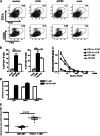
TLR7, BCR, and IFNγR signaling synergize in the up-regulation of T-bet expression in B cells in vitro. (AC) Unseparated splenocytes from WT mice were cultured for 48 h under the indicated conditions. After incubation, T-bet expression in B cells was analyzed. (D and E) IFNγR1−/− or WT splenocytes were cultured for 48 h under the indicated conditions, and T-bet expression in B cells was then analyzed. (FH) Purified WT B cells were incubated under the indicated conditions for 48 h, and T-bet expression in B cells was then analyzed. (AH) Histograms and titrations represent the levels of T-bet expressed in B cells, gated as live/B220+/CD4/CD8/CD19+. (I) Unseparated WT splenocytes were incubated under the indicated conditions for 4 d. The supernatants were then analyzed by ELISA for the presence of IFNγ. In all relevant sections of the figure, bars represent the means ± SEM of n = 3 mice per group. *P < 0.05, **P < 0.001, ***P < 0.0001 (t test). All data are representative of at least three independent experiments.
Fig. 2.
Fig. 2.
BCR and TLR7 synergize in T-bet induction in vivo driving CD11c and CD11b expression on B cells. MD4 transgenic B cells were transferred into B6.SJL recipients followed by immunization of recipient mice with HEL, a TLR7 agonist (R848), or a mixture of both i.p. 24 h after cell transfer. (A and B) Splenocytes were analyzed for T-bet expression in B cells (gated as B220+, CD19+, CD4, CD8) or (C and D) the presence of CD11c+ B cells in donor MD4 B cells (black bars) and host WT B cells (white bars). Bars represent the means ± SEM of n = 3 mice per group. *P < 0.05, **P < 0.001, ***P < 0.0001 (t test). Data are representative of three independent experiments.
Fig. 3.
Fig. 3.
T-bet expression in B cells is sufficient for their differentiation into CD11c+/CD11b+ B cells. Immature B cells from T-bet−/− mice were generated by culture of bone marrow-derived cells for 5 d in IL-7 and transduced with retorviruses expressing GFP alone or GFP plus T-bet. The cells were transferred into sublethally irradiated congenic hosts, and B cells in the spleens of the mice were analyzed for expression of GFP and CD11b and CD11c 10 d later. (A and B) Sample histograms and bar graphs show the expression of CD11c (A) and CD11b (B) on splenic donor B cells. The B cells were gated as CD4/CD8/CD19+/CD45.1+. Bars represent means ± SEM of B cells from n = 5 mice per group. (C) Donor GFP+ B cells were subsequently gated as GFP high, intermediate, and low. The mean fluorescent intensity (MFI) of CD11c was analyzed for each population. Bars represent the means ± SEM of cells from n = 5 mice per group. Data are representative of two independent experiments.
Fig. 4.
Fig. 4.
T-bet induction in B cells by TLR7, BCR, and IFNγR leads to IgG2a production. Purified B cells from young C57BL/6 mice were cultured under the indicated conditions, and supernatants were analyzed for the presence of different IgG isotypes by ELISA at day 7. Bars represent the means ± SEM of n = 3 samples per group. Data are representative of three independent experiments.
Fig. 5.
Fig. 5.
T-bet+/ CD11c+/CD11b+ B cells appearing at the peak of antiviral response secrete antiviral IgG2a and essential for effective viral clearance. C57BL/6 mice were infected with gHV68 or mock infected for 14 d or alternatively infected with LCMV or vaccinia or mock infected for 10 d. (A) The appearance of T-bet+/CD11c+/CD11b+ B cells in spleen was analyzed by FACS. FACS plots present the data for B cells gated as CD19+/B220+/CD4/CD8. (B) The percentages of splenic T-bet positive B cells in infected mice is shown. Bars represent the means ± SEM for n = 5 mice per group. (C) CD11c+ and follicular B cells from gHV68-infected mice were sorted at day 14 postinfection with gHV68 and stimulated in vitro for 7 d with LPS or TLR7 agonist. Supernatants were analyzed at day 7 for the presence of antiviral antibodies by ELISA (n = 3 samples per group). (D and E) WT mice were lethally irradiated and reconstituted with mixtures of bone marrow cells from WT and μMT or T-bet−/− and μMT mice, as indicated. Seven weeks later, the chimeras were infected with gHV68 or mock infected. (D) Serum levels of antiviral IgG of different isotypes were analyzed by ELISA 18 dpi. Bars represent the means ± SEM of n = 5 mice per group. (E) Spleens from the bone marrow chimeric mice were analyzed for the viral loads by real-time PCR at 18 dpi. The viral genome copy number per 100 ng of DNA is shown. P = 0.02 (t test). All data are representative of three independent experiments.

References

    1. Markine-Goriaynoff D, Coutelier JP. Increased efficacy of the immunoglobulin G2a subclass in antibody-mediated protection against lactate dehydrogenase-elevating virus-induced polioencephalomyelitis revealed with switch mutants. J Virol. 2002;76(1):432–435. - PMC - PubMed
    1. Coutelier JP, van der Logt JT, Heessen FW, Vink A, van Snick J. Virally induced modulation of murine IgG antibody subclasses. J Exp Med. 1988;168(6):2373–2378. - PMC - PubMed
    1. Schmitz N, et al. Universal vaccine against influenza virus: Linking TLR signaling to anti-viral protection. Eur J Immunol. 2012;42(4):863–869. - PubMed
    1. Snapper CM, Paul WE. Interferon-gamma and B cell stimulatory factor-1 reciprocally regulate Ig isotype production. Science. 1987;236(4804):944–947. - PubMed
    1. Gerth AJ, Lin L, Peng SL. T-bet regulates T-independent IgG2a class switching. Int Immunol. 2003;15(8):937–944. - PubMed

Publication types

MeSH terms