

Sonographic evaluation of proximal gastric accommodation in patients with functional dyspepsia
- PMID: 23922476
- PMCID: PMC3732851
- DOI: 10.3748/wjg.v19.i29.4774
Sonographic evaluation of proximal gastric accommodation in patients with functional dyspepsia
Abstract
Aim: To assess the value of ultrasonography (US) in evaluation of proximal gastric accommodation disorder in patients with functional dyspepsia (FD).
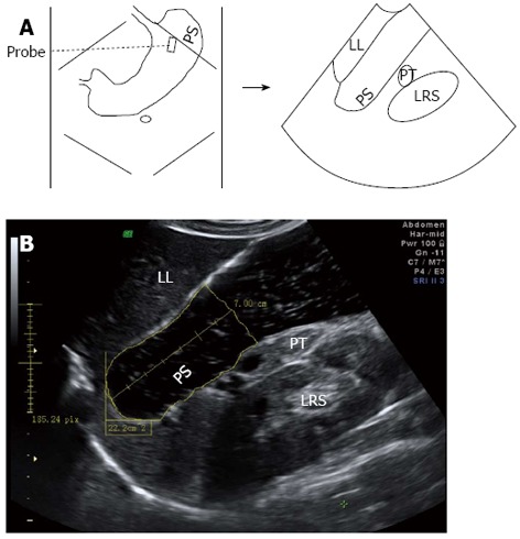
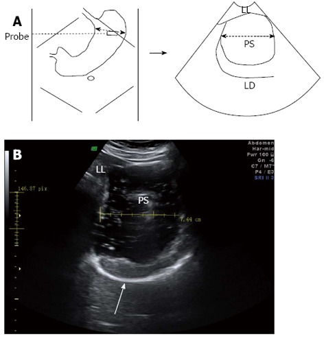
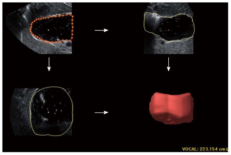
Methods: Between April 2011 and March 2012, 45 patients with FD and 27 healthy volunteers were enrolled in this study. Two-dimensional ultrasound (2DUS) and 3-dimensional ultrasound (3DUS) were performed sequentially to measure proximal gastric area (PGA), maximal proximal gastric diameter (MPGD), and proximal gastric volume (PGV). These values were measured separately in the two groups every other 5 min for a duration of 25 min after the beginning of ingestion of a test meal. Air pocket grading was done separately for images of 2DUS and blocks of 3DUS obtained at five scanning time points.
Results: Both PGA and PGV of patients were significantly smaller than healthy controls (P = 0.000 and 0.002, respectively). Comparing the two parameters between the groups at each time point, the differences were also statistically significant (P = 0.000-0.013), except at 10 min for the PGV (P = 0.077). However, no overall difference was found between the groups in the MPGD measurements (P = 0.114), though it was statistically significant at a 20-minute examination point (P = 0.026). A total of 360 sets or blocks of images were obtained for both 2DUS and 3DUS. For the images analyzed by 2DUS, none were excluded because of gastric gas, and 50 (13.9%) and 310 (86.1%) sets were determined as air pockets grades 1 and 2, respectively. For the images analyzed by 3DUS, 23 (6.4%) blocks were excluded from the measurement due to presence of a large fundus air pocket (grade 3); fifty (13.9%) and 287 (79.7%) blocks were also graded as 1 and 2, respectively.
Conclusion: Measurement of both PGA and PGV by 2DUS and 3DUS could be useful for assessment of the proximal gastric accommodation.
Keywords: 2-dimensional ultrasound; 3-dimensional ultrasound; Diagnosis; Functional dyspepsia; Gastric accommodation; Ultrasonography.
Figures
Comment in
-
Assessment of proximal gastric accommodation in patients with functional dyspepsia.World J Gastroenterol. 2013 Dec 21;19(47):9137-8. doi: 10.3748/wjg.v19.i47.9137. World J Gastroenterol. 2013. PMID: 24379642 Free PMC article.
Similar articles
-
Assessment of proximal gastric accommodation in patients with functional dyspepsia.World J Gastroenterol. 2013 Dec 21;19(47):9137-8. doi: 10.3748/wjg.v19.i47.9137. World J Gastroenterol. 2013. PMID: 24379642 Free PMC article.
-
Fundal dysaccommodation in functional dyspepsia: head-to-head comparison between the barostat and three-dimensional ultrasonographic technique.Gut. 2006 Dec;55(12):1725-30. doi: 10.1136/gut.2004.062836. Epub 2006 Jan 26. Gut. 2006. PMID: 16439420 Free PMC article.
-
Impaired accommodation of proximal stomach to a meal in functional dyspepsia.Dig Dis Sci. 1996 Apr;41(4):689-96. doi: 10.1007/BF02213124. Dig Dis Sci. 1996. PMID: 8674389
-
Abdominal Ultrasound for the Evaluation of Gastric Emptying Revisited.J Gastrointestin Liver Dis. 2015 Sep;24(3):329-38. doi: 10.15403/jgld.2014.1121.243.mur. J Gastrointestin Liver Dis. 2015. PMID: 26405705 Review.
-
Gastric accommodation assessed by ultrasonography.World J Gastroenterol. 2006 May 14;12(18):2825-9. doi: 10.3748/wjg.v12.i18.2825. World J Gastroenterol. 2006. PMID: 16718805 Free PMC article. Review.
Cited by
-
Impaired gastric accommodation in patients with postprandial distress syndrome type of functional dyspepsia assessed by 2D ultrasonography.Indian J Gastroenterol. 2023 Dec;42(6):824-832. doi: 10.1007/s12664-023-01436-7. Epub 2023 Oct 9. Indian J Gastroenterol. 2023. PMID: 37814116
-
Assessment of proximal gastric accommodation in patients with functional dyspepsia.World J Gastroenterol. 2013 Dec 21;19(47):9137-8. doi: 10.3748/wjg.v19.i47.9137. World J Gastroenterol. 2013. PMID: 24379642 Free PMC article.
-
Can Ultrasonographic Measurements of Gastric Motility Identify Pathophysiological Abnormalities of Functional Dyspepsia and Irritable Bowel Syndrome?J Neurogastroenterol Motil. 2020 Jan 30;26(1):1-3. doi: 10.5056/jnm19228. J Neurogastroenterol Motil. 2020. PMID: 31917911 Free PMC article. No abstract available.
-
Transabdominal Ultrasound of the Stomach in Patients with Functional Dyspepsia: A Review.Diagnostics (Basel). 2024 Sep 30;14(19):2193. doi: 10.3390/diagnostics14192193. Diagnostics (Basel). 2024. PMID: 39410597 Free PMC article. Review.
References
-
- Tack J, Talley NJ, Camilleri M, Holtmann G, Hu P, Malagelada JR, Stanghellini V. Functional gastroduodenal disorders. Gastroenterology. 2006;130:1466–1479. - PubMed
-
- Tack J, Piessevaux H, Coulie B, Caenepeel P, Janssens J. Role of impaired gastric accommodation to a meal in functional dyspepsia. Gastroenterology. 1998;115:1346–1352. - PubMed
-
- Miwa H, Watari J, Fukui H, Oshima T, Tomita T, Sakurai J, Kondo T, Matsumoto T. Current understanding of pathogenesis of functional dyspepsia. J Gastroenterol Hepatol. 2011;26 Suppl 3:53–60. - PubMed
Publication types
MeSH terms
LinkOut - more resources
Full Text Sources
Other Literature Sources
Medical
Miscellaneous
