. 2013 Jul 26;8(7):e69099.
doi: 10.1371/journal.pone.0069099. Print 2013.

Loss of NAC1 expression is associated with defective bony patterning in the murine vertebral axis

Affiliations

Loss of NAC1 expression is associated with defective bony patterning in the murine vertebral axis

Kai Lee Yap et al. PLoS One. .

Abstract

NAC1 encoded by NACC1 is a member of the BTB/POZ family of proteins and participates in several pathobiological processes. However, its function during tissue development has not been elucidated. In this study, we compared homozygous null mutant Nacc1(-/-) and wild type Nacc1(+/+) mice to determine the consequences of diminished NAC1 expression. The most remarkable change in Nacc1(-/-) mice was a vertebral patterning defect in which most knockout animals exhibited a morphological transformation of the sixth lumbar vertebra (L6) into a sacral identity; thus, the total number of pre-sacral vertebrae was decreased by one (to 25) in Nacc1(-/-) mice. Heterozygous Nacc1(+/-) mice had an increased tendency to adopt an intermediate phenotype in which L6 underwent partial sacralization. Nacc1(-/-) mice also exhibited non-closure of the dorsal aspects of thoracic vertebrae T10-T12. Chondrocytes from Nacc1(+/+) mice expressed abundant NAC1 while Nacc1(-/-) chondrocytes had undetectable levels. Loss of NAC1 in Nacc1(-/-) mice was associated with significantly reduced chondrocyte migratory potential as well as decreased expression of matrilin-3 and matrilin-4, two cartilage-associated extracellular matrix proteins with roles in the development and homeostasis of cartilage and bone. These data suggest that NAC1 participates in the motility and differentiation of developing chondrocytes and cartilaginous tissues, and its expression is necessary to maintain normal axial patterning of murine skeleton.


Conflict of interest statement

Competing Interests: The authors have declared that no competing interests exist.

Figures

Figure 1
Figure 1. Strategy used in generation of Nacc1 knockout allele.
A. Schematic illustration of gene targeting and Southern strategies. Black lines and blue lines indicate the expected sizes of fragments detected by Southern hybridization of genomic DNAs digested by HindIII/EcoRV and XbaI, respectively. The black bar indicates the probe for Southern hybridization. The fragments within the square on the top are the expected bands from cross-hybridization of the probe to the genomic DNA in Chr. 6. Note Nacc1 is located in Chr. 8. B. Representative clones from Southern hybridization as shown in panel A. Two positive clones, c72 and c156, were identified. Clones c73 and c74 are partially targeted because they lack the first LoxP site as shown in panel A. C. Quantitative real-time PCR to measure neo levels revealed that both positive clones, c72 and c156, likely have a single copy insertion of the neo cassette. The housekeeping gene control used in this experiment was Rpp30, and the data was normalized to single neo copy controls (T/O +/- and TET2 k-in) to calculate relative neo levels.
Figure 2
Figure 2. Reduced NAC1 expression alters axial skeletal patterning.
The images show representative whole-body radiographs comparing the skeletal organization of adult Nacc1+/+ (wild type, A), Nacc1+/- (heterozygous, B), and Nacc1-/- (knockout, C) mice. The major defects are an absence of the sixth lumbar vertebra (L6) in the Nacc1-/- mouse and an intermediate phenotype showing partial transformation of L6 into a sacral identity (B, red box). The numbers denote the number of vertebrae in the thoracic (T), lumbar (L), and sacral (S) regions of the vertebral column.
Figure 3
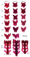
Figure 3. Reduced NAC1 expression ablates or changes the conformation of the sixth lumbar vertebra 6 (L6).
Comparison of representative Alizarin red-stained lumbar (L) and sacral (S) vertebrae of adult Nacc1+/+ (wild type), Nacc1+/- (heterozygous), and Nacc1-/- (knockout) mice. In the Nacc1-/- animal, L6 is missing, and the processes of all sacral vertebrae (S1-4) are fused; indeed, caudal extensions of the broad wings of S3 in this genotype resemble the profile of vertebra S2 rather than the usual contours of S3 in Nacc1+/+ mice. In the Nacc1+/- counterpart, L6 has undergone partial (unilateral) transformation into a sacral identity (“sacralization,” red box). The lumbar vertebrae were disarticulated to better showcase their characteristic shapes.
Figure 4
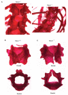
Figure 4. Loss of NAC1 expression is associated with dorsal non-closure of the vertebral arches in thoracic vertebrae 10 and 11.
A. Vertebral fissures affecting adult, male (left) and female (right) Nacc1-/- mice at T10 and T11. Disarticulated, Alizarin red-stained T10 vertebrae from a Nacc1+/+ (B) and Nacc1-/- (C) demonstrating the presence of an observable dorsal fissure in the null mutant animals.
Figure 5
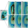
Figure 5. Reduced NAC1 expression is associated with altered specification of cartilage primordia for vertebral patterning.
Comparison of representative Alcian blue-stained, whole mount mouse embryos at embryonic day 15 (E15) reveals the typical vertebral formulas related to the Nacc1+/+ (wild type, formula T13/L6), Nacc1+/- (heterozygous, formula T13/L5), and Nacc1-/- (knockout, formula T12/L6) genotypes. The numbers denote the number of vertebrae in the thoracic (T, top row) and lumbar (L, bottom row) regions of the vertebral column.
Figure 6
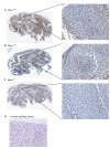
Figure 6. NAC1 expression in the chondrocytes of wild type mouse embryos.
Comparison of representative para-sagittal sections of Nacc1+/+ (wild type, A), Nacc1+/- (heterozygous, B), and Nacc1-/- (knockout, C) mouse embryos at embryonic day 16 (E16) shows the widespread distribution of NAC1 in various tissues, including chondrocytes (boxed regions). The wild type expression pattern exhibits widespread immunoreactivity including such axial tissues as vertebral primordia and the peri-ventricular germinal zones in the brain. D. Chondrocytes in cartilaginous tissues of a human embryo (estimated gestational age, 7 weeks) also exhibit extensive NAC1 immunoreactivity. Anti-NAC1 immunohistochemistry (using species-specific primary antibodies) with hematoxylin as the counterstain.
Figure 7
Figure 7. Reduced NAC1 expression decreases chondrocyte migration but does not affect chondrocyte proliferation or the balance of NAC2 expression.
Comparison of cultured costal chondrocytes derived from Nacc1+/+ (wild type), Nacc1+/- (heterozygous), and Nacc1-/- (knockout) mouse pups at postnatal day 1 (P1) with respect to their migratory (A) and proliferative (B) properties and their complements of NAC1 and NAC2 (a protein closely related to NAC1, C). A: ** denotes a significant difference in cell migration, p < 0.01). B: Nacc1 depletion did not affect chondrocyte division. C: Real-time quantitative PCR analysis showed that levels of Nacc2 in chondrocytes of Nacc1-/- mice were not altered to compensate for the reduction in Nacc1.
Figure 8
Figure 8. Reduced NAC1 expression is associated with alterations in chondrocytic expression of matrilin-3 and matrilin-4.
Quantitative real-time PCR analysis performed on cultured costal chondrocytes derived from Nacc1+/+ (wild type) and Nacc1-/- (knockout) mouse pups at postnatal day 1 (P1) to measure the relative expression levels of the different matrilin (MATN) proteins. Only MATN3 and MATN4 are expressed at a significantly different level (** denotes p < 0.01) between the two genotypes.

References

    1. Stead MA, Carr SB, Wright SC (2009) Structure of the human Nac1 POZ domain. Acta Crystallogr Sect Struct Biol Cryst Commun 65: 445-449. doi:10.1107/S0108768109024161. PubMed: 19407373. - DOI - PMC - PubMed
    1. Cha XY, Pierce RC, Kalivas PW, Mackler SA (1997) NAC-1, a rat brain mRNA, is increased in the nucleus accumbens three weeks after chronic cocaine self-administration. J Neurosci 17: 6864-6871. PubMed: 9278521. - PMC - PubMed
    1. Korutla L, Wang PJ, Lewis DM, Neustadter JH, Stromberg MF et al. (2002) Differences in expression, actions and cocaine regulation of two isoforms for the brain transcriptional regulator NAC1. Neuroscience 110: 421-429. doi:10.1016/S0306-4522(01)00518-8. PubMed: 11906783. - DOI - PubMed
    1. Mackler S, Pacchioni A, Degnan R, Homan Y, Conti AC et al. (2008) Requirement for the POZ/BTB protein NAC1 in acute but not chronic psychomotor stimulant response. Behav Brain Res 187: 48-55. doi:10.1016/j.bbr.2007.08.036. PubMed: 17945361. - DOI - PMC - PubMed
    1. Mancini C, Messana E, Turco E, Brussino A, Brusco A (2011) Gene-targeted embryonic stem cells: real-time PCR assay for estimation of the number of neomycin selection cassettes. Biol Proced Online 13: 10. doi:10.1186/1480-9222-13-10. PubMed: 22035318. - DOI - PMC - PubMed

Publication types

MeSH terms

LinkOut - more resources