. 2013 Jul 29;8(7):e69668.
doi: 10.1371/journal.pone.0069668. Print 2013.

Modulation of the Akt pathway reveals a novel link with PERK/eIF2α, which is relevant during hypoxia

Affiliations

Modulation of the Akt pathway reveals a novel link with PERK/eIF2α, which is relevant during hypoxia

Matías Blaustein et al. PLoS One. .

Abstract

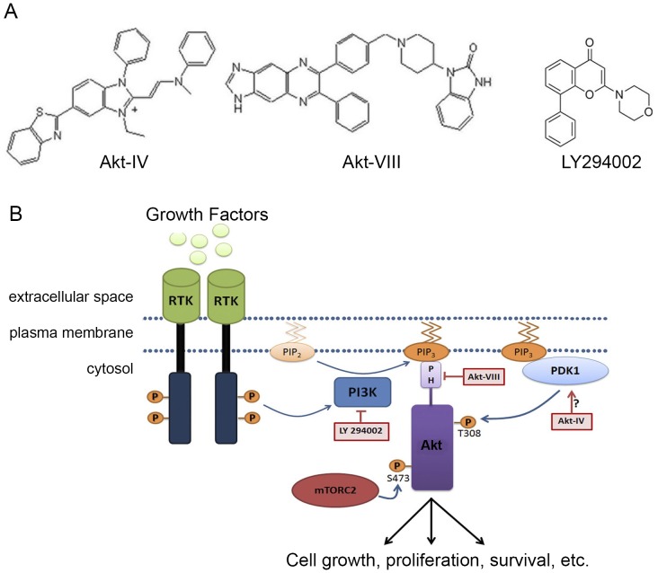
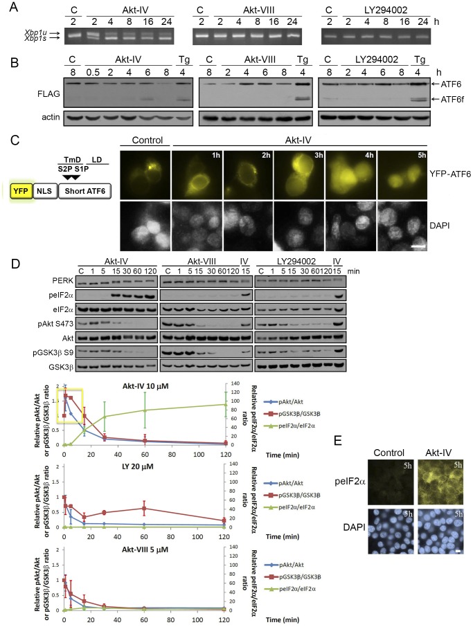
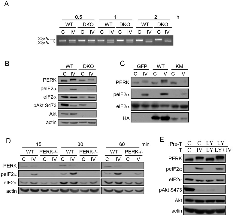
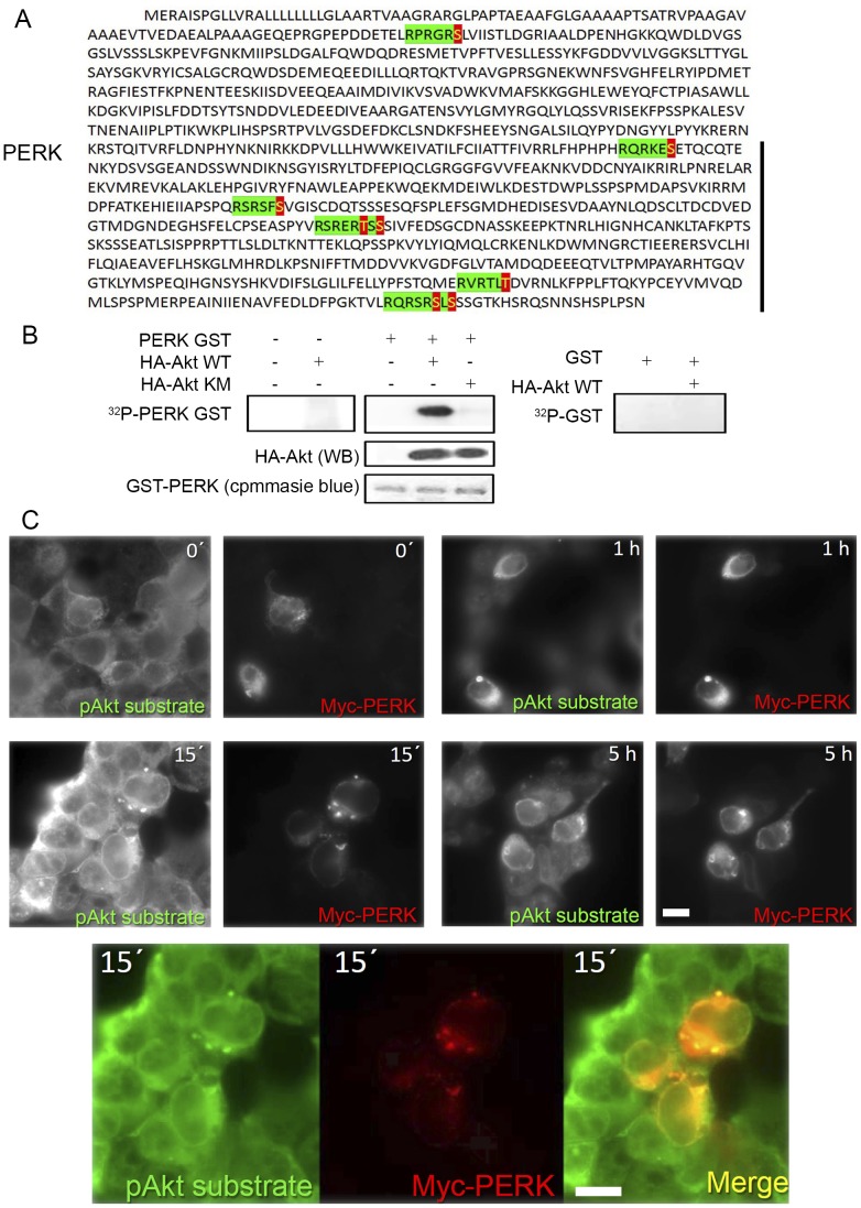
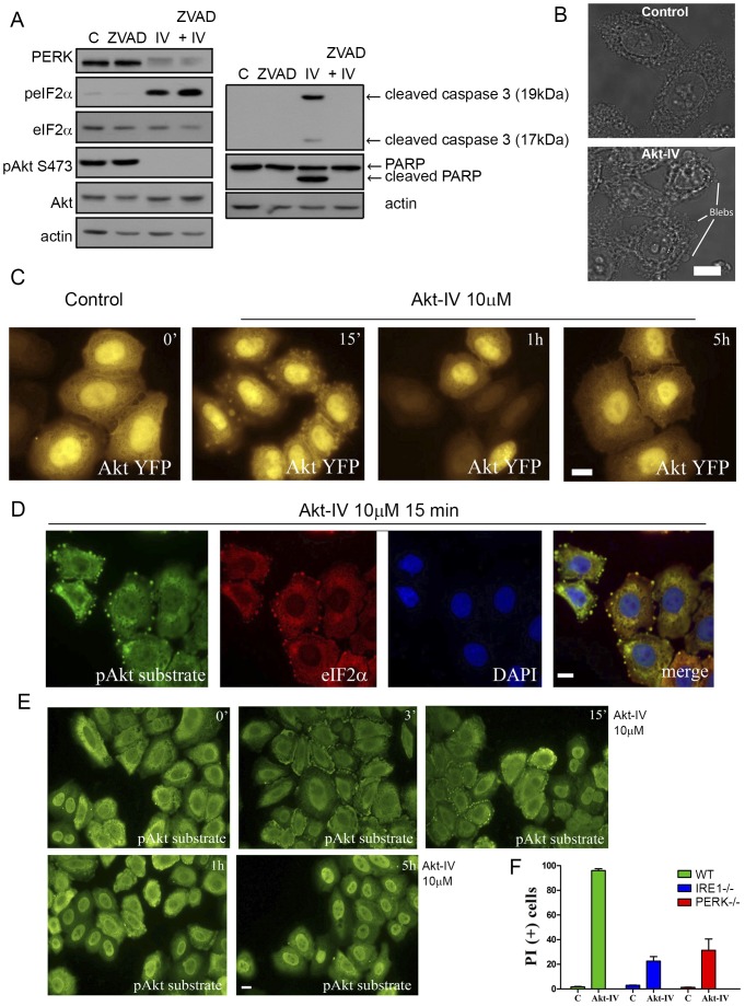
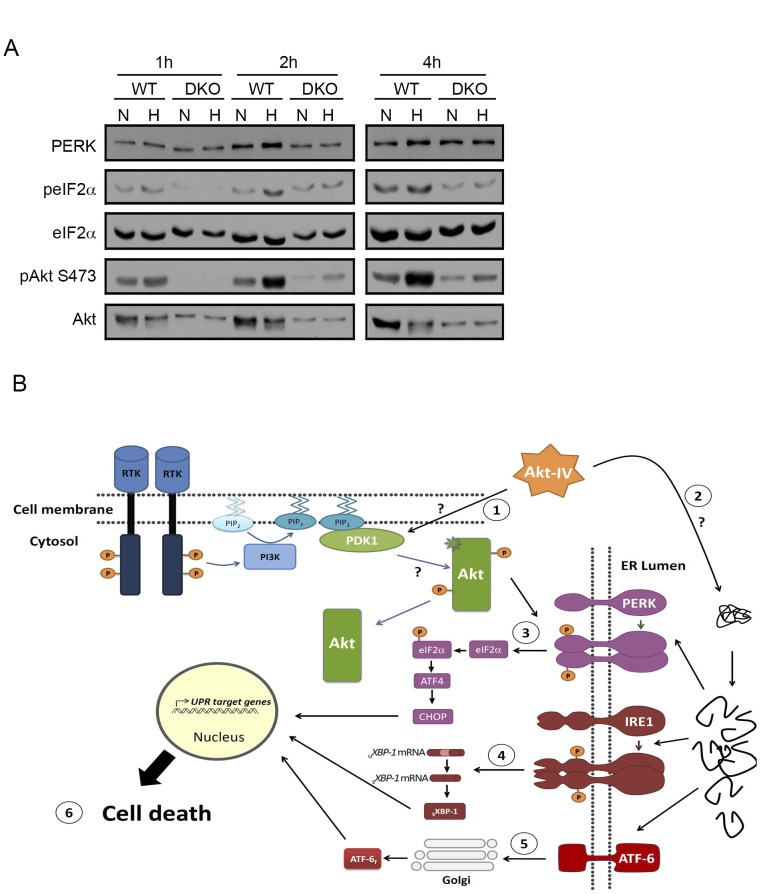
The unfolded protein response (UPR) and the Akt signaling pathway share several regulatory functions and have the capacity to determine cell outcome under specific conditions. However, both pathways have largely been studied independently. Here, we asked whether the Akt pathway regulates the UPR. To this end, we used a series of chemical compounds that modulate PI3K/Akt pathway and monitored the activity of the three UPR branches: PERK, IRE1 and ATF6. The antiproliferative and antiviral drug Akt-IV strongly and persistently activated all three branches of the UPR. We present evidence that activation of PERK/eIF2α requires Akt and that PERK is a direct Akt target. Chemical activation of this novel Akt/PERK pathway by Akt-IV leads to cell death, which was largely dependent on the presence of PERK and IRE1. Finally, we show that hypoxia-induced activation of eIF2α requires Akt, providing a physiologically relevant condition for the interaction between Akt and the PERK branch of the UPR. These data suggest the UPR and the Akt pathway signal to one another as a means of controlling cell fate.


Conflict of interest statement

Competing Interests: The authors have declared that no competing interests exist.

Figures

Figure 1
Figure 1. Strategy.
(A) Chemical structure of the compounds targeting the PI3K/Akt pathway used in this study. (B) Scheme of Akt signaling pathway, which regulates cell survival, showing the point of action of the drugs shown in A. Akt phosphorylation and activation result from its recruitment to PIP3 at plasma membrane, after which it exerts cytoplasmic and nuclear functions. Accumulation of PIP3 classically follows ligand (L) binding to tyrosin kinase cell-surface receptors (RTK), adapter proteins (AP) recruitment to RTK and finally, PI3K activation to phosphorylate PIP2 to PIP3. While Akt can be activated by the UPR it is not known if Akt can also regulate the UPR.
Figure 2
Figure 2. Pharmacological modulation of Akt with Akt-IV activates all three UPR branches: PERK responds first.
(A) When activated, IRE1 processes Xbp1 mRNA by a non-conventional cytoplasmic splicing reaction, changing Xbp1 open reading frame. HEK293T cells were treated with Akt-IV (10 µM), Akt-VIII (5 µM) or LY294002 (20 µM) for the indicated times. Xbp1 mRNA splicing was detected by RT-PCR. Xbp1s: spliced form (activated IRE1); Xbp1u: unspliced form (inactive IRE1). (B) When activated, ATF6 translocates to the Golgi apparatus where it is cleaved to release a fragment that enters the nucleus where it functions as a transcription factor. HEK293 cells were transfected with ATF6-Flag plasmid and 24 h later they were treated with Akt-IV (10 µM), Akt-VIII (5 µM), LY294002 (20 µM), or thapsigargin (Tg; 100 nM) for the indicated times. Western blots (WB) using antibodies against FLAG and actin are shown for every case (B, upper panel). ATF6: uncleaved protein; ATF6f: cleaved form. (C) HEK293T cells were transfected with a plasmid that expresses the YFP-NLS-mATF6short reporter (top, see Fig. S1A for details). Forty-eight hours post-transfection cells were treated for the indicated times with Akt-IV and then fixed, DNA was stained with DAPI and cells were imaged (lower panel); Yellow, YFP-ATF6; Blue, DNA; scale bar, 5 µm. For all cases cells treated with DMSO were used as a control (Control). (D) When activated, PERK is autophosphorylated at multiple residues and activated to phosphorylate eIF2α. HEK293T cells were treated with Akt-IV (10 µM), Akt-VIII (5 µM) or LY294002 (20 µM) for the indicated times. Protein extracts were analyzed by WB using the indicated antibodies. Data in the plot corresponds to ratio of phosphorylated total abundance of each of the indicated proteins (normalized to the initial value) in cells treated with the indicated drugs for different times. Error bars correspond to the standard error of three independent experiments. (E) HEK293T cells were treated for 5 h with Akt-IV. peIF2α abundance was detected by immunofluorescence. Green, peIF2α; Blue, DNA; scale bar, 5 µm. Data are representative of at least three independent experiments.
Figure 3
Figure 3. Akt-IV stimulation of eIF2α phosphorylation is Akt- and PERK- dependent but PI3K-independent.
(A) MEF WT or Akt DKO cells were treated with Akt-IV (IV; 10 µM) for the indicated times. Xbp1 mRNA splicing was detected by RT-PCR. Xbp1s: spliced form (activated IRE1); Xbp1u: unspliced form (inactive IRE1). (B) MEF WT or Akt DKO cells were treated with Akt-IV (IV; 10 µM) for 1 h. Protein extracts were analyzed by WB using the indicated antibodies. The fold change in peIF2α/eIF2α ratio induced by Akt-IV was quantified for three independent experiments. On average, this fold change was reduced to 15% of the original effect in MEF Akt DKO cells compared to WT cells (11.0 vs 2.6). (C) MEF WT cells were transfected with HA-Akt or HA-Akt KM plasmids. Forty-eight hours post-transfection cells were treated with DMSO (C) or with Akt-IV (IV; 10 µM) for 1 h. A GFP expressing plasmid was used as a transfection control. Protein extracts were analyzed by WB using the indicated antibodies. (D) MEFs WT or PERK−/− were treated with Akt-IV (IV; 10 µM) for the indicated times. Protein extracts were analyzed by WB using the indicated antibodies. (E) HEK293T cells were pretreated with DMSO (C) or LY294002 (LY; 20 µM) for 30 min and then treated for 1 h with DMSO, or Akt-IV (without removing the corresponding pre-treatment). Protein extracts were analyzed by WB using the indicated antibodies. Data are representative of at least three independent experiments.
Figure 4
Figure 4. Akt is a PERK kinase.
(A) Human PERK gene containing eight sequences that match the Akt consensus (RxRxxS/T) seven of which are located in its cytosolic domain (black bar). Green: Akt consensus sequence. Red/Yellow: Akt phosphorylation site. (B) WT and KM HA-Akt mutant protein was incubated with 10 µCi of [γ-32P] ATP for 30 min and with 1 µg of GST or PERK GST protein as substrate. Phosphate incorporation was analyzed by SDS-PAGE and autoradiography. Akt levels were determined by WB while GST-PERK levels were revealed by Coommassie blue staining. (C) HEK293T cells were transfected with pCDNA-Myc-PERK and treated for 15 min with Akt-IV (10 µM). Cells were fixed and immunostained for pAkt substrate and Myc tag. Green, pAkt substrate; Red, Myc-PERK; scale bar, 5 µm. Data are representative of at least three independent experiments.
Figure 5
Figure 5. Akt-IV induces cell blebbing and a UPR dependent cell death.
(A) HEK293T cells were treated with DMSO (C), 100 µM of the caspase inhibitor ZVAD, 10 µM Akt-IV (IV) or both. PERK mobility, eIF2α phosphorylation, Akt phosphorylation on Ser473, caspase 3 cleavage and PARP cleavage were detected by WB. (B) Transmission images of HeLa cells treated for 15 min with DMSO (15 min) or with Akt-IV (10 µM), with blebs indicated. Bleb formation was clearly observed in HeLa and MEF cells but could not be detected in HEK293T cells. (C) YFP channel images of HeLa cells transfected with pAkt1-YFP plasmid and then treated for the indicated times with Akt-IV (10 µM). Akt1-YFP can be detected in blebs after 15 min of treatment. (D) HeLa cells were treated for 15 min with Akt-IV. Cells were fixed and immunostained against pAkt substrate/Alexa Fluor® 488 and total eIF2α/Alexa Fluor® 594. Green, pAkt substrate; Red, eIF2α; Blue, DNA. scale bar, 5 µm. (E) HeLa cells were treated for different times with Akt-IV (10 µM) and then cells were fixed and immunostained for pAkt substrate/Alexa Fluor® 488; scale bar, 5 µm. (F) MEF WT, IRE1−/− or PERK−/− were treated with Akt-IV (10 µM) for 12 h. Cell viability was measured by flow cytometry using propidium iodide. Data are representative of at least three independent experiments.
Figure 6
Figure 6. A physiological link between Akt and PERK/eIF2α.
(A) WT or Akt DKO MEF cells were subjected to normoxia (C) or hypoxia (0.1%±0.1 O2) (H) for the indicated times. Protein extracts were analyzed by WB using the indicated antibodies. The fold change in peIF2α/eIF2α ratio induced by hypoxia was quantified for two independent experiments. On average, this fold change was reduced to 0, 40 or 60% of the original effect in MEF Akt DKO cells compared to WT cells (1 h, 2 h and 4 h, respectively). (B) A model summarizing our results. Akt-IV (or other stimuli, such as hypoxia) targets an unknown kinase, possibly PDK1, triggering apoptotic cell blebbing and activating Akt in a PI3K-independent manner (1). Subsequently, UPR is activated (2). Akt presence and activity are necessary for eIF2α phosphorylation due to the existence of a connection between Akt and PERK/eIF2α signaling pathways. IRE1 (4) and ATF6 (5) are activated are later times. At the end, activation of IRE1 and PERK and dephosphorylation of Akt and GSK3β are associated with cell death (6).

References

    1. Bellacosa A, Kumar CC, Di Cristofano A, Testa JR (2005) Activation of AKT kinases in cancer: implications for therapeutic targeting. Adv Cancer Res 94: 29–86. - PubMed
    1. Hay N, Sonenberg N (2004) Upstream and downstream of mTOR. Genes Dev 18: 1926–1945. - PubMed
    1. Doble BW, Woodgett JR (2003) GSK-3: tricks of the trade for a multi-tasking kinase. J Cell Sci 116: 1175–1186. - PMC - PubMed
    1. Alessi DR, James SR, Downes CP, Holmes AB, Gaffney PR, et al. (1997) Characterization of a 3-phosphoinositide-dependent protein kinase which phosphorylates and activates protein kinase Balpha. Curr Biol 7: 261–269. - PubMed
    1. Sarbassov DD, Guertin DA, Ali SM, Sabatini DM (2005) Phosphorylation and regulation of Akt/PKB by the rictor-mTOR complex. Science 307: 1098–1101. - PubMed

Publication types

LinkOut - more resources