. 2013 Jul 29;8(7):e69882.
doi: 10.1371/journal.pone.0069882. Print 2013.

Schiff base switch II precedes the retinal thermal isomerization in the photocycle of bacteriorhodopsin

Affiliations

Schiff base switch II precedes the retinal thermal isomerization in the photocycle of bacteriorhodopsin

Ting Wang et al. PLoS One. .

Abstract

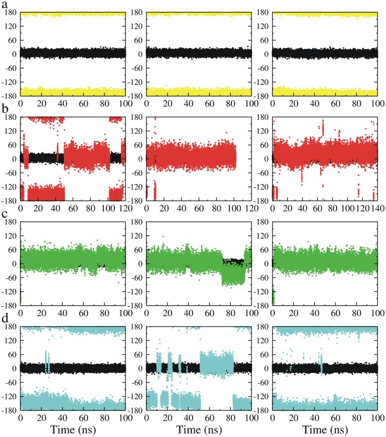
In bacteriorhodopsin, the order of molecular events that control the cytoplasmic or extracellular accessibility of the Schiff bases (SB) are not well understood. We use molecular dynamics simulations to study a process involved in the second accessibility switch of SB that occurs after its reprotonation in the N intermediate of the photocycle. We find that once protonated, the SB C15 = NZ bond switches from a cytoplasmic facing (13-cis, 15-anti) configuration to an extracellular facing (13-cis, 15-syn) configuration on the pico to nanosecond timescale. Significantly, rotation about the retinal's C13 = C14 double bond is not observed. The dynamics of the isomeric state transitions of the protonated SB are strongly influenced by the surrounding charges and dielectric effects of other buried ions, particularly D96 and D212. Our simulations indicate that the thermal isomerization of retinal from 13-cis back to all-trans likely occurs independently from and after the SB C15 = NZ rotation in the N-to-O transition.


Conflict of interest statement

Competing Interests: The authors have declared that no competing interests exist.

Figures

Figure 1
Figure 1. Schematic view of the photocycle of bacteriorhodopsin.
The accessibility and orientation of the SB nitrogen atom undergoes two switches. In switch I, de-protonated SB (SB0) changes its orientation from extracellular to cytoplasmic; in switch II, re-protonated SB (SBH+) changes its orientation from cytoplasmic to extracellular. We aim to understand whether switch II occurs before the thermal isomerization of retinal (the red route) or after (the green route).
Figure 2
Figure 2. Time development of the dihedral angles C14-C15 = NZ-CE (color symbols) and C12-C13 = C14-C15 (black symbols) in the simulations starting from the N’ structure (1P8U).
a) The SB was un-protonated and D96 was protonated, mimicking the M state; b) The SB was protonated and D96 was deprotonated, mimicking the rising of the N state; c) The SB was protonated and D96 was protonated, mimicking the decay of the N state; d) The SB, D96 and D212 were protonated. The black symbols in each of the plots depict the isomeric state of the retinal C13 = C14 double bond, which remained its initial 13-cis configuration in all simulations.
Figure 3
Figure 3. Structural snapshots of the protonated SB (SBH+) in different simulations.
a) D96 was protonated. SBH+ was 15-syn, pointing to the EC side and it formed a hydrogen bond with D212 which also formed a hydrogen bond with the protonated D85. One water molecule was in the close vicinity of both D212 and D85 at the EC side; b-c) D96 was deprotonated. The cytoplasmic proton uptake pathway opened and formed a water channel (transparent pink pipe). SBH+ was able to take two configurations: 15-syn/EC-pointing as shown in b) and 15-anti/CP-pointing as shown in c). When CP-pointing, SBH+ formed a hydrogen bond with one water molecule at the CP side and in the same time another water molecule bridged D85 and D212 at the EC side. For clarity, non-polar hydrogen atoms are not shown in b) and c).
Figure 4
Figure 4. Time development of the number of the water molecules in the D96-K216 cavity.
Left column: three simulations with un-protonated SB (SB0) and protonated D96; middle column: three simulations with protonated SB (SBH+) and deprotonated D96; right column: three simulations with protonated SB (SBH+) and protonated D96.see also Figure S5.
Figure 5
Figure 5. Percent occupancies of the 15-anti/CP-pointing (lower panel) and 15-syn/EC-pointing (upper panel) configurations of the SB C15 = NZ bond in each of 48 simulations based on crystal structure of 1p8u, 1kg8, 1f4z and 1c8s.
Simulations starting from a same crystal structure are boxed in one column. Yellow symbols represent the simulations in which the SB is unprotonated and D96 is protonated; red symbols represent the simulations in which the SB is protonated and D96 is deprotonated; green symbols represent the simulations in which both the SB and D96 are protonated; cyan symbols represent the simulations in which the SB, D96 and D212 are protonated; For each of the protonation state variations, three runs are performed and they are represented by a same color but placed separately along the x-axis in a crystal structure box. See also Figures S2, S3 and S4.

References

    1. Lozier RH, Bogomolni RA, Stoeckenius W (1975) Bacteriorhodopsin: a light-driven proton pump in Halobacterium Halobium. Biophys J 15: 955–962. - PMC - PubMed
    1. Oesterhelt D, Stoeckenius W (1971) Rhodopsin-like protein from the purple membrane of Halobacterium halobium. Nat New Biol 233: 149–152. - PubMed
    1. Haupts U, Tittor J, Oesterhelt D (1999) Closing in on bacteriorhodopsin: progress in understanding the molecule. Annu Rev Biophys Biomol Struct 28: 367–399. - PubMed
    1. Heberle J (2000) Proton transfer reactions across bacteriorhodopsin and along the membrane. BBA Bioenergetics 1458: 135–147. - PubMed
    1. Lanyi JK (2004) Bacteriorhodopsin. Annu Rev Physiol 66: 665–688. - PubMed

Publication types

MeSH terms