. 2013 Jul 29;8(7):e69972.
doi: 10.1371/journal.pone.0069972. Print 2013.

Biliverdin protects against liver ischemia reperfusion injury in swine

Affiliations

Biliverdin protects against liver ischemia reperfusion injury in swine

Barbara Andria et al. PLoS One. .

Abstract

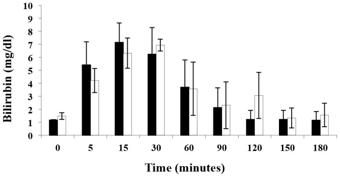
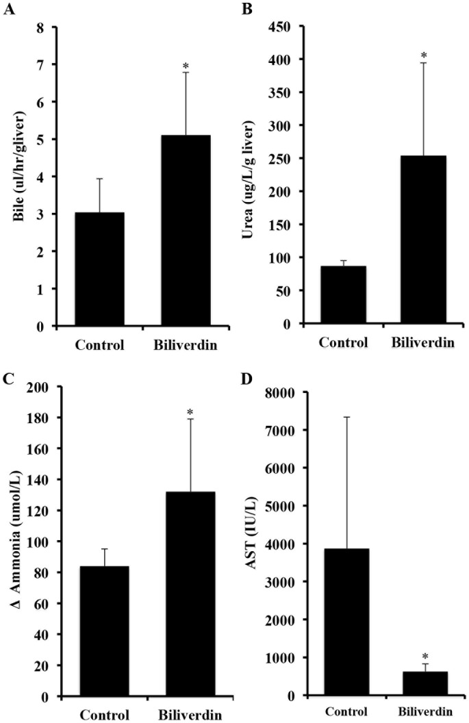
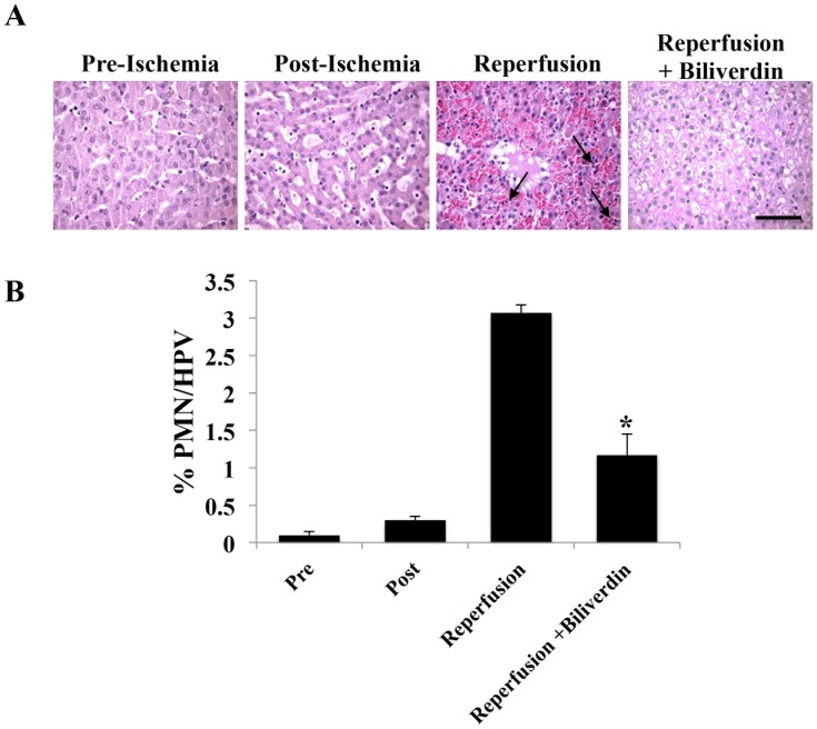
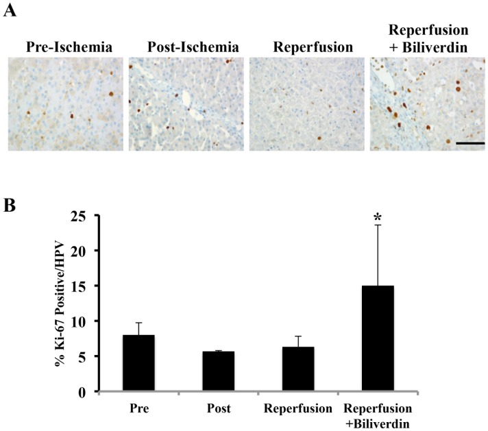
Ischemia reperfusion injury (IRI) in organ transplantation remains a serious and unsolved problem. Organs that undergo significant damage during IRI, function less well immediately after reperfusion and tend to have more problems at later times when rejection can occur. Biliverdin has emerged as an agent that potently suppress IRI in rodent models. Since the use of biliverdin is being developed as a potential therapeutic modality for humans, we tested the efficacy for its effects on IRI of the liver in swine, an accepted and relevant pre-clinical animal model. Administration of biliverdin resulted in rapid appearance of bilirubin in the serum and significantly suppressed IRI-induced liver dysfunction as measured by multiple parameters including urea and ammonia clearance, neutrophil infiltration and tissue histopathology including hepatocyte cell death. Taken together, our findings, in a large animal model, provide strong support for the continued evaluation of biliverdin as a potential therapeutic in the clinical setting of transplantation of the liver and perhaps other organs.


Conflict of interest statement

Competing Interests: The authors have declared that non competing interests exist.

Figures

Figure 1
Figure 1. Schematic of the liver perfusion circuit.
Representation of the ex vivo liver perfusion circuit as described in methods. HA: hepatic artery; PV: portal vein; ICV: inferior cava vein; Tb: blood temperature; Tw: water temperature; P: pressure transducer.
Figure 2
Figure 2. Kinetics of serum bilirubin levels in pigs in response to intravenous biliverdin administration.
Biliverdin was administered as a single i.v. bolus of 50 µmol/kg. Results are mean ± SD of 3 pigs/treatment group. The black bars correspond to the donors and the open bars to the recipient.
Figure 3
Figure 3. Effects of biliverdin on IRI-induced liver dysfunction.
A. Biliverdin was administered separately to both donors and recipients before surgery. Bile production as a measure of liver function was collected throughout the experiment and expressed as µl/hr/g liver. Control pigs show very little bile production during 12 hours of reperfusion. Note that biliverdin significantly improved bile production and thus is indicative of better liver function. Results are expressed as mean ± SD from 3 pigs/group. The increase in bile production is statistically significant comparing biliverdin vs Ctrl *p = 0.03. B. Effects biliverdin on urea production. Urea synthesis is expressed as the difference (Δ) of the urea concentration as µg/L/g liver. Results are expressed as means ± SD of 3 pigs/group. *p = 0.022 C. Effects of biliverdin on ammonia clearance. Biliverdin was administered as described above. Ammonia was measured in the serum and the clearance is expressed as a difference (Δ) in ammonia in µmol/L between the inflow and outflow ports of the perfused liver. Results are expressed as mean ± SD of 3 pigs/group. The Δ ammonia clearance is statistically significant between the biliverdin treated vs Ctrl groups, *p = 0.027. D. Effects of biliverdin on serum AST levels. Venous blood samples were taken before and every 2 hours after graft reperfusion and expressed as calculation of the total amount of serum AST released throughout the 12 hr experiment. Livers from untreated controls showed a significant increase in AST levels indicating severe liver damage. Administration of biliverdin prevented the damage and release of AST into the serum vs Ctrl *p = 0.021. Results are mean ± SD of 3 pigs/group.
Figure 4
Figure 4. Effects of biliverdin on hepatocyte cell death.
A. Representative immunostained liver sections for TUNEL and caspase 3 from liver sections harvested pre-ischemia, immediately post-ischemia and 12 hrs after reperfusion ± BV treatment. B. Quantitation of the number of positive cells in each stained sections as described in the methods. The degree of apoptosis and caspase 3 positivity was quantified by counting the number of positive cells among the total cells present in at least 10 selected fields with a minimum of 500 total positive cells counted. There is a statistically significant difference after 12 hrs of reperfusion *p = 0.01 versus ischemia alone and between biliverdin preconditioned animals after 12 hrs of reperfusion compared to controls (*p = 0.017). Results represent mean ± SD of 10 fields from 3 pigs/group where a total number of cells counted was at least 500. Magnification = 400×, Bar represents 50 µm.
Figure 5
Figure 5. Effects of Biliverdin to Reduce Neutrophil Influx into the Liver.
A. Representative H&E staining of liver sections harvested pre-ischemia, immediately post-ischemia and 12 hrs after reperfusion ± BV treatment. B. Neutrophils were counted based on morphology and expressed as a percentage of neutrophils among the total number of cells present in each field. Data are expressed as mean ± SD of n = 3 pigs/group where a total number of cells counted was at least 500. Magnification = 400×, Bar represents 50 µm. (*p = 0.013).
Figure 6
Figure 6. Biliverdin Treatment Increases Hepatocyte Proliferation After IRI.
A. Representative immunostaining images for Ki-67 as a marker of cell proliferation in liver sections harvested pre-ischemia, immediately post-ischemia and 12 hrs after reperfusion ± BV treatment. Effects of biliverdin on Ki-67 expression as an indicator of cell (primarily hepatocyte) proliferation. B. Ki-67 positive cells were counted and expressed as a percentage of positive cells among the total number of cells present in each field. Data are expressed as mean ± SD of n = 3 pigs/group where a total number of cells counted was at least 500. Magnification = 400×, Bar represents 50 µm. (*p = 0.046).

References

    1. Scuderi V, Ceriello A, Maida P, Aragiusto G, Arenga G, et al. (2006) The marginal donor: a single center experience in orthotopic liver transplantation. Transplant Proc 38: 1069–1073. - PubMed
    1. Busuttil RW, Tanaka K (2003) The utility of marginal donors in liver transplantation. Liver Transpl 9: 651–663. - PubMed
    1. Fondevila C, Busuttil RW, Kupiec-Weglinski JW (2003) Hepatic ischemia/reperfusion injury-a fresh look. Exp Mol Pathol. 74: 86–93. - PubMed
    1. Selzner N, Rudiger H, Graf R, Clavien PA (2003) Protective strategies against ischemic injury of the liver. Gastroenterology 125: 917–936. - PubMed
    1. Jaeschke H, Farhood A (1991) Neutrophil and Kupffer cell-induced oxidant stress and ischemia-reperfusion injury in rat liver. Am J Physiol 260: 355–362. - PubMed

Publication types

LinkOut - more resources