

Sequence analysis of the genome of piscine orthoreovirus (PRV) associated with heart and skeletal muscle inflammation (HSMI) in Atlantic salmon (Salmo salar)
- PMID: 23922911
- PMCID: PMC3726481
- DOI: 10.1371/journal.pone.0070075
Sequence analysis of the genome of piscine orthoreovirus (PRV) associated with heart and skeletal muscle inflammation (HSMI) in Atlantic salmon (Salmo salar)
Erratum in
- PLoS One. 2013;8(7). doi: 10.1371/annotation/746a9036-0e54-4a9a-ab16-adb108a3a227 doi: 10.1371/annotation/746a9036-0e54-4a9a-ab16-adb108a3a227
Abstract
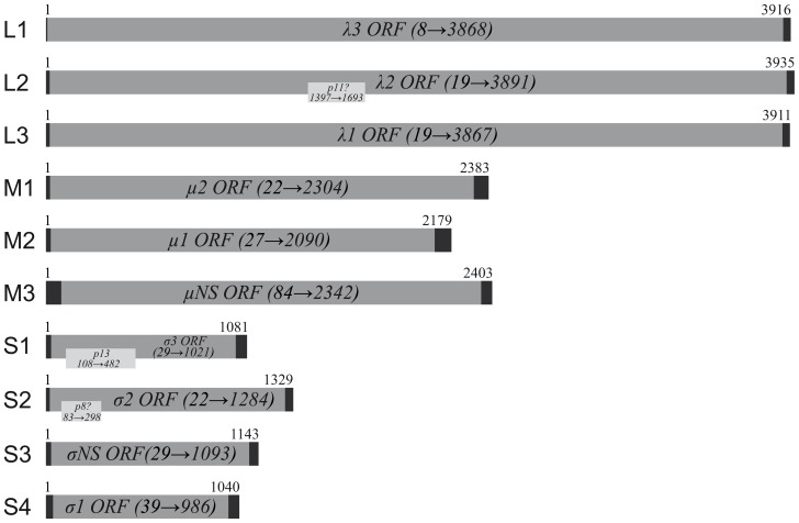
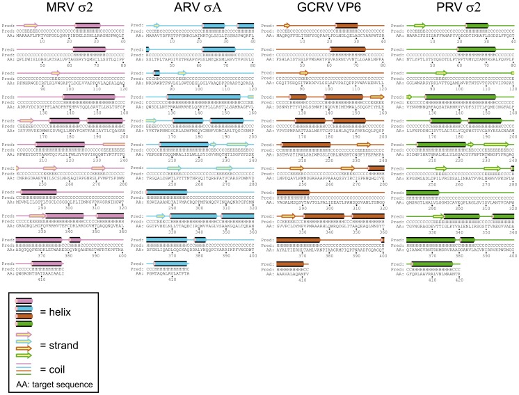
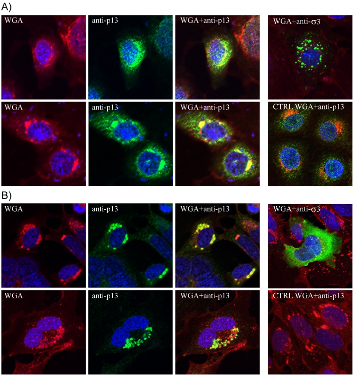
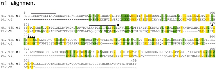
Piscine orthoreovirus (PRV) is associated with heart- and skeletal muscle inflammation (HSMI) of farmed Atlantic salmon (Salmo salar). We have performed detailed sequence analysis of the PRV genome with focus on putative encoded proteins, compared with prototype strains from mammalian (MRV T3D)- and avian orthoreoviruses (ARV-138), and aquareovirus (GCRV-873). Amino acid identities were low for most gene segments but detailed sequence analysis showed that many protein motifs or key amino acid residues known to be central to protein function are conserved for most PRV proteins. For M-class proteins this included a proline residue in μ2 which, for MRV, has been shown to play a key role in both the formation and structural organization of virus inclusion bodies, and affect interferon-β signaling and induction of myocarditis. Predicted structural similarities in the inner core-forming proteins λ1 and σ2 suggest a conserved core structure. In contrast, low amino acid identities in the predicted PRV surface proteins μ1, σ1 and σ3 suggested differences regarding cellular interactions between the reovirus genera. However, for σ1, amino acid residues central for MRV binding to sialic acids, and cleavage- and myristoylation sites in μ1 required for endosomal membrane penetration during infection are partially or wholly conserved in the homologous PRV proteins. In PRV σ3 the only conserved element found was a zinc finger motif. We provide evidence that the S1 segment encoding σ3 also encodes a 124 aa (p13) protein, which appears to be localized to intracellular Golgi-like structures. The S2 and L2 gene segments are also potentially polycistronic, predicted to encode a 71 aa- (p8) and a 98 aa (p11) protein, respectively. It is concluded that PRV has more properties in common with orthoreoviruses than with aquareoviruses.
Conflict of interest statement
Figures
References
-
- Kongtorp RT, Taksdal T, Lyngøy A (2004) Pathology of heart and skeletal muscle inflammation (HSMI) in farmed Atlantic salmon Salmo salar. Dis Aquat Organ 59: 217–224. - PubMed
-
- Attoui H, Fang Q, Mohd JF, Cantaloube JF, Biagini P, et al. (2002) Common evolutionary origin of aquareoviruses and orthoreoviruses revealed by genome characterization of Golden shiner reovirus, Grass carp reovirus, Striped bass reovirus and golden ide reovirus (genus Aquareovirus, family Reoviridae). J Gen Virol 83: 1941–1951. - PubMed
Publication types
MeSH terms
LinkOut - more resources
Full Text Sources
Other Literature Sources
