. 2013 Jul 26;8(7):e70636.
doi: 10.1371/journal.pone.0070636. Print 2013.

Distinct functional interactions between actin isoforms and nonsarcomeric myosins

Affiliations

Distinct functional interactions between actin isoforms and nonsarcomeric myosins

Mirco Müller et al. PLoS One. .

Abstract

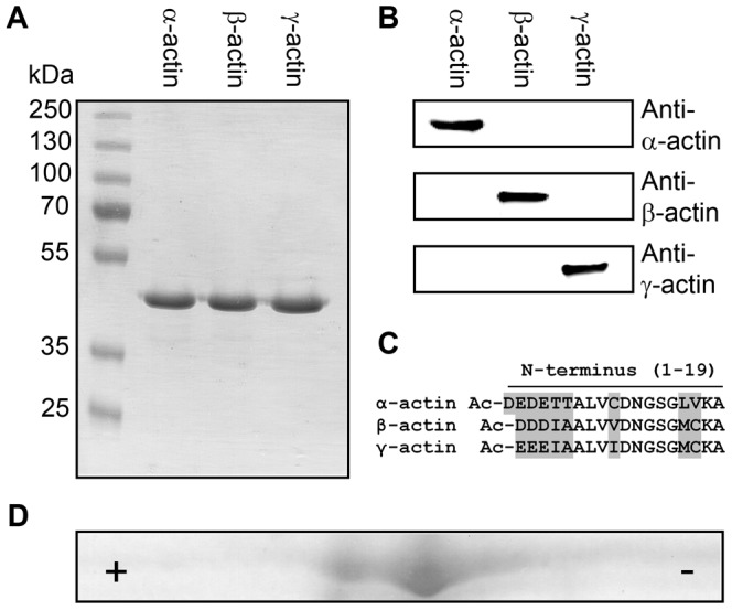
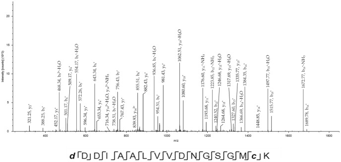
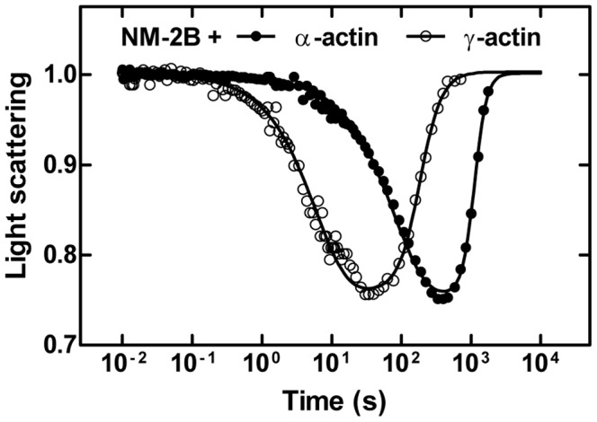
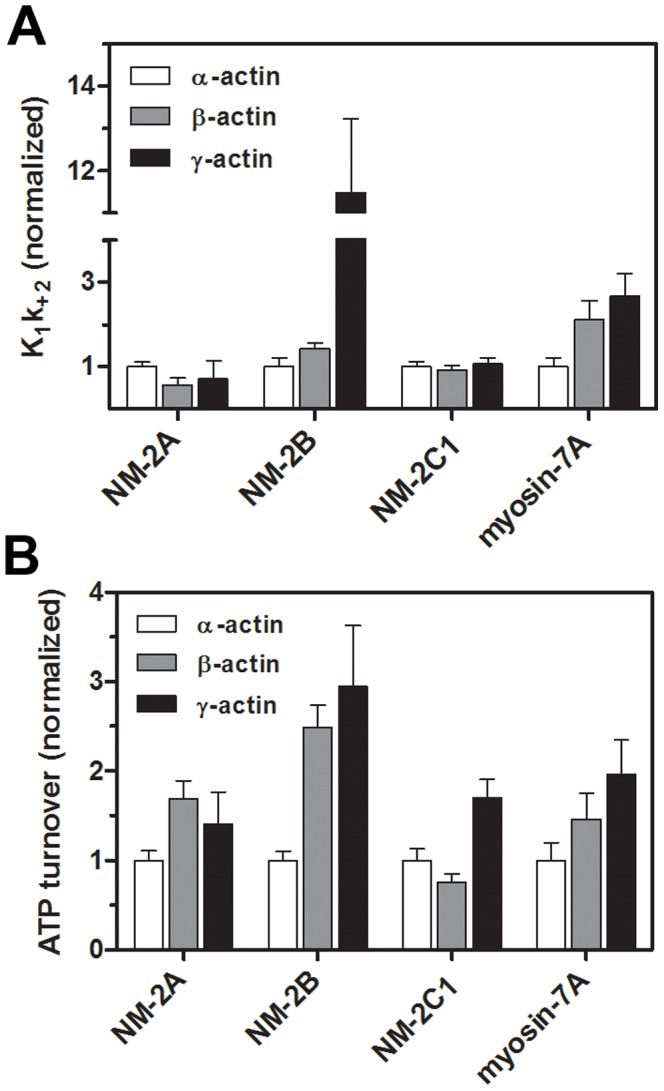
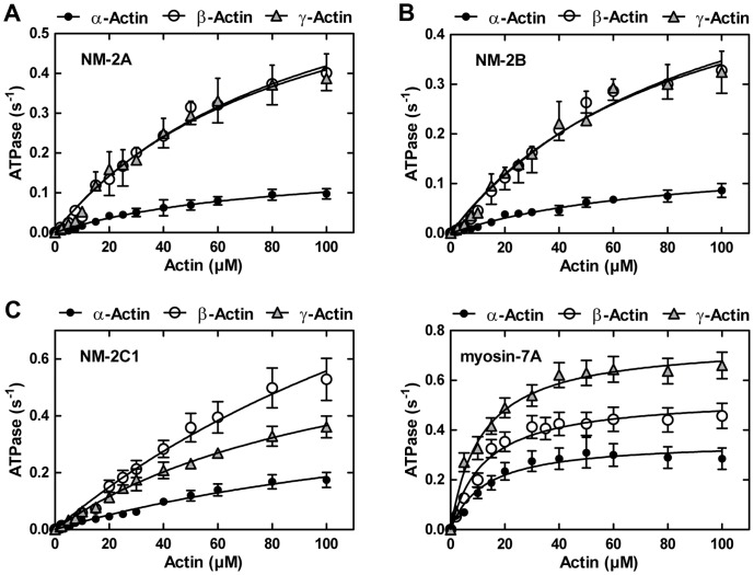
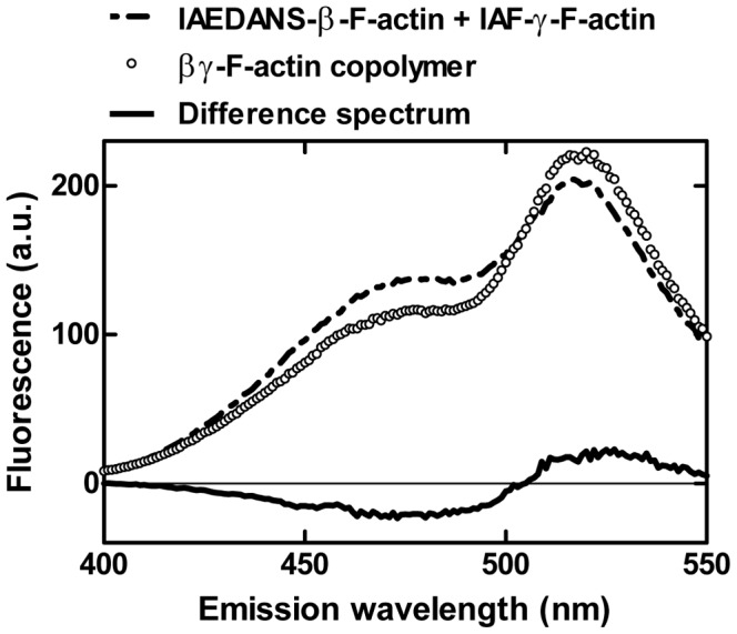
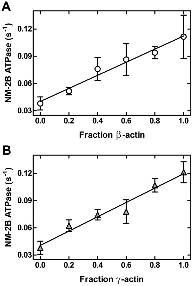
Despite their near sequence identity, actin isoforms cannot completely replace each other in vivo and show marked differences in their tissue-specific and subcellular localization. Little is known about isoform-specific differences in their interactions with myosin motors and other actin-binding proteins. Mammalian cytoplasmic β- and γ-actin interact with nonsarcomeric conventional myosins such as the members of the nonmuscle myosin-2 family and myosin-7A. These interactions support a wide range of cellular processes including cytokinesis, maintenance of cell polarity, cell adhesion, migration, and mechano-electrical transduction. To elucidate differences in the ability of isoactins to bind and stimulate the enzymatic activity of individual myosin isoforms, we characterized the interactions of human skeletal muscle α-actin, cytoplasmic β-actin, and cytoplasmic γ-actin with human myosin-7A and nonmuscle myosins-2A, -2B and -2C1. In the case of nonmuscle myosins-2A and -2B, the interaction with either cytoplasmic actin isoform results in 4-fold greater stimulation of myosin ATPase activity than was observed in the presence of α-skeletal muscle actin. Nonmuscle myosin-2C1 is most potently activated by β-actin and myosin-7A by γ-actin. Our results indicate that β- and γ-actin isoforms contribute to the modulation of nonmuscle myosin-2 and myosin-7A activity and thereby to the spatial and temporal regulation of cytoskeletal dynamics. FRET-based analyses show efficient copolymerization abilities for the actin isoforms in vitro. Experiments with hybrid actin filaments show that the extent of actomyosin coupling efficiency can be regulated by the isoform composition of actin filaments.


Conflict of interest statement

Competing Interests: The authors have declared that no competing interests exist.

Figures

Figure 1
Figure 1. Purification of actin isoforms.
(A) SDS-PAGE of purified actin isoforms. (B) Immunoblot-based identification of actin isoforms. The actin antibodies used show no cross-reactivity. (C) Actin isoforms display subtle differences primarily located at their N-terminus as shown in the protein sequence alignment. Sequence variations at the N-terminus of α-, β- and γ-actin are marked in grey. Mature actin isoforms are acetylated (Ac) at their N-terminus (D) 2D-gelelectrophoresis of purified γ-actin (IEP 5.31) containing ∼10% co-purified insect actin (IEP 5.29). The acidic (+) and the basic end (−) are marked.
Figure 2
Figure 2. β- and γ-actin are N-terminally acetylated. Mass spectrum of the N-terminal peptide of β-actin.
The protonated molecular ion (MH+) of the identified peptide has a mass of 1835.8 Da and is modified by removal of Met1 (m = 1722.9 Da), acetylation of Asp1 (indicated as d, Δm = +42 Da) and due to sample processing by an artificial propionamidation of cysteine (indicated as c, Δm = +71 Da). Similar results were obtained with γ-actin (spectrum not shown).
Figure 3
Figure 3. Time-dependent dissociation and association of NM-2B from filamentous α- and γ-actin.
The light scattering signal normalized to the initial value shows the time-dependent dissociation and association of the actomyosin complex formed by NM-2B and α- or γ-actin. The initial drop in the light scattering signal follows the release of ATP from caged-ATP by a flash of UV-laser. The single exponential reduction in scattering intensity monitors the ATP-induced dissociation of myosin from F-actin. The following restoration of the light scattering signal is caused by rebinding of myosin to the actin filament after ATP hydrolysis is completed. As shown for NM-2B, γ-actin causes faster dissociation and association compared to α-actin. Note the logarithmic time scale on the graph.
Figure 4
Figure 4. Flash photolysis experiments with nonmuscle myosin and actin isoforms.
(A) The graphs show the relative changes of the apparent second order rate constants K1k+2. P-values of unpaired t-tests were <0.05 for the combinations NM-2A (α- and β-actin), NM-2B (α- and β-/γ-actin), and myosin-7A (α- and β-/γ-actin). (B) ATP turnover rates of nonmuscle myosin isoforms interacting with β- or γ-actin. P-values of unpaired t-tests were <0.05 for the combinations NM-2A (α- and β-/γ-actin), NM-2B (α- and β-/γ-actin), NM-2C1 (γ- and α-/β-actin), and myosin-7A (all combinations). Rates were normalized using α-actin as a reference. The ATP-induced dissociation is accelerated for the complexes NM-2B/β-/γ-actin and myosin-7A/β-/γ-actin. The interaction of cytoplasmic actins and NM-2 or myosin-7A isoforms leads to 2- to 3-fold increased catalytic activities. Reference values for K1k+2 (µM−1 s−1) as determined with α-actin are as follows: NM-2A, 0.14±0.003; NM-2B, 0.24±0.02; NM-2C1, 0.89±0.01; myosin-7A, 0.44±0.01 (see also [32], [48]).
Figure 5
Figure 5. Cytoplasmic actins enhance nonmuscle myosin-2 and myosin-7A efficiency.
Steady-state ATPase activities of (A) NM-2A, (B) NM-2B, (C) NM-2C1 and (D) myosin-7A measured as a function of actin concentration (α-, β-, and γ-F-actin). The ATPase activity in the absence of F-actin was subtracted from the actin-activated data. Values for Kactin and kcat were calculated from a hyperbolic fit (ATPase activity = (kcat [F-actin])/(Kactin+[F-actin])) to the data (see Table 1).
Figure 6
Figure 6. FRET copolymerization analysis of actin isoforms.
Fluorescence emission spectra of IAEDANS-β-actin and IAF-γ-actin, both separately polymerized and after copolymerization. The donor fluorescence (IAEDANS) peaks around 490 nm and decreases significantly upon copolymerization whereas the fluorescence of the acceptor (IAF) at 520 nm apparently increases as shown in the difference spectrum.
Figure 7
Figure 7. Activation of NM-2B ATPase rate by αβ- and αγ-actin copolymers.
Copolymers with different mixing ratios were used to stimulate the ATPase activity of NM-2B. Measurements were performed using a final concentration of 20 µM F-actin.

References

    1. Vandekerckhove J, Weber K (1978) At least six different actins are expressed in a higher mammal: an analysis based on the amino acid sequence of the amino-terminal tryptic peptide. J Mol Biol 126: 783–802. - PubMed
    1. Tondeleir D, Vandamme D, Vandekerckhove J, Ampe C, Lambrechts A (2009) Actin isoform expression patterns during mammalian development and in pathology: insights from mouse models. Cell Motil Cytoskeleton 66: 798–815. - PubMed
    1. Dugina V, Zwaenepoel I, Gabbiani G, Clement S, Chaponnier C (2009) Beta and gamma-cytoplasmic actins display distinct distribution and functional diversity. J Cell Sci 122: 2980–2988. - PubMed
    1. Hoock TC, Newcomb PM, Herman IM (1991) Beta actin and its mRNA are localized at the plasma membrane and the regions of moving cytoplasm during the cellular response to injury. J Cell Biol 112: 653–664. - PMC - PubMed
    1. von Arx P, Bantle S, Soldati T, Perriard JC (1995) Dominant negative effect of cytoplasmic actin isoproteins on cardiomyocyte cytoarchitecture and function. J Cell Biol 131: 1759–1773. - PMC - PubMed

Publication types

LinkOut - more resources