. 2013 Jul 26;8(7):e70949.
doi: 10.1371/journal.pone.0070949. Print 2013.

Neural correlates of central inhibition during physical fatigue

Affiliations

Neural correlates of central inhibition during physical fatigue

Masaaki Tanaka et al. PLoS One. .

Abstract

Central inhibition plays a pivotal role in determining physical performance during physical fatigue. Classical conditioning of central inhibition is believed to be associated with the pathophysiology of chronic fatigue. We tried to determine whether classical conditioning of central inhibition can really occur and to clarify the neural mechanisms of central inhibition related to classical conditioning during physical fatigue using magnetoencephalography (MEG). Eight right-handed volunteers participated in this study. We used metronome sounds as conditioned stimuli and maximum handgrip trials as unconditioned stimuli to cause central inhibition. Participants underwent MEG recording during imagery of maximum grips of the right hand guided by metronome sounds for 10 min. Thereafter, fatigue-inducing maximum handgrip trials were performed for 10 min; the metronome sounds were started 5 min after the beginning of the handgrip trials. The next day, neural activities during imagery of maximum grips of the right hand guided by metronome sounds were measured for 10 min. Levels of fatigue sensation and sympathetic nerve activity on the second day were significantly higher relative to those of the first day. Equivalent current dipoles (ECDs) in the posterior cingulated cortex (PCC), with latencies of approximately 460 ms, were observed in all the participants on the second day, although ECDs were not identified in any of the participants on the first day. We demonstrated that classical conditioning of central inhibition can occur and that the PCC is involved in the neural substrates of central inhibition related to classical conditioning during physical fatigue.


Conflict of interest statement

Competing Interests: The authors have declared that no competing interests exist.

Figures

Figure 1
Figure 1. Experimental paradigm.
(A) Experimental design. On the first day, neural activities during the imagery of handgrips guided by the handgrip cues of metronome sounds were measured using magnetoencephalography (MEG) for 10 min (First MEG session). Thereafter, 10-min fatigue-inducing maximum handgrip trials (Conditioning session) were performed, in which metronome sounds were started 5 min after the beginning of the handgrip trials as conditioned stimuli. On the next day, neural activities during the imagery of handgrips guided by the handgrip cues of metronome sounds were measured using MEG for 10 min (Second MEG session). (B) Metronome sounds. Each MEG session consisted of 150 blocks, and each block consisted of three pacing cues followed by one handgrip cue. During the MEG session, participants heard the sound cues every 1 s with their eyes closed, and during the handgrip cue period, they were requested to imagine that they were gripping a soft ball with their right hand at a maximal voluntary contraction level every 4 s for 1 s.
Figure 2
Figure 2. Visual analogue scale (VAS) value of the right (A) and left (B) hands for fatigue immediately before (open columns) and after (closed columns) the 10-min fatigue-inducing handgrip trials.
Data are presented as mean and SD. **P<0.01, significantly different from the corresponding values before handgrip trials (paired t-test).
Figure 3
Figure 3. Visual analogue scale (VAS) values of right (A) and left (B) hands for fatigue immediately after the first and second magnetoencephalography (MEG) sessions.
Data are presented as mean and SD. *P<0.05, significantly different from the corresponding values of the first MEG session (paired t-test).
Figure 4
Figure 4. Autonomic nerve activities assessed by frequency analyses of electrocardiography (ECG).
High-frequency power (HF; A) and low-frequency power (LF)/HF ratio (B) were assessed during the first and second magnetoencephalography (MEG) sessions. Data are presented as means and SD. **P<0.01, significantly different from the corresponding values of the first MEG session (paired t-test).
Figure 5
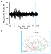
Figure 5. Typical example of magnetic fields (A) and an isofield contour map (B) caused by the cue sounds and imagery of handgrips (participant No. 5).
The equivalent current dipole (ECD) in the posterior cingulate cortex is shown at the peak latency of 471 ms. R: right side; L: left side.
Figure 6
Figure 6. Locations of equivalent current dipoles (ECDs) in the posterior cingulate cortex induced by cue sounds and imagery of handgrips during the second magnetoencephalography (MEG) sessions.
Closed red circles indicate the locations of ECDs and short red lines radiating from closed circles indicate the orientation of ECDs. ECDs are superimposed on individual magnetic resonance images (sagittal view) (A: anterior side; P: posterior side). The peak latencies are presented in parentheses.

Similar articles

Cited by

References

    1. Chaudhuri A, Behan PO (2004) Fatigue in neurological disorders. Lancet 363: 978–988. - PubMed
    1. Bigland-Ritchie B, Woods JJ (1984) Changes in muscle contractile properties and neural control during human muscular fatigue. Muscle Nerve 7: 691–699. - PubMed
    1. Pedersen TH, Nielsen OB, Lamb GD, Stephenson DG (2004) Intracellular acidosis enhances the excitability of working muscle. Science 305: 1144–1147. - PubMed
    1. Allen DG, Westerblad H (2004) Physiology. Lactic acid-the latest performance-enhancing drug. Science 305: 1112–1113. - PubMed
    1. Allen DG, Lamb GD, Westerblad H (2008) Skeletal muscle fatigue: cellular mechanisms. Physiol Rev 88: 287–332. - PubMed

Publication types