

TcTASV-C, a protein family in Trypanosoma cruzi that is predominantly trypomastigote-stage specific and secreted to the medium
- PMID: 23923058
- PMCID: PMC3726618
- DOI: 10.1371/journal.pone.0071192
TcTASV-C, a protein family in Trypanosoma cruzi that is predominantly trypomastigote-stage specific and secreted to the medium
Abstract
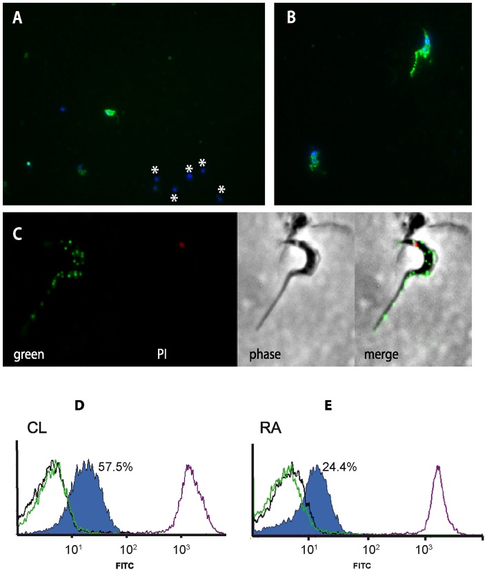
Among the several multigene families codified by the genome of T. cruzi, the TcTASV family was the latest discovered. The TcTASV (Trypomastigote, Alanine, Serine, Valine) family is composed of ∼40 members, with conserved carboxi- and amino-termini but with a variable central core. According to the length and sequence of the central region the family is split into 3 subfamilies. The TcTASV family is conserved in the genomes of - at least - lineages TcI and TcVI and has no orthologues in other trypanosomatids. In the present work we focus on the study of the TcTASV-C subfamily, composed by 16 genes in the CL Brener strain. We determined that TcTASV-C is preferentially expressed in trypomastigotes, but it is not a major component of the parasite. Both immunoflourescence and flow cytometry experiments indicated that TcTASV-C has a clonal expression, i.e. it is not expressed by all the parasites of a certain population at the same time. We also determined that TcTASV-C is phosphorylated and glycosylated. TASV-C is attached to the parasite surface by a GPI anchor and is shed spontaneously into the medium. About 30% of sera from infected hosts reacted with TcTASV-C, confirming its exposition to the immune system. Its superficial localization and secretory nature suggest a possible role in host-parasite interactions.
Conflict of interest statement
Figures


Similar articles
-
The protein family TcTASV-C is a novel Trypanosoma cruzi virulence factor secreted in extracellular vesicles by trypomastigotes and highly expressed in bloodstream forms.PLoS Negl Trop Dis. 2018 May 4;12(5):e0006475. doi: 10.1371/journal.pntd.0006475. eCollection 2018 May. PLoS Negl Trop Dis. 2018. PMID: 29727453 Free PMC article.
-
TcTASV: a novel protein family in trypanosoma cruzi identified from a subtractive trypomastigote cDNA library.PLoS Negl Trop Dis. 2010 Oct 5;4(10):e841. doi: 10.1371/journal.pntd.0000841. PLoS Negl Trop Dis. 2010. PMID: 20957201 Free PMC article.
-
The TcTASV proteins are novel promising antigens to detect active Trypanosoma cruzi infection in dogs.Parasitology. 2016 Sep;143(11):1382-9. doi: 10.1017/S0031182016000822. Epub 2016 May 13. Parasitology. 2016. PMID: 27173912
-
Genetic structure and expression of the surface glycoprotein GP82, the main adhesin of Trypanosoma cruzi metacyclic trypomastigotes.ScientificWorldJournal. 2013;2013:156734. doi: 10.1155/2013/156734. Epub 2013 Feb 4. ScientificWorldJournal. 2013. PMID: 23431251 Free PMC article. Review.
-
The mucin-like glycoprotein super-family of Trypanosoma cruzi: structure and biological roles.Mol Biochem Parasitol. 2001 May;114(2):143-50. doi: 10.1016/s0166-6851(01)00245-6. Mol Biochem Parasitol. 2001. PMID: 11378194 Review.
Cited by
-
Sialic Acid Glycobiology Unveils Trypanosoma cruzi Trypomastigote Membrane Physiology.PLoS Pathog. 2016 Apr 8;12(4):e1005559. doi: 10.1371/journal.ppat.1005559. eCollection 2016 Apr. PLoS Pathog. 2016. PMID: 27058585 Free PMC article.
-
Towards High-throughput Immunomics for Infectious Diseases: Use of Next-generation Peptide Microarrays for Rapid Discovery and Mapping of Antigenic Determinants.Mol Cell Proteomics. 2015 Jul;14(7):1871-84. doi: 10.1074/mcp.M114.045906. Epub 2015 Apr 28. Mol Cell Proteomics. 2015. PMID: 25922409 Free PMC article.
-
Exoproteome profiling of Trypanosoma cruzi during amastigogenesis early stages.PLoS One. 2019 Nov 22;14(11):e0225386. doi: 10.1371/journal.pone.0225386. eCollection 2019. PLoS One. 2019. PMID: 31756194 Free PMC article.
-
Interactions between Trypanosoma cruzi Secreted Proteins and Host Cell Signaling Pathways.Front Microbiol. 2016 Mar 31;7:388. doi: 10.3389/fmicb.2016.00388. eCollection 2016. Front Microbiol. 2016. PMID: 27065960 Free PMC article. Review.
-
The protein family TcTASV-C is a novel Trypanosoma cruzi virulence factor secreted in extracellular vesicles by trypomastigotes and highly expressed in bloodstream forms.PLoS Negl Trop Dis. 2018 May 4;12(5):e0006475. doi: 10.1371/journal.pntd.0006475. eCollection 2018 May. PLoS Negl Trop Dis. 2018. PMID: 29727453 Free PMC article.
References
-
- TDR Disease Reference Group, WHO (2012) Research Priorities for Chagas Disease, Human African Trypanosomiasis and Leishmaniasis. WHO Technical Report Series 975. Switzerland. - PubMed
-
- Anez N, Crisante G, da Silva FM, Rojas A, Carrasco H, et al. (2004) Predominance of lineage I among Trypanosoma cruzi isolates from Venezuelan patients with different clinical profiles of acute Chagas' disease. Trop Med Int Health 9: 1319–1326. - PubMed
-
- Zingales B, Miles MA, Campbell DA, Tibayrenc M, Macedo AM, et al. (2012) The revised Trypanosoma cruzi subspecific nomenclature: rationale, epidemiological relevance and research applications. Infect Genet Evol 12: 240–253. - PubMed
-
- Cunha-Neto E, Teixeira PC, Nogueira LG, Kalil J (2011) Autoimmunity. Adv Parasitol 76: 129–152. - PubMed
Publication types
MeSH terms
Substances
LinkOut - more resources
Full Text Sources
Other Literature Sources
