Altered islet morphology but normal islet secretory function in vitro in a mouse model with microvascular alterations in the pancreas
- PMID: 23923060
- PMCID: PMC3726616
- DOI: 10.1371/journal.pone.0071277
Altered islet morphology but normal islet secretory function in vitro in a mouse model with microvascular alterations in the pancreas
Abstract
Background: Our previous studies have shown that signal transducer and activator of transcription 3 (STAT3) signaling is important for the development of pancreatic microvasculature via its regulation of vascular endothelial growth factor-A (VEGF-A). Pancreas-specific STAT3-KO mice exhibit glucose intolerance and impaired insulin secretion in vivo, along with microvascular alterations in the pancreas. However, the specific role of STAT3 signaling in the regulation of pancreatic islet development and function is not entirely understood.
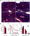
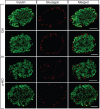
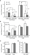
Methodology/principal findings: To investigate the role of STAT3 signaling in the formation and maintenance of pancreatic islets, we studied pancreas-specific STAT3-KO mice. Histological analysis showed that STAT3 deficiency affected pancreatic islet morphology. We found an increased proportion of small-sized islets and a reduced fraction of medium-sized islets, indicating abnormal islet development in STAT3-KO mice. Interestingly, the islet area relative to the whole pancreas area in transgenic and control mice was not significantly different. Immunohistochemical analysis on pancreatic cryosections revealed abnormalities in islet architecture in STAT3-KO mice: the pattern of peripheral distribution of glucagon-positive α-cells was altered. At the same time, islets belonging to different size categories isolated from STAT3-KO mice exhibited normal glucose-stimulated insulin secretion in perifusion experiments in vitro when compared to control mice.
Conclusions: Our data demonstrate that STAT3 signaling in the pancreas is required for normal islet formation and/or maintenance. Altered islet size distribution in the KO mice does not result in an impaired islet secretory function in vitro. Therefore, our current study supports that the glucose intolerance and in vivo insulin secretion defect in pancreas-specific STAT3-KO mice is due to altered microvasculature in the pancreas, and not intrinsic beta-cell function.
Conflict of interest statement
Figures


Similar articles
-
Glucose intolerance and impaired insulin secretion in pancreas-specific signal transducer and activator of transcription-3 knockout mice are associated with microvascular alterations in the pancreas.Endocrinology. 2010 May;151(5):2050-9. doi: 10.1210/en.2009-1199. Epub 2010 Mar 9. Endocrinology. 2010. PMID: 20215569 Free PMC article.
-
Insulin secretory defects and impaired islet architecture in pancreatic beta-cell-specific STAT3 knockout mice.Biochem Biophys Res Commun. 2004 Jul 9;319(4):1159-70. doi: 10.1016/j.bbrc.2004.05.095. Biochem Biophys Res Commun. 2004. PMID: 15194489
-
CART knock out mice have impaired insulin secretion and glucose intolerance, altered beta cell morphology and increased body weight.Regul Pept. 2005 Jul 15;129(1-3):203-11. doi: 10.1016/j.regpep.2005.02.016. Regul Pept. 2005. PMID: 15927717
-
The Eye as a Transplantation Site to Monitor Pancreatic Islet Cell Plasticity.Front Endocrinol (Lausanne). 2021 Apr 23;12:652853. doi: 10.3389/fendo.2021.652853. eCollection 2021. Front Endocrinol (Lausanne). 2021. PMID: 33967961 Free PMC article. Review.
-
Status of ghrelin as an islet hormone and paracrine/autocrine regulator of insulin secretion.Peptides. 2022 Feb;148:170681. doi: 10.1016/j.peptides.2021.170681. Epub 2021 Oct 30. Peptides. 2022. PMID: 34728253 Review.
Cited by
-
Intra-islet endothelial cell and β-cell crosstalk: Implication for islet cell transplantation.World J Transplant. 2017 Apr 24;7(2):117-128. doi: 10.5500/wjt.v7.i2.117. World J Transplant. 2017. PMID: 28507914 Free PMC article. Review.
-
STAT3 modulates β-cell cycling in injured mouse pancreas and protects against DNA damage.Cell Death Dis. 2016 Jun 23;7(6):e2272. doi: 10.1038/cddis.2016.171. Cell Death Dis. 2016. PMID: 27336716 Free PMC article.
-
Hypothyroidism Affects Vascularization and Promotes Immune Cells Infiltration into Pancreatic Islets of Female Rabbits.Int J Endocrinol. 2015;2015:917806. doi: 10.1155/2015/917806. Epub 2015 Jun 14. Int J Endocrinol. 2015. PMID: 26175757 Free PMC article.
-
The Price of Surviving on Adrenaline: Developmental Programming Responses to Chronic Fetal Hypercatecholaminemia Contribute to Poor Muscle Growth Capacity and Metabolic Dysfunction in IUGR-Born Offspring.Front Anim Sci. 2021 Dec;2:769334. doi: 10.3389/fanim.2021.769334. Epub 2021 Dec 9. Front Anim Sci. 2021. PMID: 34966907 Free PMC article.
-
Hyperglycemic Conditions Promote Rac1-Mediated Serine536 Phosphorylation of p65 Subunit of NFκB (RelA) in Pancreatic Beta Cells.Cell Physiol Biochem. 2022 Aug 19;56(4):367-381. doi: 10.33594/000000557. Cell Physiol Biochem. 2022. PMID: 35981264 Free PMC article.
References
-
- Ballian N, Brunicardi FC (2007) Islet vasculature as a regulator of endocrine pancreas function. World journal of surgery 31: 705–714. - PubMed
-
- Gustavsson N, Han W (2009) Calcium-sensing beyond neurotransmitters: functions of synaptotagmins in neuroendocrine and endocrine secretion. Bioscience reports 29: 245–259. - PubMed
-
- Bonner-Weir S, Orci L (1982) New perspectives on the microvasculature of the islets of Langerhans in the rat. Diabetes 31: 883–889. - PubMed
-
- Brunicardi FC, Stagner J, Bonner-Weir S, Wayland H, Kleinman R, et al. (1996) Microcirculation of the islets of Langerhans. Long Beach Veterans Administration Regional Medical Education Center Symposium. Diabetes 45: 385–392. - PubMed
Publication types
MeSH terms
Substances
LinkOut - more resources
Full Text Sources
Other Literature Sources
Medical
Molecular Biology Databases
Research Materials
Miscellaneous

