. 2013 Jul 15;6(8):1467-80.
Print 2013.

Histopathological and functional effects of antimony on the renal cortex of growing albino rat

Affiliations

Histopathological and functional effects of antimony on the renal cortex of growing albino rat

Ahmed H Rashedy et al. Int J Clin Exp Pathol. .

Abstract

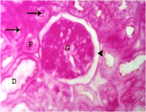
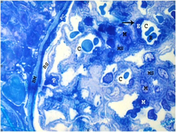
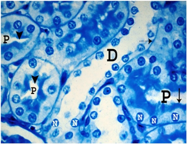
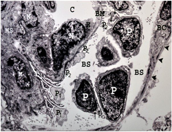
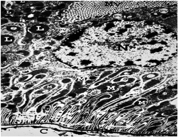
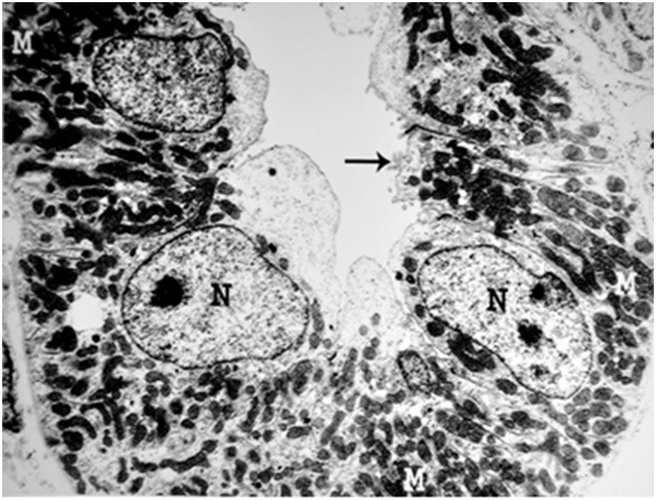
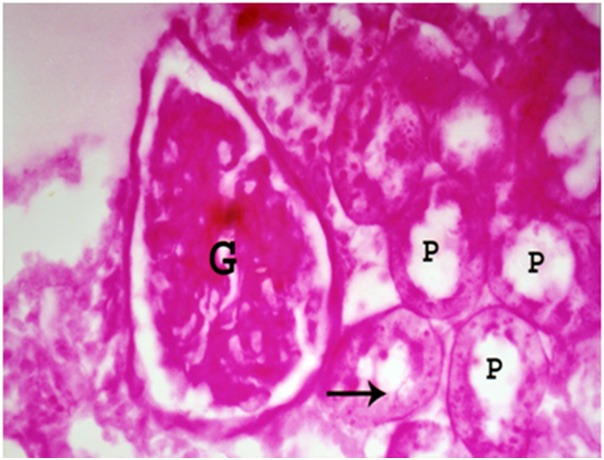
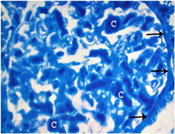
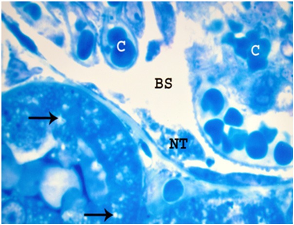
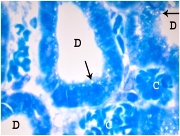
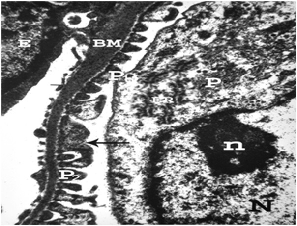
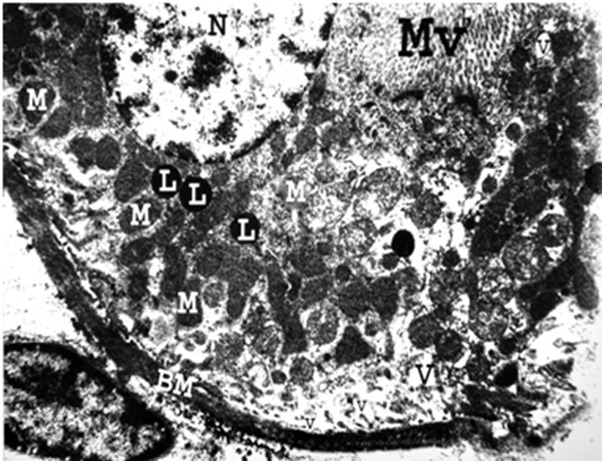
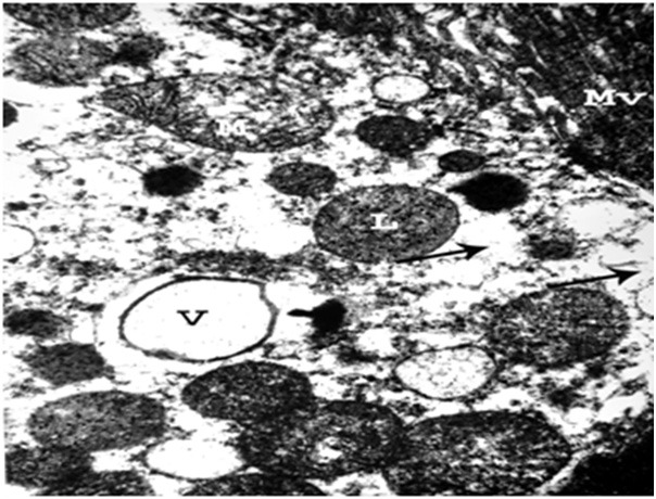
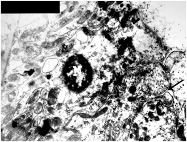
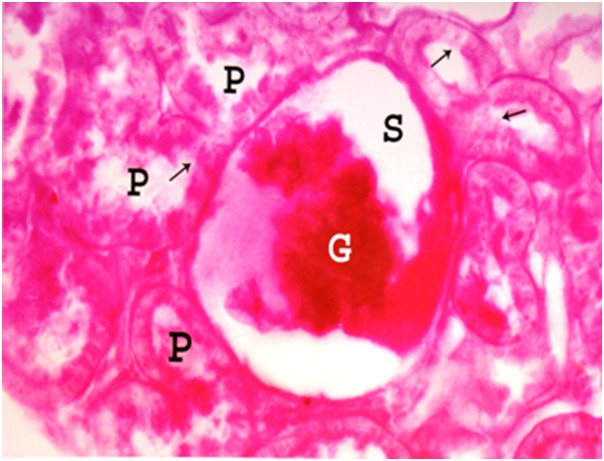
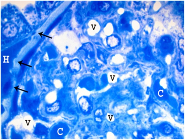
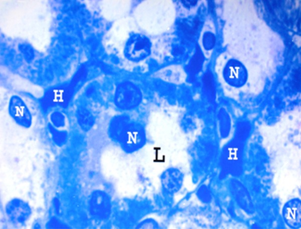
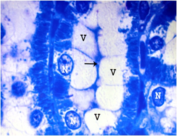
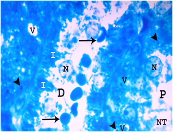
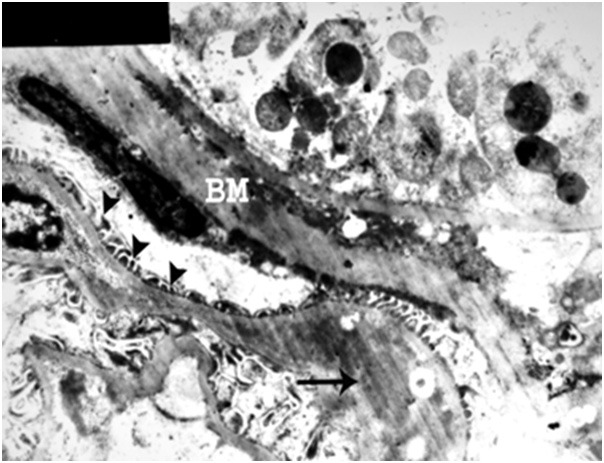
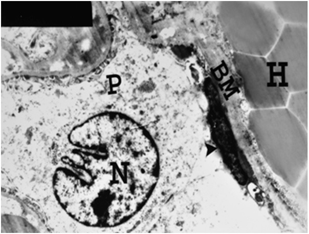
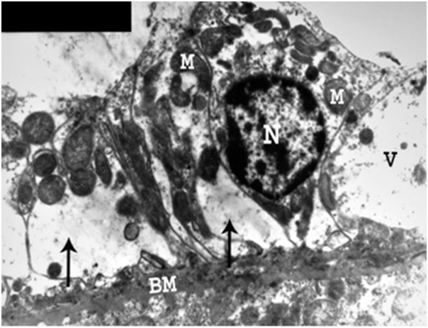
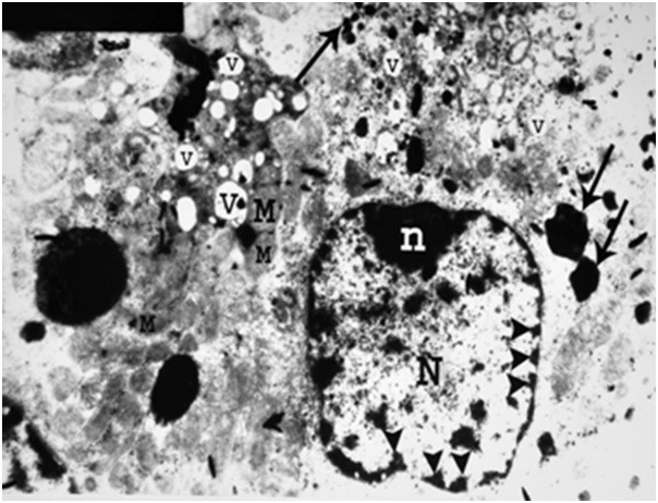
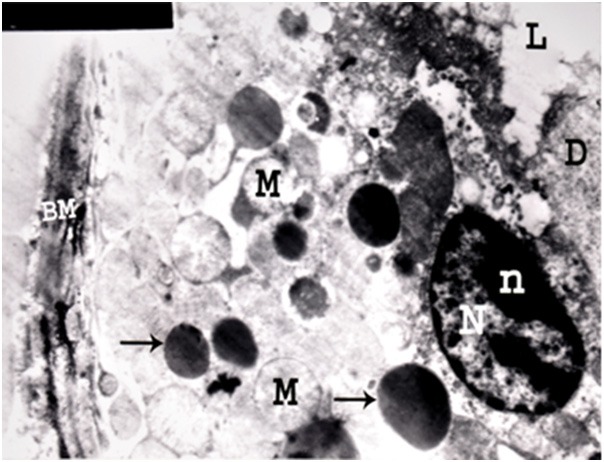
Contamination of the environment with antimony compounds may affect human health through the persistent exposure to small doses over a long period. Sixty growing male albino rats, weighing 43-57 grams, utilized in this study. The animals were divided into 3 groups; each of 20 rats: animals of group I served as control, animals of group II received 6 mg/kg body weight antimony trisulfide daily for 8 weeks with drinking water, and those of group III received the same dose by the same route for 12 weeks. The Malpighian renal corpuscles showed distortion, destruction and congestion of glomerular tuft, vacuoles in the glomeruli, peritubular haemorrhage, obliteration of Bowman's space, and thickening with irregularity of Bowman's membrane. The proximal convoluted tubules demonstrated patchy loss of their brush border, thickening of the basement membrane with loss of its basal infoldings, disarrangement of the mitochondria, pleomorphic vacuoles in the cytoplasm, apical destruction of the cells, apical migration of the nuclei, and absence of microvilli. On the other hand, peri-tubular hemorrhage, apical vacuolation, small atrophic nuclei, swelling of mitochondria, obliteration of the lumina, destruction of cells, and presence of tissue debris in the lumina, were observed in the distal convoluted tubules. The present work demonstrated the hazardous effect of antimony on the renal function as evidenced by the significant increase of the level of blood urea, serum creatinine, and serum sodium and potassium. In conclusion, this study proposed that continuous oral administration of antimony for 8 and 12 weeks has hazardous toxic effect on the structure and function of the kidney in growing albino rat. Based on the results of the present study, it is recommended to avoid the use of any drinking water contaminated with antimony compounds and forbidden its use in infants and children foods.

Keywords: Antimony; blood chemistry; growing albino rat; histopathology; renal cortex.


Figures

Figure 1
Figure 1
A photomicrograph of a section of a renal cortex of a control albino rat, showing normal structure of the glomerulus (G), proximal (P) and distal (D) convoluted tubules with PAS positive brush border (arrows). Normal Bowman’s space (arrow head) can be seen (PAS reaction, X 400).
Figure 2
Figure 2
A photomicrograph of a semithin section of a renal cortex of a control albino rat, showing glomerular capillaries (C), some of which containing red blood cells (R). Capillary endothelial cells (E) are occasionally seen bulging into the capillary lumen and resting on thin basement membrane (arrow). Mesangium which consists of mesangial cells (M) and extracellular substance called mesangial substance (MS), can be observed. The surface of the glomerular capillaries exposed to Bowman’s space (BS) is invested by visceral layer of Bowman’s capsule i.e. podocytes (P). The parietal cells (PC) of Bowman’s capsule are also seen resting on thin Bowman’s membrane (BM) (Toluidine blue, X 1000).
Figure 3
Figure 3
A photomicrograph of a semithin section of a renal cortex of a control albino rat, showing the cells of proximal convoluted tubules (P) with their rounded basal nuclei (N), indistinct cell boundaries and well defined luminal brush border (arrow). Note the narrow lumen (arrow head) of the tubules. The distal convoluted tubule (D) revealed wider lumen and are lined by a single layer of cuboidal cells laying on clear basement membrane (dotted arrow) with indistinct cell boundaries. Note rounded nuclei (N) and indistinct luminal brush border (Toluidine blue; X 1000).
Figure 4
Figure 4
An electron micrograph of a renal cortex of a control albino rat showing glomerular capillary (C) which is lined by endothelial cells having a thin layer of cytoplasm (E) and a large nucleus (N) bulging into the capillary lumen. Note the mesangial cells (M) and mesangial substance (MS) support the capillary wall. The large nuclei of several podocytes (P), are surrounded by cytoplasm containing rough endoplasmic reticulum (arrows) and their primary processes (P1) giving rise to numerous secondary foot processes (P2) which rest on the glomerular capillary basement membrane (BM). Part of parietal layer of Bowman’s capsule (BC), resting on regular Bowman’s membrane (arrow heads) and Bowman’s space (BS) are also seen (Uranyl acetate and lead citrate X 5000).
Figure 5
Figure 5
An electron micrograph of a renal cortex of a control albino rat, showing a proximal convoluted tubule with thin regular basement membrane (BM) which exhibits basal infoldings (arrows) closely related to columns of parallel elongated mitochondria (M). The basement membrane (BM) is separating the cells of the tubule from the delicate capillary (C). The cytoplasm immediately beneath the prominent microvilli (MV) contains many pinocytic vesicles (PV) and lysosomes (L). Note the large nucleus (N) with peripheral chromatin condensations (Uranyl acetate and lead citrate X 5000).
Figure 6
Figure 6
An electron micrograph of a renal cortex of a control albino rat, showing a distal convoluted tubule which is lined by cubical cells with few microvilli (arrow) and large nuclei (N) close to their luminal surface. Large numbers of mitochondria (M) are situated in the basal parts of the cells (Uranyl acetate and lead citrate: X 5000).
Figure 7
Figure 7
A photomicrograph a section of renal cortex of albino rat from group III showing deformity of glomerular tuft (G). The proximal convoluted tubules (P) reveal patchy loss of brush border (arrow) (PAS reaction; X 400).
Figure 8
Figure 8
A photomicrograph of a semithin section of a renal cortex of albino rat from group III, showing dilatation and congestion of glomerular capillaries (C). Note obliteration of the Bowman’s space and thickening with irregularity of Bowman’s basement membrane (arrows) (Toluidine blue X 1000).
Figure 9
Figure 9
A photomicrograph of a semithin section of a renal cortex of albino rat from group III, showing dilated and congested glomerular capillaries (C). Bowman’s space (BS) is dilated and containing necrotic tissues (NT). The proximal convoluted tubules reveal pleomorphic vacuoles (arrows) in the cytoplasm (Toluidine blue X 1000).
Figure 10
Figure 10
A photomicrograph of a semithin section of a renal cortex of albino rat from group II showing distal convoluted tubules (D) with marked peritubular congestion (C) and apical vacuolation (arrows) (Toluidine blue X 1000).
Figure 11
Figure 11
An electron micrograph of a renal cortex of albino rat from group III, showing part of a glomerulus with endothelial cell (E) resting on thick glomerular basement membrane (BM). Note podocytes (P) with its rough endoplasmic reticulum (ER), large nucleus (N) with its peripheral nucleolus (n) normal major process (P1) and fusion (arrow) of some secondary foot processes (P2) (Uranyl acetate and lead citrate X 12000).
Figure 12
Figure 12
An electron micrograph of a renal cortex of albino rat from group III, showing part of a cell lining the proximal convoluted tubule that has a thick tubular basement membrane (BM) with absence of its normal basal infoldings and loss of parallel arrangement of mitochondria (M). Pleomorphic cytoplasmic vacuoles (V) are seen. Note the presence of normal nucleus (N), lysosomes (L) and microvilli (MV) (Uranyl acetate and lead citrate X 5000).
Figure 13
Figure 13
An electron micrograph of a renal cortex of albino rat from group III showing the apical part of a cell lining the proximal convoluted tubule with large vacuole (V) and irregular destruction (arrows) of the cytoplasm. Mitochondrial swelling (M) with destruction of its cristae and lysosomal swelling (L) are seen. Normal microvilli (MV) are noticed (Uranyl acetate and lead citrate; X 10,000).
Figure 14
Figure 14
An electron micrograph of a renal cortex of albino rat from group III showing a cell lining the distal convoluted tubule with small atrophic nucleus (N), swelling of mitochondria (M) and obliteration of tubular lumen (arrow) (Uranyl acetate and lead citrate X 5000).
Figure 15
Figure 15
A photomicrograph of a section of a renal cortex of albino rat from group IV, showing distorsion, destruction and shrinkage of glomerular tuft (G) with wide irregular Bowman’s space (S). There is extensive loss of the brush border (arrows) of proximal convoluted tubules (P) (PAS reaction; X 400).
Figure 16
Figure 16
A photomicrograph of a semithin section of a renal cortex of albino rat from group IV, showing part of a glomerulus with obliteration of Bowman’s space as well as congestion and dilatation of glomerular capillaries (C). Multiple vacuoles (V) in the glomerulus, and periglomerular haemorrhage (H) are also noticed. Note irregular thickening in the Bowman’s membrane (arrows) (Toluidine blue; X 1000).
Figure 17
Figure 17
A photomicrograph of a semithin section of a renal cortex of albino rat from group IV, showing proximal convoluted tubules with apical destruction of the tubular cells and migration of the nuclei (N) towards the wide lumen (L). Note the peritubular haemorrhage (H) (Toluidine blue X 1000).
Figure 18
Figure 18
A photomicrograph of a semithin section of a renal cortex of albino rat from group IV, showing distal convoluted tubule with apical vacuolation (V), obliteration of the lumen (arrow) and basal location of the nuclei (N) (Toluidine blue; X 1000).
Figure 19
Figure 19
A photomicrograph of a semithin section of a renal cortex of albino rat from group IV, showing parts of proximal (P) and distal (D) convoluted tubules with destruction of tubular cells, apical direction of the nuclei (N) , necrotic tissues (NT) and red blood cells (arrows) in the lumen. Irregular basal invaginations (I) into the cytoplasm, pleomorphic vacuoles (V) and dense bodies (arrow heads) are noticed (Toluidine blue X 1000).
Figure 20
Figure 20
An electron micrograph of a renal cortex of albino rat from group IV showing part of a glomerulus with marked irregular thickening of the capillary basement membrane (arrow), marked thickening of Bowman’s membrane (BM) and few small atrophic secondary foot processes (arrow heads) (Uranyl acetate and lead citrate; X 4000).
Figure 21
Figure 21
An electron micrograph of a renal cortex of albino rat from group IV, showing part of a glomerulus with marked thickening of Bowman’s membrane (BM), atrophy of parietal cell (arrow head) and obliteration of Bowman’s space. Note the podocyte (P) with its irregular nucleus (N). Periglomerular haemorrhage (H) is also noticed (Uranyl acetate and lead citrate; X 4000).
Figure 22
Figure 22
An electron micrograph of a renal cortex of albino rat from group IV, showing a cell lining a proximal convoluted tubule with marked thickening of basement membrane (BM) and absence of its basal infolding. There are destruction of basal cytoplasm (arrows), marked vacuolation (V) and migration of mitochondria (M) towards the lumen with loss of their parallel arrangement. Note absence of luminal microvilli and presence of a normal nucleus (N) when compared to the control (Uranyl acetate and lead citrate; X 5,000).
Figure 23
Figure 23
An electron micrograph of a renal cortex of albino rat from group IV, showing a cell lining a proximal convoluted tubule with few swollen mitochondria (M), pleomorphic vacuolation (V) and heterogenous irregular dense bodies mostly lysosomes (arrows). Note the nucleus (N) and marginal nucleolus (n) with fragmentation and migration of chromatin with areas of karyolysis (arrow heads) (Uranyl acetate and lead citrate; X 5000).
Figure 24
Figure 24
An electron micrograph of a renal cortex of albino rat from group IV showing a cell lining a distal convoluted tubule with destruction of its apical part, apical direction of its nucleus (N), prominent peripheral nucleolus (n), swelling of mitochondria (M) and thickening of its basement membrane (BM). Pleomorphic dense bodies mostly lysosomes (arrows) are seen in the cytoplasm. Tissue debris (D) is noticed in the lumen (L) (Uranyl acetate and lead citrate; X 5,000).

Similar articles

Cited by

References

    1. Abadin HG, Murray HE, Wheeler JS. The use of hematological effects in the development of minimal risk levels. Regul Toxicol Pharmacol. 1998;28:61–66. - PubMed
    1. Kale RM. Genotoxic effects of antimony on rat and mice. J Appl Toxicol. 2011;25:17–20.
    1. Marc SB, Gonzalez HP, Obregon ER. Mechanisms of selective action of heavy metal toxicity. Ann Rev Pharmacol Toxicol. 2012;63:321–327.
    1. Aboel-Zahab H, el-Khyat Z, Sidhom G, Awadallah R, Abdel-al W, Mahdy K. Physiological effects of some synthetic food colouring additives on rats. Boll Chim Farm. 1997;136:615–627. - PubMed
    1. Adda J, Roger S, Dumont J. Modulating effect of antimony and vanillin in a rat medium term multi-organ carcinogenesis model. Cancer Letters. 2003;104:U3–121.

Publication types

LinkOut - more resources