. 2013 Aug 7:13:25.
doi: 10.1186/1471-2342-13-25.

Imaging features of primary sarcomas of the great vessels in CT, MRI and PET/CT: a single-center experience

Imaging features of primary sarcomas of the great vessels in CT, MRI and PET/CT: a single-center experience

Christian von Falck et al. BMC Med Imaging. .

Abstract

Background: To investigate the imaging features of primary sarcomas of the great vessels in CT, MRI and (18)F-FDG PET/CT.

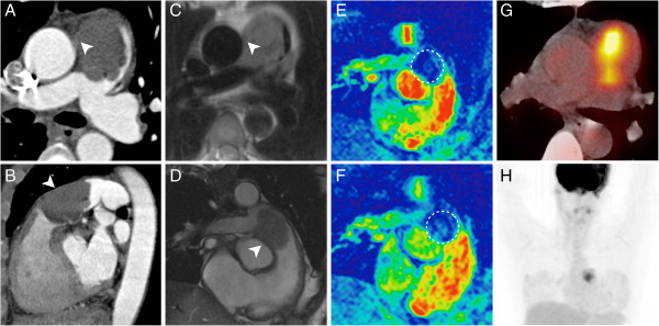
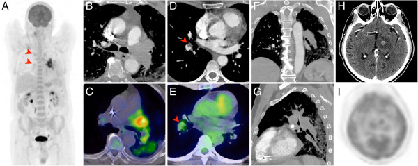
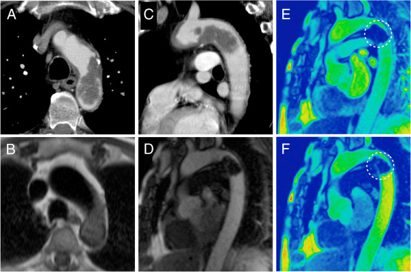
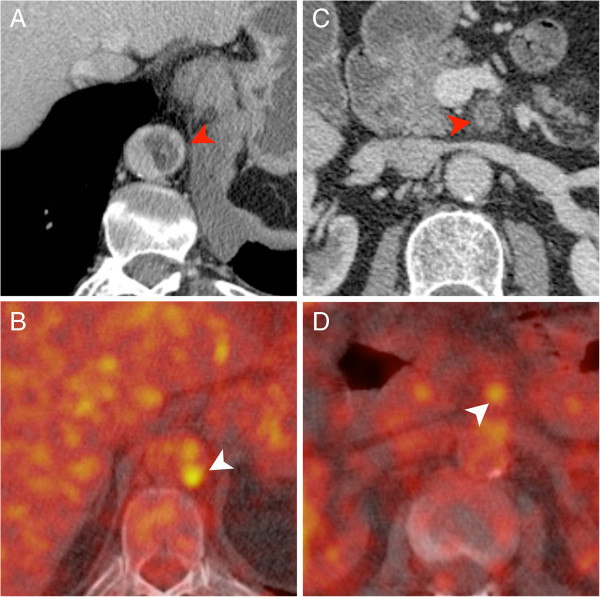
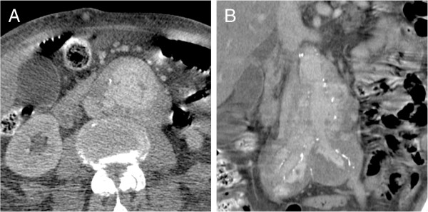
Methods: Thirteen patients with a primary sarcoma of the great vessels were retrospectively evaluated. All available images studies including F-18 FDG PET(/CT) (n = 4), MDCT (n = 12) and MRI (n = 6) were evaluated and indicative image features of this rare tumor entity were identified.

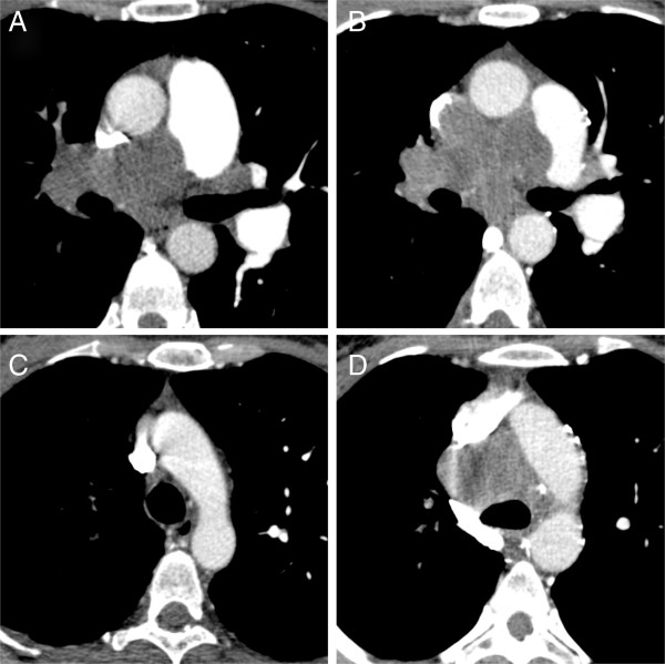
Results: The median interval between the first imaging study and the final diagnosis was 11 weeks (0-12 weeks). The most frequently observed imaging findings suggestive of malignant disease in patients with sarcomas of the pulmonary arteries were a large filling defect with vascular distension, unilaterality and a lack of improvement despite effective anticoagulation. In patients with aortic sarcomas we most frequently observed a pedunculated appearance and an atypical location of the filling defect. The F-18 FDG PET(/CT) examinations demonstrated an unequivocal hypermetabolism of the lesion in all cases (4/4). MRI proved lesion vascularization in 5/6 cases.

Conclusion: Intravascular unilateral or atypically located filling defects of the great vessels with vascular distension, a pedunculated shape and lack of improvement despite effective anticoagulation are suspicious for primary sarcoma on MDCT or MRI. MR perfusion techniques can add information on the nature of the lesion but the findings may be subtle and equivocal. F-18 FDG PET/CT may have a potential role in these patients and may be considered as part of the imaging workup.


Figures

Figure 1
Figure 1
CT pulmonary angiography (CTPA) of a 49-year-old female patient with a myxofibrosarcoma of the right pulmonary artery. Suspicious imaging features were already present in the primary imaging (A, C). However, the patient was treated conservatively for suspected pulmonary embolism. A follow-up exam was acquired three month later and demonstrated massive disease progression with tumor expansion beyond the vessel wall and local lymph node metastases (B, D)
Figure 2
Figure 2
This figure illustrates an in-depth preoperative work-up in a 46-year-old female patient with an undifferentiated intimal sarcoma of the central pulmonary artery. Both, the MDCT (A, B) and the MRI (C, D) clearly demonstrate a subtotal occlusion of the pulmonary trunk due to an intraluminal process. Imaging findings are suggestive of a focal expansion beyond the vessel wall (A-D, arrowheads). The MR first-pass perfusion sequence (F) demonstrates a subtle perfusion of the lesion as compared to the non-enhanced control scan (E, circle). The F-18 FDG PET/CT proves a high metabolic activity (SUVmax = 7.8) within the lesion (G, H) and strengthens the suspicion of malignant disease.
Figure 3
Figure 3
This figure illustrates the imaging findings in a 74-year-old male patient with an extensive undifferentiated intimal sarcoma of the left pulmonary artery (A-C, F, G). Both, the MDCT and the F-18 FDG PET/CT readily demonstrate the local embolic spread in the pulmonary vasculature of the right lung (A, D, E; arrowheads) as well as a distant metastasis to the brain (H, I).
Figure 4
Figure 4
This figure shows an example of an aortic angiosarcoma of the distal aortic arch in a 71-year-old male patient who was treated for congestive heart failure. The MDCT and MRI images (A-D) nicely illustrate the typical pedunculated appearance of the lesion in a location that is atypical for atheromatous thrombi. The first pass perfusion sequence (F) suggests vascularisation of the lesion as compared to the unenhanced control acquisition (E, circle).
Figure 5
Figure 5
MDCT and PET/CT of a 71-year-old male patient with an undifferentiated intimal sarcoma of the descending aorta and extension into the superior mesenteric artery (A, C). The PET/CT adds valuable information about the metabolic status of vascular filling defect and strengthens the suspicion of malignant disease (B, D).
Figure 6
Figure 6
MDCT images in axial (A) and coronal (B) orientation of a 81-year-old patient with an angiosarcoma of the abdominal aorta who had previously undergone open aortic surgery. The extensive extramural tumor formation is clearly visualized and may be confounded with chronic inflammatory disease and contained rupture.

References

    1. Mayer F, Aebert H, Rudert M, Königsrainer A, Horger M, Kanz L, Bamberg M, Ziemer G, Hartmann JT. Primary malignant sarcomas of the heart and great vessels in adult patients—a single-center experience. Oncologist. 2007;12:1134–1142. doi: 10.1634/theoncologist.12-9-1134. - DOI - PubMed
    1. Burke AP, Virmani R. Sarcomas of the great vessels. A clinicopathologic study. Cancer. 1993;71:1761–1773. doi: 10.1002/1097-0142(19930301)71:5<1761::AID-CNCR2820710510>3.0.CO;2-7. - DOI - PubMed
    1. Bendel EC, Maleszewski JJ, Araoz PA. Imaging sarcomas of the great vessels and heart. Semin Ultrasound CT MR. 2011;32:377–404. doi: 10.1053/j.sult.2011.06.001. - DOI - PubMed
    1. Restrepo CS, Betancourt SL, Martinez-Jimenez S, Gutierrez FR. Tumors of the pulmonary artery and veins. Semin Ultrasound CT MR. 2012;33:580–590. doi: 10.1053/j.sult.2012.04.002. - DOI - PubMed
    1. Choong CK, Lawton JS, Moon MR, Damiano RJ Jr. Failure of medical therapy for pulmonary “thromboembolic” disease: beware the unsuspected primary sarcoma of the pulmonary artery. J Thorac Cardiovasc Surg. 2004;128:763–765. doi: 10.1016/j.jtcvs.2004.03.049. - DOI - PubMed

Publication types

Substances