

HIV type 1 infection of plasmacytoid and myeloid dendritic cells is restricted by high levels of SAMHD1 and cannot be counteracted by Vpx
- PMID: 23924154
- PMCID: PMC3910455
- DOI: 10.1089/AID.2013.0119
HIV type 1 infection of plasmacytoid and myeloid dendritic cells is restricted by high levels of SAMHD1 and cannot be counteracted by Vpx
Abstract
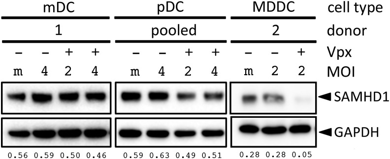
Dendritic cells are professional antigen-presenting cells of the immune system and are major producers of type-I interferon. Their role in HIV-1 infection is not well understood. They express CD4 and CCR5 yet appear to be resistant to infection. In culture, infection of the cells with HIV-1 is inhibited by the host cell restriction factor SAMHD1. Lentiviruses such as HIV-2/SIVmac counteract the restriction by encoding Vpx, a virion-packaged accessory protein that induces the proteasomal degradation of SAMHD1. In this study we investigated SAMHD1-mediated restriction in the two major dendritic cell subsets: plasmacytoid dendritic cells (pDC) and myeloid dendritic cells (mDC). The cells were highly resistant to HIV-1 and expressed high levels of SAMHD1. SAMHD1 amino acid residue T592, a target of CDK1 phosphorylation, was unphosphorylated, corresponding to the antiviral form of the enzyme. The resistance to infection was not counteracted by Vpx and SAMHD1 was not degraded in these cells. Treatment of pDCs with a cocktail of antibodies that blocked type-I interferon signaling partially restored the ability of Vpx to induce SAMHD1 degradation and caused the cells to become partially permissive to infection. pDCs and mDCs responded to HIV-1 virions by inducing an innate immune response but did not appear to sense newly produced Gag protein. The findings suggest that in vivo, dendritic cells serve as sentinels to alert the immune system to the virus but do not themselves become infected by virtue of high levels of SAMHD1.
Figures


Similar articles
-
The efficiency of Vpx-mediated SAMHD1 antagonism does not correlate with the potency of viral control in HIV-2-infected individuals.Retrovirology. 2013 Mar 5;10:27. doi: 10.1186/1742-4690-10-27. Retrovirology. 2013. PMID: 23497283 Free PMC article.
-
SAMHD1 is the dendritic- and myeloid-cell-specific HIV-1 restriction factor counteracted by Vpx.Nature. 2011 May 25;474(7353):654-7. doi: 10.1038/nature10117. Nature. 2011. PMID: 21613998 Free PMC article.
-
Degradation of SAMHD1 by Vpx Is Independent of Uncoating.J Virol. 2015 May;89(10):5701-13. doi: 10.1128/JVI.03575-14. Epub 2015 Mar 11. J Virol. 2015. PMID: 25762741 Free PMC article.
-
SAMHD1: a new insight into HIV-1 restriction in myeloid cells.Retrovirology. 2011 Jul 8;8:55. doi: 10.1186/1742-4690-8-55. Retrovirology. 2011. PMID: 21740548 Free PMC article. Review.
-
SAMHD1 host restriction factor: a link with innate immune sensing of retrovirus infection.J Mol Biol. 2013 Dec 13;425(24):4981-94. doi: 10.1016/j.jmb.2013.10.022. Epub 2013 Oct 23. J Mol Biol. 2013. PMID: 24161438 Review.
Cited by
-
Vpx rescue of HIV-1 from the antiviral state in mature dendritic cells is independent of the intracellular deoxynucleotide concentration.Retrovirology. 2014 Feb 1;11:12. doi: 10.1186/1742-4690-11-12. Retrovirology. 2014. PMID: 24485168 Free PMC article.
-
The role of human dendritic cells in HIV-1 infection.J Invest Dermatol. 2015 May;135(5):1225-1233. doi: 10.1038/jid.2014.490. Epub 2014 Nov 19. J Invest Dermatol. 2015. PMID: 25407434 Review.
-
Increased SAMHD1 transcript expression correlates with interferon-related genes in HIV-1-infected patients.Med Microbiol Immunol. 2019 Oct;208(5):679-691. doi: 10.1007/s00430-018-0574-x. Epub 2018 Dec 18. Med Microbiol Immunol. 2019. PMID: 30564919
-
Intertwined: SAMHD1 cellular functions, restriction, and viral evasion strategies.Med Microbiol Immunol. 2019 Aug;208(3-4):513-529. doi: 10.1007/s00430-019-00593-x. Epub 2019 Mar 16. Med Microbiol Immunol. 2019. PMID: 30879196 Review.
-
SAMHD1 transcript upregulation during SIV infection of the central nervous system does not associate with reduced viral load.Sci Rep. 2016 Mar 3;6:22629. doi: 10.1038/srep22629. Sci Rep. 2016. PMID: 26936683 Free PMC article.
References
Publication types
MeSH terms
Substances
Grants and funding
LinkOut - more resources
Full Text Sources
Other Literature Sources
Molecular Biology Databases
Research Materials
Miscellaneous
