. 2013 Aug 28;135(34):12540-3.
doi: 10.1021/ja4059553. Epub 2013 Aug 14.

A genetically encoded fluorescent probe in mammalian cells

Affiliations

A genetically encoded fluorescent probe in mammalian cells

Abhishek Chatterjee et al. J Am Chem Soc. .

Abstract

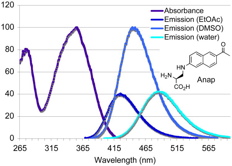
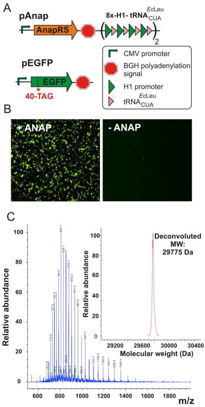
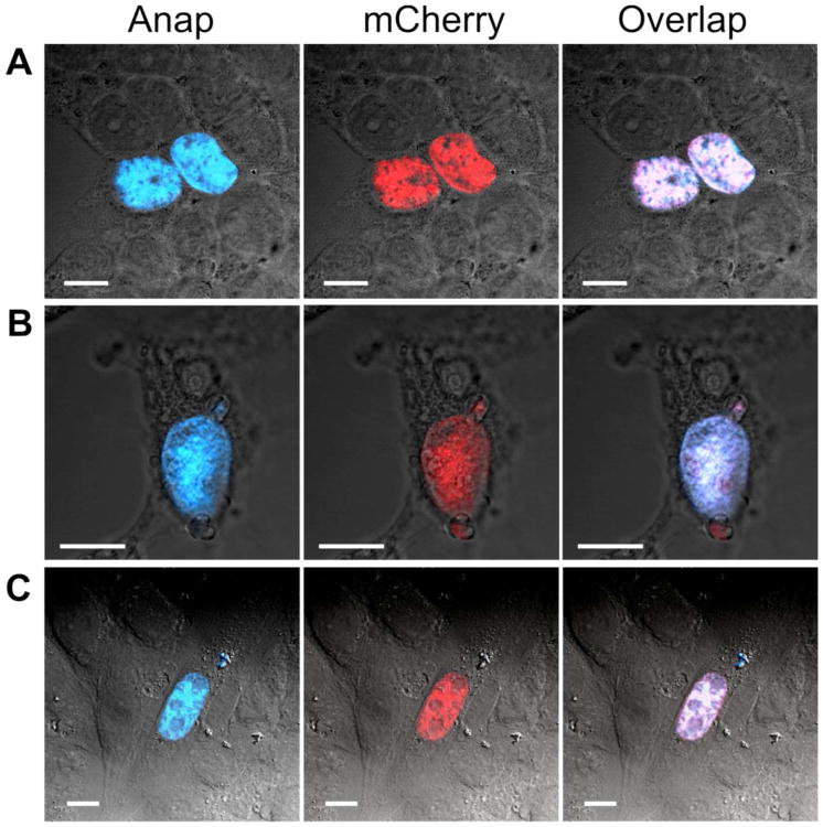
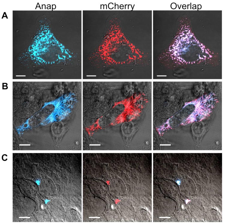
Fluorescent reporters are useful in vitro and in vivo probes of protein structure, function, and localization. Here we report that the fluorescent amino acid, 3-(6-acetylnaphthalen-2-ylamino)-2-aminopropanoic acid (Anap), can be site-specifically incorporated into proteins in mammalian cells in response to the TAG codon with high efficiency using an orthogonal amber suppressor tRNA/aminoacyl-tRNA synthetase (aaRS) pair. We further demonstrate that Anap can be used to image the subcellular localization of proteins in live mammalian cells. The small size of Anap, its environment-sensitive fluorescence, and the ability to introduce Anap at specific sites in the proteome by simple mutagenesis make it a unique and valuable tool in eukaryotic cell biology.


Figures

Figure 1
Figure 1
Structure and normalized absorption (purple) and fluorescence (Deep blue, in EtOAc; light blue, in DMSO; Cyan, in water) spectra of Anap.
Figure 2
Figure 2
Evaluation of the activity of Anap specific tRNAC-UAEcLeu/AnapRS in mammalian cells. (A) The construction of pAnap and pEGFP plamids. Identities of different genetic elements used in these constructs are shown. (B) Expression of EGFP-40-Anap in HEK293 cells in the presence (left) and absence (right) of 0.5 mM Anap, achieved by co-transfection of pAnap and pEGFP, analyzed by fluorescence microscopy. (B) ESI-MS analysis of purified EGFP-40-Anap mutant. Expected molecular weight: 29773 Da; observed: 29775 Da.
Figure 3
Figure 3
Nuclear localization of Histone-H3 (Thr59-Anap)-mCherry (C-terminal) fusion protein in HEK293 (panel A), HeLa (panel B) and CHO (panel C) cells, visualized using Anap (first column, pseudo-colored in blue, superimposed with the bright field image) or mCherry (second column, pseudo-colored in red, superimposed with the bright field image) as fluorescent reporters by laser scanning confocal microscopy. A composite image of these is shown in the third column. Scale bars, 10 μm.
Figure 4
Figure 4
Subcellular localization of endoplasmic reticulum (ER) and golgi-resident proteins in human cell-lines visualized using Anap (first column, pseudo-colored in blue, superimposed with the bright field image) or mCherry (second column, pseudo-colored in red, superimposed with the bright field image) as fluorescent reporters by confocal microscopy: (A) ER-resident Grp94 (Ser127-Anap)-mCherry (C-terminal) in HeLa cells. (B) ER-resident Grp94 (Leu7-Anap)-mCherry (C-terminal) in HeLa cells. (C) Golgi resident GalT1 (Ser3-Anap)-mCherry (C-terminal) in HEK293 cells. Scale bars: 10 μm.

References

    1. Giepmans BNG, Adams SR, Ellisman MH, Tsien RY. Science. 2006;312:217–224. - PubMed
    1. Tsien RY. Annu Rev Biochem. 1998;67:509–544. - PubMed
    1. Zhang J, Campbell RE, Ting AY, Tsien RY. Nat Rev Mol Cell Biol. 2002;3:906–918. - PubMed
    1. Stadler C, Rexhepaj E, Singan VR, Murphy RF, Pepperkok R, Uhlén M, Simpson JC, Lundberg E. Nat Methods. 2013;10:315–23. - PubMed
    1. Hinner MJ, Johnsson K. Curr Opin Biotechnol. 2010;21:766–76. - PubMed

Publication types

Substances