. 2013 Oct 1;188(7):820-30.
doi: 10.1164/rccm.201212-2297OC.

Role of lung pericytes and resident fibroblasts in the pathogenesis of pulmonary fibrosis

Affiliations

Role of lung pericytes and resident fibroblasts in the pathogenesis of pulmonary fibrosis

Chi Hung et al. Am J Respir Crit Care Med. .

Abstract

Rationale: The origin of cells that make pathologic fibrillar collagen matrix in lung disease has been controversial. Recent studies suggest mesenchymal cells may contribute directly to fibrosis.

Objectives: To characterize discrete populations of mesenchymal cells in the normal mouse lung and to map their fate after bleomycin-induced lung injury.

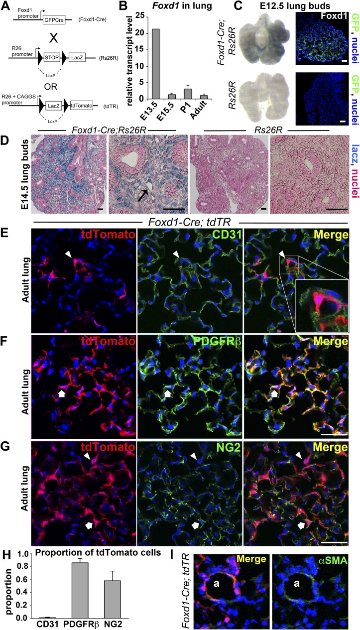
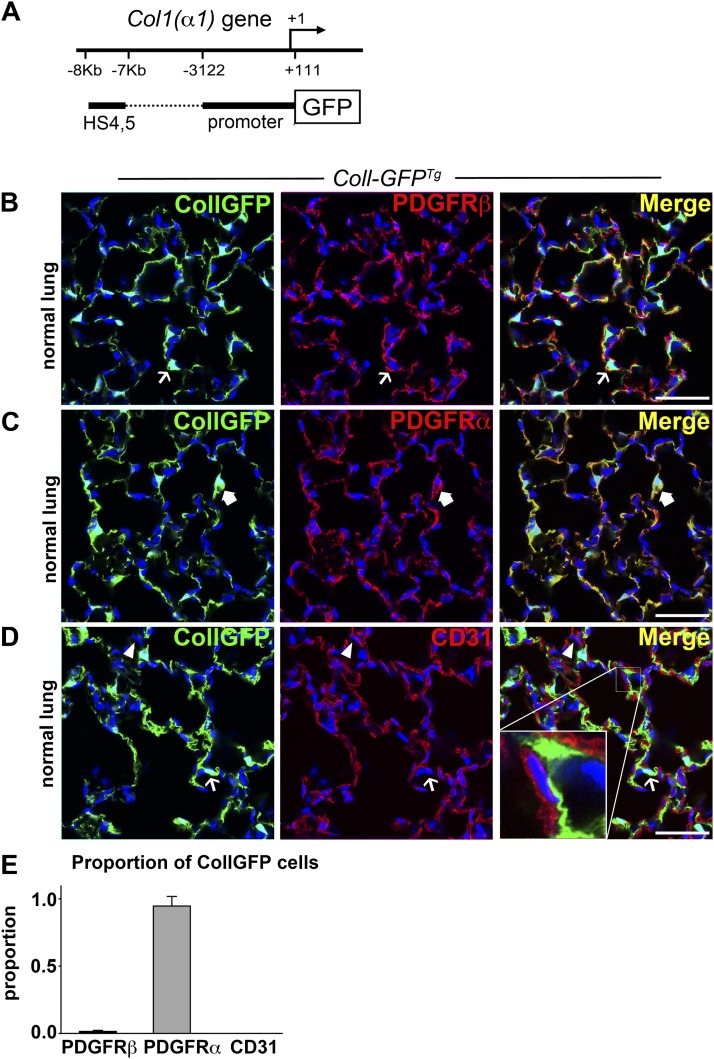
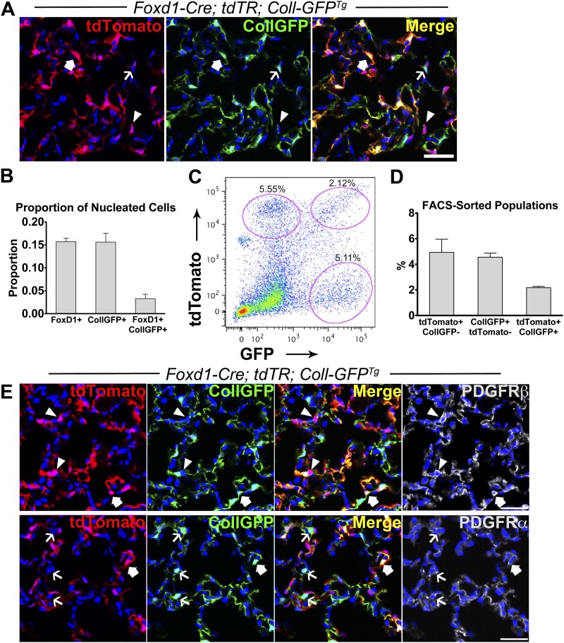
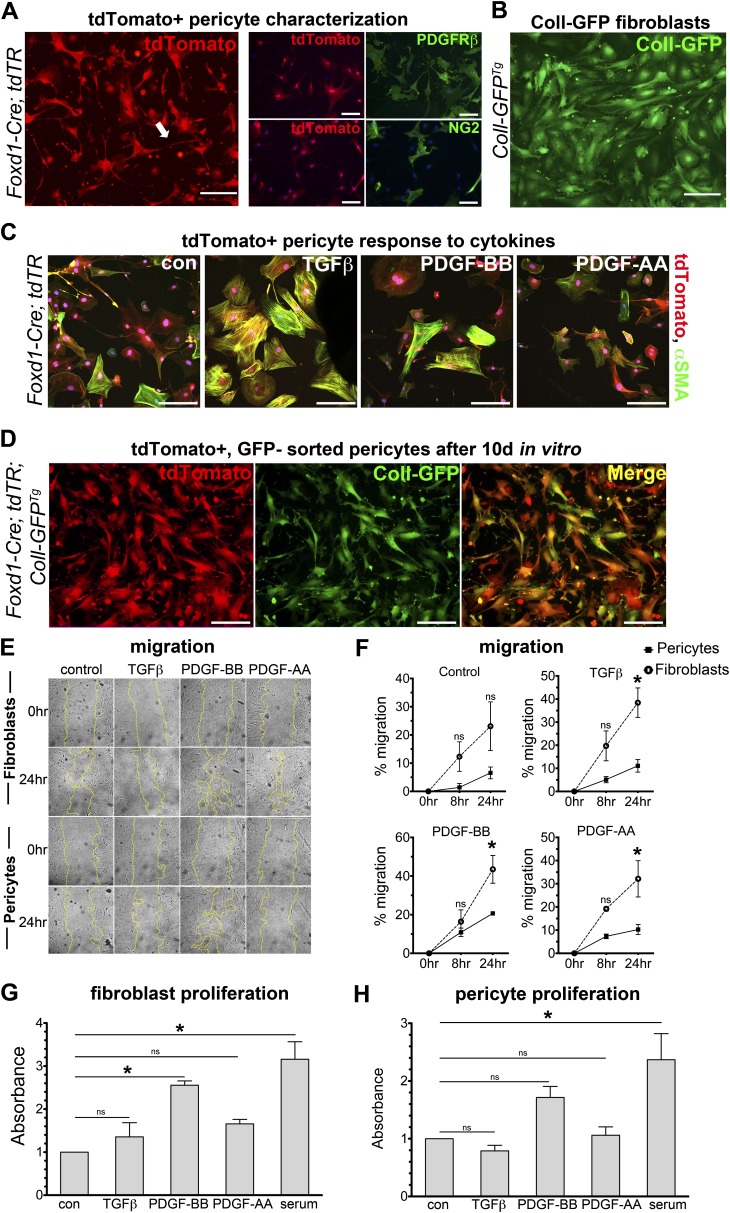
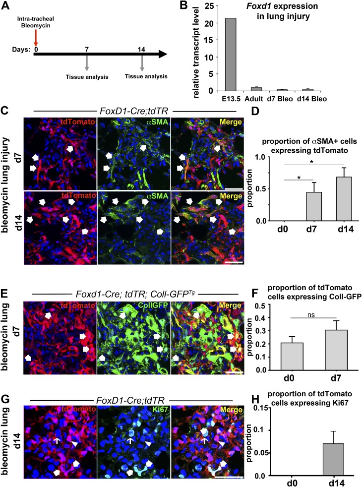
Methods: We mapped the fate of Foxd1-expressing embryonic progenitors and their progeny during lung development, adult homeostasis, and after fibrosing injury in Foxd1-Cre; Rs26-tdTomato-R mice. We studied collagen-I(α)1-producing cells in normal and diseased lungs using Coll-GFP(Tg) mice.

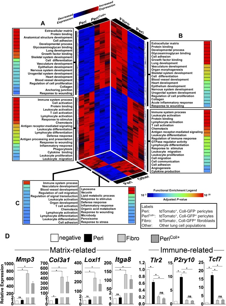
Measurements and main results: Foxd1-expressing embryonic progenitors enter lung buds before 13.5 days post-conception, expand, and form an extensive lineage of mesenchymal cells that have characteristics of pericytes. A collagen-I(α)1-expressing mesenchymal population of distinct lineage is also found in adult lung, with features of a resident fibroblast. In contrast to resident fibroblasts, Foxd1 progenitor-derived pericytes are enriched in transcripts for innate immunity, vascular development, WNT signaling pathway, and cell migration. Foxd1 progenitor-derived pericytes expand after bleomycin lung injury, and activate expression of collagen-I(α)1 and the myofibroblast marker αSMA in fibrotic foci. In addition, our studies suggest a distinct lineage of collagen-I(α)1-expressing resident fibroblasts that also expands after lung injury is a second major source of myofibroblasts.

Conclusions: We conclude that the lung contains an extensive population of Foxd1 progenitor-derived pericytes that are an important lung myofibroblast precursor population.


Figures

<i>Figure 1.</i>
Figure 1.
Foxd1-expressing progenitors enter early lung buds and differentiate into lung mesenchyme, which matures to form mural cells of the adult lung. (A) Bigenic Foxd1-Cre; Rs26R mice or Foxd1-Cre; tdTR mice activate GFPCre fusion protein expression in lung progenitor cells present in early lung buds and differentiate into a population of lung mesenchyme. The GFPCre recombinase results in removal of the loxP-STOP-loxP sequence in genomic DNA of these mesenchymal cells, leading to permanent, heritable expression of lacz or tdTomato in Foxd1 progenitor–derived cells. (B) Real-time polymerase chain reaction analysis of Foxd1 mRNA expression during lung development. Data were normalized to hypoxanthine-guanine phosphoribosyltransferase expression. Y-axis represents fold increase compared with adult. Mean value ± SD is indicated. n = 3–4 per time point. (C) Whole-mount heart and lung buds from Foxd1-Cre; Rs26R mice show presence of blue-stained mesenchymal cells derived from Foxd1 progenitors by E12.5. Specificity of this blue stain is shown by lack of lacz expression in control lung (left). Foxd1-expressing progenitors are seen predominantly in the posterior portion of the whole lung bud by detection of CreGFP expression under regulation of the Foxd1 locus (right). (D) E14.5 lung buds from Foxd1-Cre; Rs26R mice show large numbers of blue-stained mesenchymal cells derived from Foxd1 progenitors (left). Arrow illustrating a lacz+ mesenchymal cell adjacent to a developing blood vessel. Specificity of this blue stain is shown by lack of expression of lacz in control lung buds (right). (E–G) Confocal images of normal adult Foxd1-Cre; tdTR lung showing heritable labeling with tdTomato fluorophore of progeny of Foxd1 progenitors. tdTomato cells lie in close apposition to alveolar endothelium labeled with CD31 (arrowhead) (E), but do not overlap with this endothelium (inset). By contrast, almost all tdTomato cells coexpress PDGFRβ (arrow) (F), and many show expression of NG2 (arrow), whereas a minority do not express this protein (arrowhead) (G). (H) Graph quantifying the proportion of tdTomato cells coexpressing the indicated markers. (I) Images showing the presence of αSMA+ vascular smooth muscle in an arteriole (a) coexpressing tdTomato. Bar = 50 μm. Mean ± SEM. n = 3 per group.
<i>Figure 2.</i>
Figure 2.
Collagen-Iα1 GFP transgene–expressing cells coexpress PDGFRα in normal lung. (A) Schema showing the Coll-GFP transgene with a 3.2-kb fragment of the Col1a1 promoter and a 1-kb enhancer fused to GFP. (B–D) Confocal images showing the expression of GFP in the adult lung of Coll-GFPTg mice and colabeling with (B) PDGFRβ, (C) PDGFRα, and (D) CD31. Example of Coll-GFP+ cell (green) lacking the marker is indicated by a thin arrow. GFP cell expressing the indicated marker (red) is indicated by an arrowhead. Coll-GFP+ cell coexpressing the indicated marker (yellow plasma membrane in merged image) is indicated by a thick arrow. Inset in D shows a space separating Coll-GFP+ cell from the endothelium. (E) Graph showing the proportion of Coll-GFP cells coexpressing the indicated markers. Bar = 50 μm. Mean ± SEM. n = 3 mice per group.
<i>Figure 3.</i>
Figure 3.
Three populations of mesenchymal cells in normal lung are identified in Foxd1-Cre; tdTR; Coll-GFPTg mice. (A) Confocal image of normal lung showing three distinct populations of mesenchymal cells: arrowheads indicate tdTomato+ cells (red), thin arrows indicate Coll-GFP+ cells (green), and block arrows indicate tdTomato+ cells that also express Coll-GFP transgene (yellow in merged image). (B) Quantification of nucleated cells in lung parenchyma that are tdTomato+, Coll-GFP+, and tdTomato+ Coll-GFP+. (C) Fluorescence-activated cell sorter plot of single-cell preparation of normal lung from Foxd1-Cre; tdTR; Coll-GFPTg mice showing three distinct populations of lung mesenchymal cells. (D) Data showing the three populations: (1) tdTomato+ CollGFP; (2) CollGFP+ tdTomato; and (3) tdTomato+ CollGFP+ as a percentage of total counted events by fluorescence-activated cell sorter. (E) Foxd1-Cre; tdTR; Coll-GFPTg mouse lung colabeled with PDGFRβ or PDGFRα (white). Arrowheads indicate tdTomato cells (red) colabeled with PDGFRβ (white), whereas thin arrows indicate Coll-GFP+ cells (green) colabeled with PDGFRα (white). Block arrows indicate tdTomato/Coll-GFP+ cells colabeled with PDGFRα or β (white). Bars = 50 μm. Mean ± SEM. n = 3 mice.
<i>Figure 4.</i>
Figure 4.
Gene expression profiling of Foxd1 progenitor–derived pericytes identifies two subpopulations with distinct transcriptional and functional characteristics. This composite figure summarizes the results of three separate pair-wise comparisons. (A) Foxd1 progenitor–derived/CollGFP pericytes (Peri) versus Foxd1 progenitor–derived CollGFP+ pericytes (PeriColl+). (B) Foxd1 progenitor–derived/CollGFP pericytes (Peri) versus resident fibroblasts (non-Foxd1/CollGFP+) (Fibro). (C) Foxd1 progenitor–derived/CollGFP- pericytes (Peri) versus other lung cell populations (Foxd1/CollGFP) (Other). For each panel, differentially expressed genes are displayed using a heatmap (red, up-regulated; blue, down-regulated) and each expression pattern is linked to its corresponding functional categories (adjusted enrichment P values are shown using a rainbow scale). Note the profound transcriptional and functional differences between the two Foxd1-derived pericyte populations (A). When compared with Foxd1 progenitor–derived/CollGFP pericytes, both the collagen-producing Foxd1 progenitor–derived/CollGFP+ pericytes and non-Foxd1/CollGFP+ fibroblasts are enriched in similar processes involved in matrix remodeling, development, and wound repair, whereas Foxd1-derived/CollGFP pericytes are characterized by up-regulated genes mapping to immune pathways (A and B). Compared with other lung cell populations (non-Foxd1/CollGFP), Foxd1 progenitor–derived/CollGFP pericytes are enriched in immune processes, vasculature development, and cell migration (C). (D) Validation of microarray data by quantitative reverse-transcriptase polymerase chain reaction. Several matrix-associated and immune-related genes identified in microarray assay were evaluated. Expression is shown relative to the tdTomato CollGFP population (Other). Mean ± SEM. n = 3.
<i>Figure 5.</i>
Figure 5.
Foxd1 progenitor–derived pericytes and Coll-GFP+ resident fibroblasts exhibit morphologic and behavioral differences in two-dimensional culture. (A) Fluorescence images of primary cultures of the Foxd1 progenitor–derived cells (pericytes) permanently expressing tdTomato, isolated from Foxd1-Cre; tdTR mice (left). Foxd1 progenitor–derived cells exhibit long foot processes characteristic of pericytes in vitro (arrow). Immunostaining of tdTomato+ cells in vitro demonstrates colabeling of PDGFRβ in tdTomato+ cells, whereas only some tdTomato+ cells colabel with NG2 (right). (B) Coll-GFP+ fibroblasts from Coll-GFPTg mice at passage 2 show a distinctly different morphology from Foxd1 progenitor–derived pericytes. (C) tdTomato+ pericytes isolated from Foxd1-Cre;tdTR mice activate αSMA (green) protein expression and stress fiber formation in response to TGFβ stimulation in vitro after 24 hours. (D) Foxd1 progenitor–derived pericytes (tdTomato+, Coll-GFP) isolated from Foxd1-Cre; tdTR; Coll-GFPTg mice shown after two passages in culture. Foxd1-derived pericytes show persistent expression of red fate marker (left, red) in vitro. Nearly all Foxd1 progenitor–derived pericytes activate high levels of Coll-GFP (middle, green) in vitro (bars = 50 μm). (E and F) Coll-GFP+ fibroblasts show increased migration compared with pericytes in response to TGFβ (1 ng/ml), PDGF-BB (50 ng/ml), and PDGF-AA (30 ng/ml) at 24 hours control: serum free (mean ± SEM, n = 3, *P < 0.05 fibroblast vs. pericyte). (G and H) PDGF-BB treatment increased proliferation in Coll-GFP+ fibroblasts compared with control (serum-free) at 72 hours, but not in pericytes. Proliferation measured by BrdU ELISA reported as absorbance relative to control. (mean ± SEM, n = 3, *P < 0.05 vs. control).
<i>Figure 6.</i>
Figure 6.
Fate mapping of Foxd1 progenitor–derived pericytes identifies them as a major source of myofibroblast precursors in lung injury. (A) Schema of bleomycin lung injury experiments. Lungs were harvested at Days 7 or 14 after intratracheal bleomycin administration. (B) Quantitative reverse-transcriptase polymerase chain reaction of Foxd1 in injured mouse lungs at Days 7 and 14 post-bleomycin injury shows no activation of Foxd1 expression compared with uninjured adult mouse lung (mean ± SD; n = 3–7 per group). (C) Confocal images showing fibrotic foci on Days 7 and 14 after bleomycin lung injury in Foxd1-Cre; tdTR mice stained for the myofibroblast marker αSMA (green).Coexpression of myofibroblast marker αSMA and the tdTomato fate marker of Foxd1-derived pericytes indicated by arrows. (D) Graph indicating proportion of tdTomato+ cells coexpressing αSMA+ in fibroblastic foci at indicated time points after bleomycin injury (mean ± SEM; n = 3). (E and F) Confocal images showing fibrotic foci on Day 7 after bleomycin lung injury in triple transgenic mouse Foxd1-Cre; tdTR; Coll-GFPTg lungs, and graph summarizing proportion of tdTomato cells coexpressing Coll-GFP in fibrotic foci (mean ± SEM; n = 3). (G and H) Confocal images and graph showing the proportion of tdTomato+ cells in fibroblastic foci in cell cycle (Ki67+) at Day 7 (mean ± SEM; n = 3). Bar = 50 μm.

Comment in

References

    1. Brewster CE, Howarth PH, Djukanovic R, Wilson J, Holgate ST, Roche WR. Myofibroblasts and subepithelial fibrosis in bronchial asthma. Am J Respir Cell Mol Biol. 1990;3:507–511. - PubMed
    1. Roche WR, Beasley R, Williams JH, Holgate ST. Subepithelial fibrosis in the bronchi of asthmatics. Lancet. 1989;1:520–524. - PubMed
    1. Raghu G, Weycker D, Edelsberg J, Bradford WZ, Oster G. Incidence and prevalence of idiopathic pulmonary fibrosis. Am J Respir Crit Care Med. 2006;174:810–816. - PubMed
    1. Luppi F, Spagnolo P, Cerri S, Richeldi L. The big clinical trials in idiopathic pulmonary fibrosis. Curr Opin Pulm Med. 2012;18:428–432. - PubMed
    1. Fernandez IE, Eickelberg O. New cellular and molecular mechanisms of lung injury and fibrosis in idiopathic pulmonary fibrosis. Lancet. 2012;380:680–688. - PubMed

Publication types

MeSH terms