. 2013 Sep;140(18):3731-42.
doi: 10.1242/dev.096560. Epub 2013 Aug 7.

Localized Fgf10 expression is not required for lung branching morphogenesis but prevents differentiation of epithelial progenitors

Affiliations

Localized Fgf10 expression is not required for lung branching morphogenesis but prevents differentiation of epithelial progenitors

Thomas Volckaert et al. Development. 2013 Sep.

Abstract

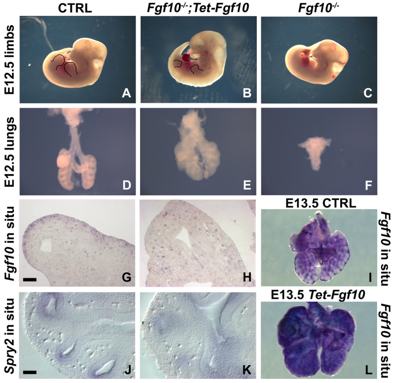
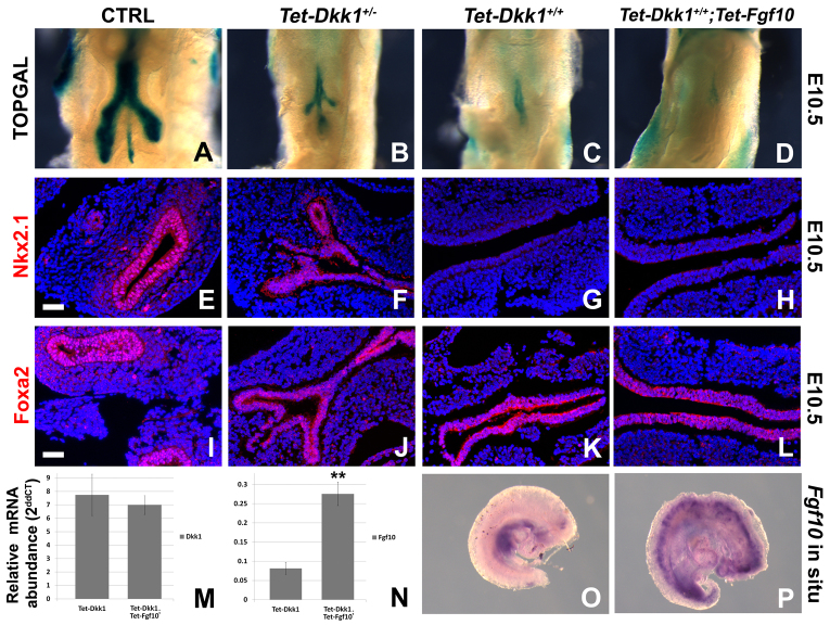
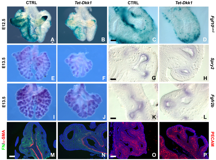
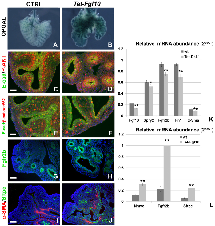
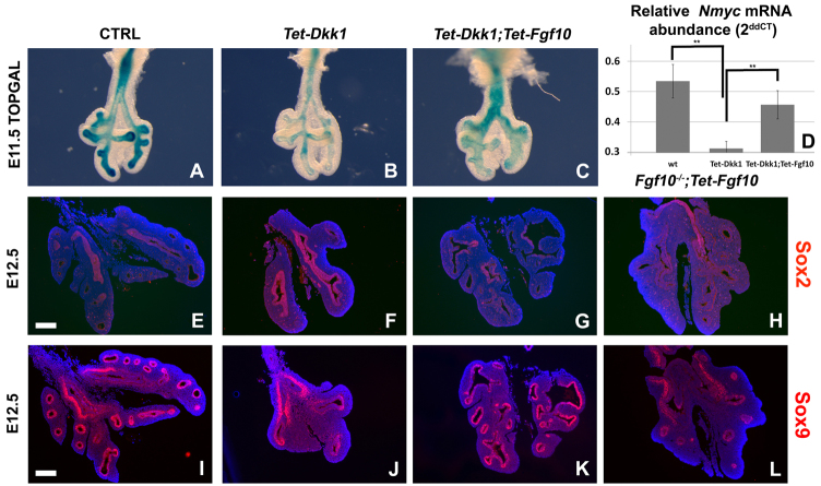
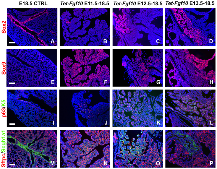
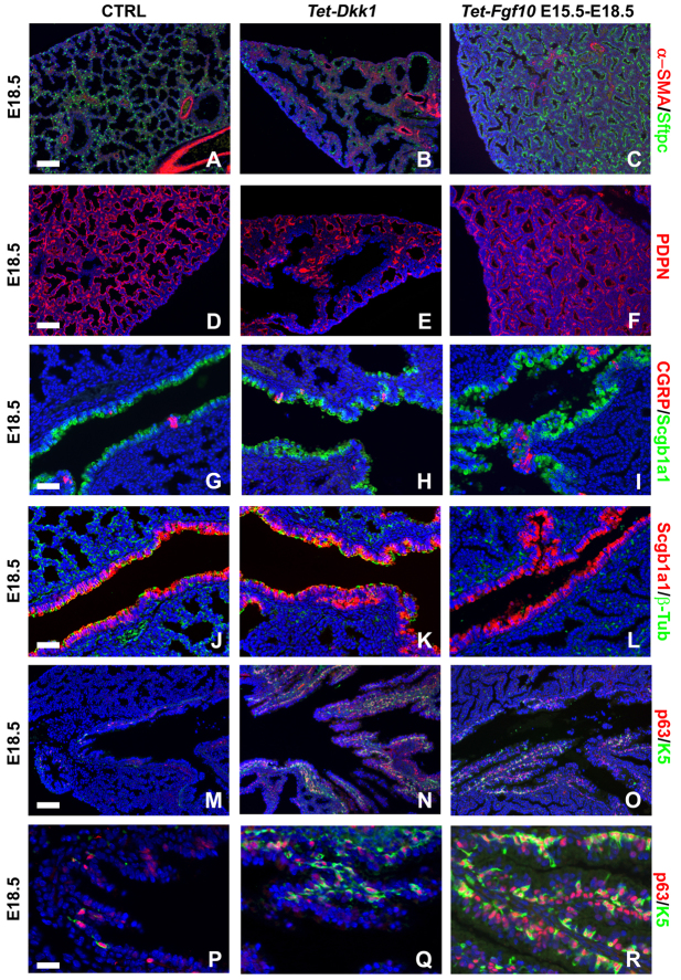
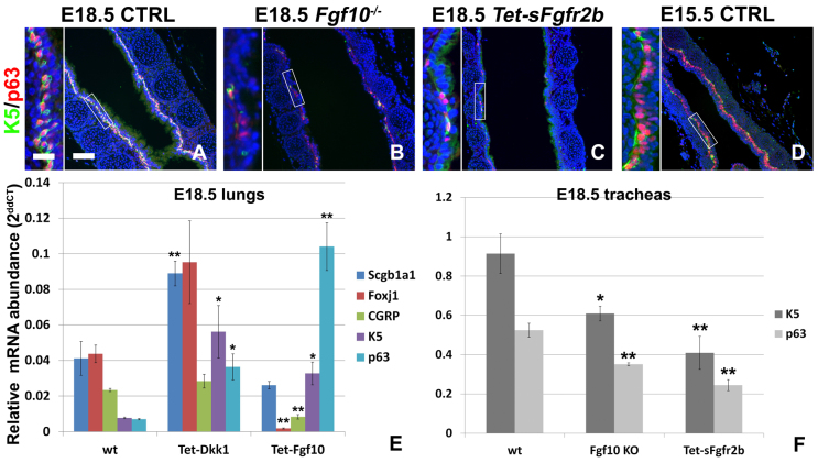
Localized Fgf10 expression in the distal mesenchyme adjacent to sites of lung bud formation has long been thought to drive stereotypic branching morphogenesis even though isolated lung epithelium branches in the presence of non-directional exogenous Fgf10 in Matrigel. Here, we show that lung agenesis in Fgf10 knockout mice can be rescued by ubiquitous overexpression of Fgf10, indicating that precisely localized Fgf10 expression is not required for lung branching morphogenesis in vivo. Fgf10 expression in the mesenchyme itself is regulated by Wnt signaling. Nevertheless, we found that during lung initiation simultaneous overexpression of Fgf10 is not sufficient to rescue the absence of primary lung field specification in embryos overexpressing Dkk1, a secreted inhibitor of Wnt signaling. However, after lung initiation, simultaneous overexpression of Fgf10 in lungs overexpressing Dkk1 is able to rescue defects in branching and proximal-distal differentiation. We also show that Fgf10 prevents the differentiation of distal epithelial progenitors into Sox2-expressing airway epithelial cells in part by activating epithelial β-catenin signaling, which negatively regulates Sox2 expression. As such, these findings support a model in which the main function of Fgf10 during lung development is to regulate proximal-distal differentiation. As the lung buds grow out, proximal epithelial cells become further and further displaced from the distal source of Fgf10 and differentiate into bronchial epithelial cells. Interestingly, our data presented here show that once epithelial cells are committed to the Sox2-positive airway epithelial cell fate, Fgf10 prevents ciliated cell differentiation and promotes basal cell differentiation.

Keywords: Basal cells; Branching; Dkk1; Fgf10; Lung development; Mouse; Wnt signaling.


Figures

Fig. 1.
Fig. 1.
Fgf10 overexpression partially rescues lung and limb agenesis in Fgf10-/- mice. (A-C) E12.5 wild-type (A), Fgf10-/-;Rosa26-rtTA;Tet-Fgf10 dox-induced at E9.5 (B) and Fgf10-/- (C) embryos. Black outlines visualize front and hind limbs. (D-F) E12.5 lungs from wild-type (D), Fgf10-/-;Rosa26-rtTA;Tet-Fgf10 dox-induced at E9.5 (E) and Fgf10-/- (F) mice. (G,H) Fgf10 in situ hybridization on sections from E12.5 wild-type lung (G) and Fgf10-/-;Rosa26-rtTA;Tet-Fgf10 lung dox-induced at E9.5 (H). (J,K) Vibratome sections from whole-mount Spry2 in situ hybridization on E12.5 wild-type lung (J) and Fgf10-/-;Rosa26-rtTA;Tet-Fgf10 lung dox-induced at E9.5 (K). (I,L) Whole-mount Fgf10 in situ hybridization on E13.5 wild-type lungs (I) and Rosa26-rtTA;Tet-Fgf10 lungs dox-induced at E10.5 (L). n≥3. Scale bars: 50 μm (G,H,J,K).
Fig. 2.
Fig. 2.
Dkk1 overexpression abrogates initial lung formation. (A-D) β-Gal staining on E10.5 control (ctrl) TOPGAL (A), Rosa26-rtTA;Tet-Dkk1+/-;TOPGAL (B), Rosa26-rtTA;Tet-Dkk1+/+;TOPGAL (C) and Rosa26-rtTA;Tet-Dkk1+/+;Tet-Fgf10;TOPGAL (D) lungs/foreguts dox-induced at E8.0. (E-H) Nkx2.1 immunostaining on E10.5 ctrl (E), Rosa26-rtTA;Tet-Dkk1+/- (F), Rosa26-rtTA;Tet-Dkk1+/+ (G) and Rosa26-rtTA;Tet-Dkk1+/+;Tet-Fgf10 (H) lungs/foreguts dox-induced at E8.0. (I-L) Foxa2 immunostaining on E10.5 ctrl (I), Rosa26-rtTA;Tet-Dkk1+/- (J), Rosa26-rtTA;Tet-Dkk1+/+ (K) and Rosa26-rtTA;Tet-Dkk1+/+;Tet-Fgf10 (L) lungs/foreguts dox-induced at E8.0. (M,N) qPCR analysis of relative Dkk1 (M) and Fgf10 (N) mRNA abundance on E10.5 Rosa26-rtTA;Tet-Dkk1+/+ and Rosa26-rtTA;Tet-Dkk1+/+;Tet-Fgf10 embryos dox-induced at E8.0. **P<0.01 (Student’s t-test); n≥3. Error bars represent standard error. (O,P) Whole-mount Fgf10 in situ hybridization on an E10.5 Rosa26-rtTA;Tet-Dkk1+/+ embryo (O) and a Rosa26-rtTA;Tet-Dkk1+/+;Tet-Fgf10 embryo dox-induced from E10.5 onwards (P). n≥3. Scale bars: 50 μm (E-L).
Fig. 3.
Fig. 3.
Dkk1 overexpression reduces Fgf10 expression and prevents amplification of distal parabronchial smooth muscle cell progenitors. (A-D) β-Gal-stained lungs (A,B) and corresponding vibratome sections through the medial lobes (C,D) of E12.5 control (ctrl) Fgf10LacZ (A,C) and Rosa26-rtTA;Tet-Dkk1;Fgf10LacZ lungs (B,D) dox-induced at E10.5. (E-L) Whole-mount in situ hybridization (E,F,I,J) and corresponding vibratome sections (G,H,K,L) for Spry2 (E-H) and Fgfr2b (I-L) on E13.5 ctrl (E,G,I,K) and Rosa26-rtTA;Tet-Dkk1 lungs (F,H,J,L) dox-induced at E10.5. (M,N) Immunostaining for fibronectin (FN) and α-SMA on E13.5 ctrl (M) and Rosa26-rtTA;Tet-Dkk1 (N) lungs dox-induced at E10.5. (O,P) Immunostaining for Pecam on E13.5 ctrl (O) and Rosa26-rtTA;Tet-Dkk1 (P) lungs dox-induced at E10.5. n≥3. Scale bars: 50 μm (C,D,G,H,K,L); 100 μm (M-P).
Fig. 4.
Fig. 4.
Fgf10 signaling prevents differentiation of distal epithelial progenitors by positively regulating β-catenin signaling. (A,B) β-Gal staining on E13.5 control (ctrl) TOPGAL (A) and Rosa26-rtTA;Tet-Fgf10;TOPGAL (B) lungs dox-induced at E10.5. (C-F) Immunostaining for E-cadherin and p-Akt-Ser473 (C,D) or β-catenin-Ser552 (E,F) on E13.5 ctrl (C,E) and Rosa26-rtTA;Tet-Fgf10 (D,F) lungs dox-induced at E10.5. (G,H) Immunostaining for Fgfr2b on E13.5 ctrl (G) and Rosa26-rtTA;Tet-Fgf10 (H) lungs dox-induced at E10.5. (I,J) Immunostaining for α-SMA and Sftpc on E13.5 ctrl (I) and Rosa26-rtTA;Tet-Fgf10 (J) lungs dox-induced at E10.5. (K) qPCR analysis of relative Fgf10, Spry2, Fgfr2b, α-Sma (Acta2) and Fn1 mRNA abundance on E11.5 (Fgf10, Spry2) or E12.5 (Fgfr2b, α-Sma, Fn1) ctrl and Rosa26-rtTA;Tet-Dkk1+/+ lungs dox-induced at E10.5. **P<0.01, *P<0.05 (Student’s t-test); n≥3. (To allow for better visualization, Fn1 expression levels presented in the graph are reduced by a factor of 2). (L) qPCR analysis of relative Nmyc, Fgfr2b and Sftpc mRNA abundance on E12.5 ctrl and Rosa26-rtTA;Tet-Fgf10 lungs dox-induced at E10.5. **P<0.01 (Student’s t-test); n≥3. Error bars represent standard error. Scale bars: 50 μm (C-F); 100 μm (G,H); 75 μm (I,J).
Fig. 5.
Fig. 5.
Reduced epithelial Wnt signaling in lungs overexpressing Dkk1 results in proximal-distal differentiation defects that can be rescued by overexpressing Fgf10. (A-C) β-Gal staining on E11.5 control (ctrl) TOPGAL (A), Rosa26-rtTA;Tet-Dkk1;TOPGAL (B) Rosa26-rtTA;Tet-Dkk1;Tet-Fgf10;TOPGAL (C) lungs dox-induced at E10.5. (D) qPCR analysis of relative Nmyc mRNA abundance on E11.5 ctrl, Rosa26-rtTA;Tet-Dkk1+/+ and Rosa26-rtTA;Tet-Dkk1+/+;Tet-Fgf10 lungs dox-induced at E10.5. **P<0.01 (Student’s t-test); n≥3. Error bars represent standard error. (E-L) Immunostaining for Sox2 (E-H) and Sox9 (I-L) on E12.5 ctrl (E,I), Rosa26-rtTA;Tet-Dkk1 (F,J), Rosa26-rtTA;Tet-Dkk1;Tet-Fgf10 (G,K) and Fgf10-/-;Rosa26-rtTA;Tet-Fgf10 (H,L) lungs dox-induced at E10.5. n≥3. Scale bars: 200 μm (E-L).
Fig. 6.
Fig. 6.
Early Fgf10 overexpression prevents proximal differentiation whereas Fgf10 overexpression after E12.5 promotes basal cell differentiation. (A-D) Immunostaining for Sox2 on E18.5 control (ctrl) (A) and Rosa26-rtTA;Tet-Fgf10 lungs induced from E11.5 (B), E12.5 (C) and E13.5 (D). (E-H) Immunostaining for Sox9 on E18.5 ctrl (E) and Rosa26-rtTA;Tet-Fgf10 lungs induced from E11.5 (F), E12.5 (G) and E13.5 (H). (I-L) Immunostaining for p63 and K5 on E18.5 ctrl (I) and Rosa26-rtTA;Tet-Fgf10 lungs induced from E11.5 (J), E12.5 (K) and E13.5 (L). (M-P) Immunostaining for Sftpc and Scgb1a1 on E18.5 ctrl (M) and Rosa26-rtTA;Tet-Fgf10 lungs induced from E11.5 (N), E12.5 (O) and E13.5 (P). n≥3. Scale bars: 200 μm (A-H); 100 μm (I-P).
Fig. 7.
Fig. 7.
Opposing and similar effects of Dkk1 and Fgf10 on lung epithelial differentiation. (A-F) Immunostaining for α-SMA and Sftpc (A-C) or for Pdpn (D-F), on E18.5 control (ctrl) lungs (A,D), E18.5 Rosa26-rtTA;Tet-Dkk1 lungs dox-induced from E10.5 onwards (B,E) and E18.5 Rosa26-rtTA;Tet-Fgf10 (C,F) lungs dox-induced from E15.5 onwards. (G-I) Immunostaining for Scgb1a1 and Cgrp on E18.5 ctrl (G), Rosa26-rtTA;Tet-Dkk1 (H) and Rosa26-rtTA;Tet-Fgf10 (I) lungs induced from E10.5 and E15.5, respectively. (J-L) Immunostaining for Scgb1a1 and β-tubulin on E18.5 ctrl (J), Rosa26-rtTA;Tet-Dkk1 (K) and Rosa26-rtTA;Tet-Fgf10 (L) lungs induced from E10.5 and E15.5, respectively. (M-R) Immunostaining for keratin5 and p63 on E18.5 ctrl (M,P), Rosa26-rtTA;Tet-Dkk1 (N,Q) and Rosa26-rtTA;Tet-Fgf10 (O,R) lungs induced from E10.5 and E15.5, respectively. P-R are higher magnifications of M-O, respectively. n≥3. Scale bars: 100 μm (A-F,M-O); 50 μm (G-L); 25 μm (P-R).
Fig. 8.
Fig. 8.
Fgf(10) signaling regulates basal cell differentiation in the trachea. (A-D) Immunostaining for p63 and K5 on E18.5 wild-type (A), Fgf10-/- (B), Rosa26-rtTA;Tet-sFgfr2b induced from E15.5 (C) and E15.5 wild-type (D) tracheas. Insets show high magnification images of boxed areas. (E) qPCR analysis of relative Scgb1a1, FoxJ1, Cgrp, keratin 5 and p63 mRNA abundance on E18.5 lungs from wild-type mice, Rosa26-rtTa;Tet-Dkk1 mice induced from E10.5 and Rosa26-rtTA-Tet-Fgf10 mice induced from E15.5. (F) qPCR analysis of relative keratin 5 and p63 mRNA abundance on E18.5 tracheas from wild-type mice, Fgf10-/- mice and Rosa26-rtTA-Tet-sFgfr2b mice induced from E15.5. **P<0.01, *P<0.05 (Student’s t-test). n≥3. (To allow for better visualization Scgb1a1, Foxj1 and Cgrp expression levels presented in the graph are reduced by a factor of 1500, 10 and 5, respectively.) Error bars represent standard error. Scale bars: 100 μm (A-D); 25 μm (insets in A-D).

References

    1. Abler L. L., Mansour S. L., Sun X. (2009). Conditional gene inactivation reveals roles for Fgf10 and Fgfr2 in establishing a normal pattern of epithelial branching in the mouse lung. Dev. Dyn. 238, 1999–2013 - PMC - PubMed
    1. Ang S. L., Wierda A., Wong D., Stevens K. A., Cascio S., Rossant J., Zaret K. S. (1993). The formation and maintenance of the definitive endoderm lineage in the mouse: involvement of HNF3/forkhead proteins. Development 119, 1301–1315 - PubMed
    1. Arman E., Haffner-Krausz R., Gorivodsky M., Lonai P. (1999). Fgfr2 is required for limb outgrowth and lung-branching morphogenesis. Proc. Natl. Acad. Sci. USA 96, 11895–11899 - PMC - PubMed
    1. Bao J., Zheng J. J., Wu D. (2012). The structural basis of DKK-mediated inhibition of Wnt/LRP signaling. Sci. Signal. 5, pe22 - PMC - PubMed
    1. Bellusci S., Grindley J., Emoto H., Itoh N., Hogan B. L. (1997). Fibroblast growth factor 10 (FGF10) and branching morphogenesis in the embryonic mouse lung. Development 124, 4867–4878 - PubMed

Publication types

MeSH terms