. 2014 Jan;8(1):164-76.
doi: 10.1038/ismej.2013.132. Epub 2013 Aug 8.

Mixotrophic haptophytes are key bacterial grazers in oligotrophic coastal waters

Affiliations

Mixotrophic haptophytes are key bacterial grazers in oligotrophic coastal waters

Fernando Unrein et al. ISME J. 2014 Jan.

Abstract

Grazing rate estimates indicate that approximately half of the bacterivory in oligotrophic oceans is due to mixotrophic flagellates (MFs). However, most estimations have considered algae as a single group. Here we aimed at opening the black-box of the phytoflagellates (PFs) <20 μm. Haptophytes, chlorophytes, cryptophytes and pigmented dinoflagellates were identified using fluorescent in situ hybridization or by standard 4',6-diamidino-2-phenylindole staining. Their fluctuations in abundance, cell size, biomass and bacterivory rates were measured through an annual cycle in an oligotrophic coastal system. On average, we were able to assign to these groups: 37% of the total pico-PFs and 65% of the nano-PFs composition. Chlorophytes were mostly picoplanktonic and they never ingested fluorescently labeled bacteria. About 50% of the PF <20 μm biomass was represented by mixotrophic algae. Pigmented dinoflagellates were the least abundant group with little impact on bacterioplankton. Cryptophytes were quantitatively important during the coldest periods and explained about 4% of total bacterivory. Haptophytes were the most important mixotrophic group: (i) they were mostly represented by cells 3-5 μm in size present year-round; (ii) cell-specific grazing rates were comparable to those of other bacterivorous non-photosynthetic organisms, regardless of the in situ nutrient availability conditions; (iii) these organisms could acquire a significant portion of their carbon by ingesting bacteria; and (iv) haptophytes explained on average 40% of the bacterivory exerted by MFs and were responsible for 9-27% of total bacterivory at this site. Our results, when considered alongside the widespread distribution of haptophytes in the ocean, indicate that they have a key role as bacterivores in marine ecosystems.


Figures

Figure 1
Figure 1
Box plot summary of the ESD (equivalent spherical diameter) distributions for the main groups of PFs in Blanes Bay. Boxes include 50% of data with the median displayed as a line. Bars indicate the range, excluding the outlier values that are represented by open circles. The number of cells measured for each group is indicated below each box.
Figure 2
Figure 2
(a) Temporal fluctuation in abundance of chlorophytes <3 μm (targeted with probe CHLO02), Micromonas (targeted with probe MICRO01) and haptophytes <3 μm (targeted with probe PRYM02). PF <3 μm represent the total picoflagellate algae enumerated by DAPI. (b) Temporal fluctuation in abundance of haptophytes 3–20 μm (targeted with probe PRYM02), chlorophytes 3–20 μm (targeted with probe CHLO02), cryptophytes <20 μm and pigmented dinoflagellates <20 μm. PF 3–20 μm represent the total nanoflagellate algae enumerated by DAPI. (c) Proportion of total PFs <3 μm and (d) 3–20 μm abundance explained by each of the four algal groups throughout the year. Avg., annual average.
Figure 3
Figure 3
Contribution of each algal group to the biomass of total PF<20 μm throughout the year. Avg., annual average.
Figure 4
Figure 4
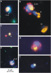
Epifluorescence micrographs of examples of the different mixotrophic groups. (a) Cryptophyte cell with an ingested FLBs. (b) Pigmented dinoflagellate with an ingested cryptophyte (D-Cr), pigmented dinoflagellate containing phycobilins pigments (D) and a cryptophyte cell (Cr). (c) Two picoplanktonic algae, the arrow indicates the haptophyte cell positively hybridized with the probe PRYM02. (d) Pigmented dinoflagellate with a haptophyte cell positively hybridized by FISH inside the food vacuole. (e) Haptophyte cell hybridized with the PRYM02 probe with an ingested FLB. (f) HF with an ingested haptophyte. Each color image is an overlay of three pictures (obtained in 8-bit grayscale) of the same cells observed under different wavelength excitations: UV radiation (showing the blue nucleus after DAPI staining), blue light excitation (red chloroplast and green FLB) and green light excitation (showing the chloroplasts of cryptophytes and dinoflagellates containing phycobilins in figures a, b, and the orange cytoplasm in haptophytes after FISH in figures cf). Pictures were obtained under × 1000 magnification; the same scale applies to all pictures.
Figure 5
Figure 5
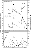
CSGR (bact. cells−1 h−1) and grazing impact (bact. ml−1 h−1) of the main mixotrophic groups along the year.
Figure 6
Figure 6
Percentage of grazing impact of mixotrophs on bacteria explained by each of the four investigated groups along the year. Avg., annual average.

References

    1. Alonso-Sáez L, Vázquez-Domínguez E, Cardelús C, Pinhassi J, Sala MM, Lekunberri I, et al. Factors controlling the year-round variability in carbon flux through bacteria in a coastal marine system. Ecosystems. 2008;11:397–409.
    1. Bell EM, Laybourn-Parry J. Mixotrophy in the antarctic phytoflagellate, Pyramimonas gelidicola (Chlorophyta: Prasinophyceae) J Phycol. 2003;39:644–649.
    1. Bielewicz S, Bell E, Kong W, Friedberg I, Priscu JC, Morgan-Kiss RM. Protist diversity in a permanently ice-covered Antarctic Lake during the polar night transition. ISME J. 2011;5:1559–1564. - PMC - PubMed
    1. Bird DF, Kalff J. Bacterial grazing by planktonic lake algae. Science. 1986;231:493–495. - PubMed
    1. Caron DA.2001Protistan herbivory and bacterivoryIn: Paul J (ed.)Marine Microbiology, Vol. 30, Methods in Microbiology Academic Press: London; 289–315.

Publication types