

Laparoscopic management or laparoscopy combined with transvaginal management of type II cesarean scar pregnancy
- PMID: 23925020
- PMCID: PMC3771793
- DOI: 10.4293/108680813X13654754535197
Laparoscopic management or laparoscopy combined with transvaginal management of type II cesarean scar pregnancy
Abstract
Objective: To evaluate the clinical effectiveness of laparoscopic management of cesarean scar pregnancy (CSP) by deep implantation.
Background: A pregnancy implanting within the scar from a previous cesarean delivery is a rare condition of ectopic pregnancy. There are two different types of CSPs. Type I is caused by implantation of the amniotic sac on the scar with progression toward either the cervicoisthmic space or the uterine cavity. Type II (CSP-II) is caused by deep implantation into a previous CS defect with infiltrating growth into the uterine myometrium and bulging from the uterine serosal surface, which may result in uterine rupture and severe bleeding during the first trimester of pregnancy. Thus, timely management with an early and accurate diagnosis of CSP-II is important. However, laparoscopic management in CSP-II has not yet been evaluated.
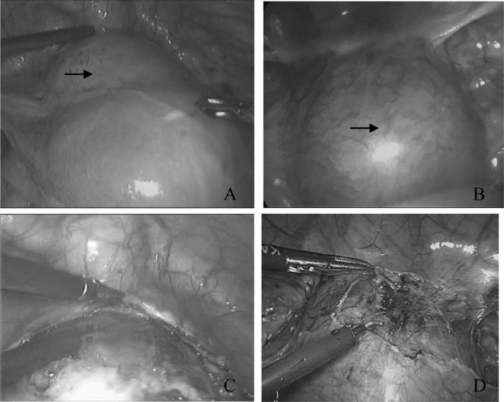
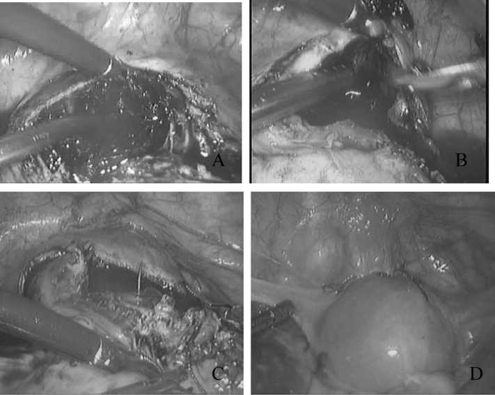
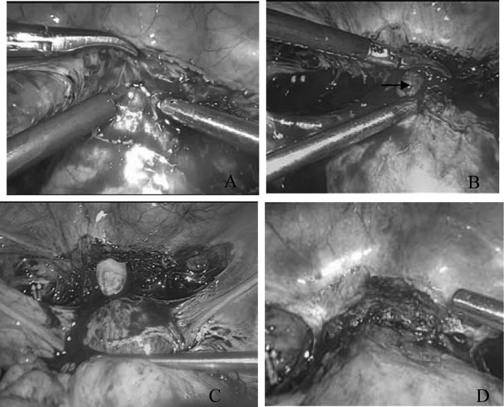
Methods: Eleven patients with CSP-II underwent conservative laparoscopic surgery or laparoscopy combined with transvaginal bilateral uterine artery ligation and resection of the scar with gestational tissue and wound repair to preserve the uterus from March 2008 to November 2011. Patients with CSP-II were diagnosed using color Doppler sonography, and the diagnosis was confirmed by laparoscopy. The operation time, the blood loss during surgery, the levels of β-human chorionic gonadotropin (β-hCG) before surgery, the time taken for serum β-hCG levels to return to <100 mIU/mL postoperatively, and the time for the uterine body to revert to its original state were retrospectively analyzed.
Results: All 11 operations were successfully performed using laparoscopy with preservation of the uterus. One patient underwent a dilation and curettage after laparoscopic bilateral uterine artery ligation. Eight patients were treated solely by laparoscopic bilateral uterine artery ligation and resection of the scar with gestational tissue and wound repair. The remaining two patients underwent laparoscopic bilateral uterine artery ligation and transvaginal resection of the CS with gestational tissue and wound repair because of dense adhesions and heavy bleeding. The average operation time was 85.5 (±17.5) minutes, and the blood loss was 250.0 (±221.4) mL. The blood serum level of β-hCG returned to <100 mIU/mL in 16.4 (±5.3) days postoperatively. Among the 10 patients who underwent resection of CS and wound repair, the time for the uterus to revert to its original state (judged by ultra-sonography) was 10.8 (±3.0) days postoperatively.
Conclusions: Laparoscopy can remove ectopic gestational tissue and allow subsequent wound repair, as well as provide diagnostic confirmation. Being a minimally invasive procedure, laparoscopic or laparoscopy combined with transvaginal bilateral uterine artery ligation and resection of the scar with gestational tissue and wound repair can become an effective alternative for the treatment of CSP-II.
Figures
Similar articles
-
Hysteroscopic removal, with or without laparoscopic assistance, of first-trimester cesarean scar pregnancy.Fertil Steril. 2022 Mar;117(3):643-645. doi: 10.1016/j.fertnstert.2021.11.027. Fertil Steril. 2022. PMID: 35219475
-
Successful Laparoscopic Management of Type I Cesarean Scar Pregnancy A Case Series.J Reprod Med. 2016 Sep;61(9-10):457-462. J Reprod Med. 2016. PMID: 30383945
-
[Clinical study on 39 cases with caesarean scar pregnancy with sonographic mass].Zhonghua Fu Chan Ke Za Zhi. 2014 Jan;49(1):10-3. Zhonghua Fu Chan Ke Za Zhi. 2014. PMID: 24694910 Chinese.
-
Uterine arteriovenous malformation or uterine artery pseudoaneurysm secondary to uterine aspiration in cesarean scar ectopic pregnancy: a case report and review of the literature.J Med Case Rep. 2025 May 23;19(1):248. doi: 10.1186/s13256-025-05312-0. J Med Case Rep. 2025. PMID: 40410916 Free PMC article. Review.
-
Cesarean scar pregnancy: Two case report and therapeutic management algorithm.J Gynecol Obstet Hum Reprod. 2021 Apr;50(4):102056. doi: 10.1016/j.jogoh.2020.102056. Epub 2021 Jan 2. J Gynecol Obstet Hum Reprod. 2021. PMID: 33401027 Review.
Cited by
-
Combination of medical and surgical management in successful treatment of caesarean scar pregnancy: a case report series.BMC Pregnancy Childbirth. 2020 Oct 13;20(1):617. doi: 10.1186/s12884-020-03237-8. BMC Pregnancy Childbirth. 2020. PMID: 33050911 Free PMC article.
-
Assessment of the necessity of uterine artery embolization during suction and curettage for caesarean scar pregnancy: a prospective cohort study.BMC Pregnancy Childbirth. 2020 Jun 29;20(1):378. doi: 10.1186/s12884-020-03062-z. BMC Pregnancy Childbirth. 2020. PMID: 32600442 Free PMC article.
-
Methotrexate-Induced Toxicity After Ultrasound-Guided Intragestational Injection in a Patient with Caesarean Scar Pregnancy-A Case Report.Medicina (Kaunas). 2024 Nov 20;60(11):1900. doi: 10.3390/medicina60111900. Medicina (Kaunas). 2024. PMID: 39597085 Free PMC article.
-
Scar Ectopic Pregnancy.J Obstet Gynaecol India. 2015 Dec;65(6):372-5. doi: 10.1007/s13224-015-0817-3. Epub 2015 Nov 21. J Obstet Gynaecol India. 2015. PMID: 26663994 Free PMC article. Review.
-
Approaches in the Treatment of Cesarean Scar Pregnancy and Risk Factors for Intraoperative Hemorrhage: A Retrospective Study.Front Med (Lausanne). 2021 Jun 24;8:682368. doi: 10.3389/fmed.2021.682368. eCollection 2021. Front Med (Lausanne). 2021. PMID: 34249974 Free PMC article.
References
-
- Godin RA, Bassil S, Donnez J. An ectopic pregnancy developing in a previous caesarian section scar. Feril Steril. 1997;67(2):398–400 - PubMed
-
- Seow KM, Huang LW, Lin YH. Cesarean scar pregnancy: issues in management. Ultrasound Obstet Gynecol. 2004;23(3): 247–253 - PubMed
-
- Vial Y, Petignat P, Hohlfeld P. Pregnancy in a cesarean scar. Ultrasound Obstet Gynecol. 2000;16(4):592–593 - PubMed
-
- Maymon R, Halperin R, Mendlovic S, et al. Ectopic pregnancies in a Caesarean scar: review of the medical approach to an iatrogenic complication. Human Reprod Update. 2004;10(6):515–523 - PubMed
-
- Rotas MA, Haberman S, Levgur M. Cesarean scar ectopic pregnancies: etiology, diagnosis, and management. Obstet Gynecol. 2006;107(6):1373–1381 - PubMed
MeSH terms
Substances
LinkOut - more resources
Full Text Sources
Other Literature Sources
Medical