. 2013 Sep;123(9):3728-39.
doi: 10.1172/JCI68182. Epub 2013 Aug 8.

Age-dependent hepatic lymphoid organization directs successful immunity to hepatitis B

Affiliations

Age-dependent hepatic lymphoid organization directs successful immunity to hepatitis B

Jean Publicover et al. J Clin Invest. 2013 Sep.

Abstract

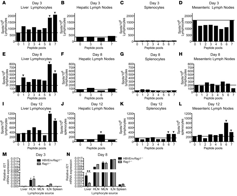
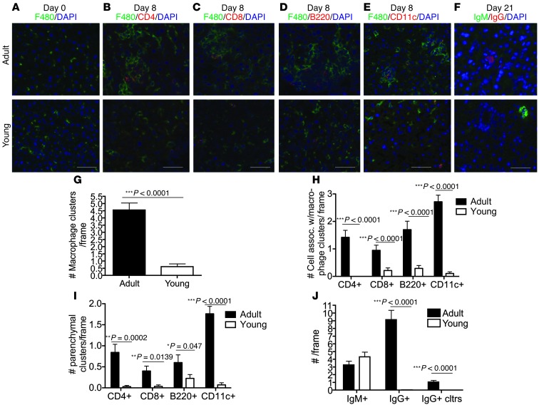
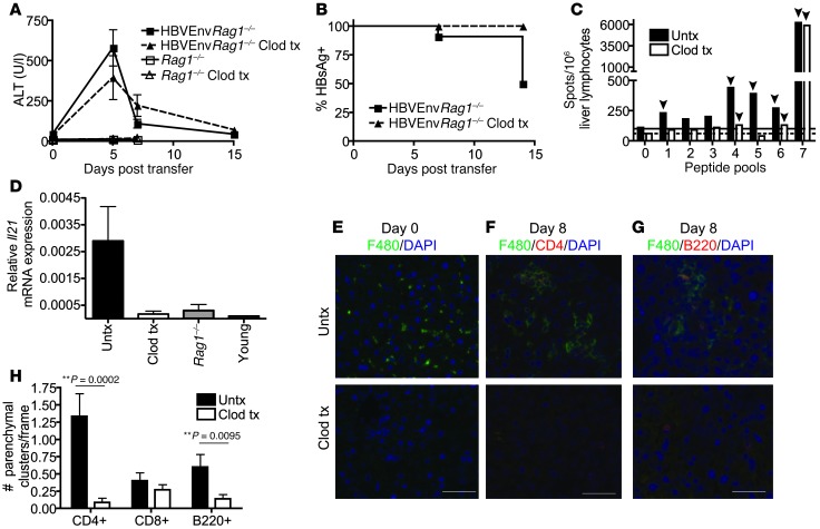
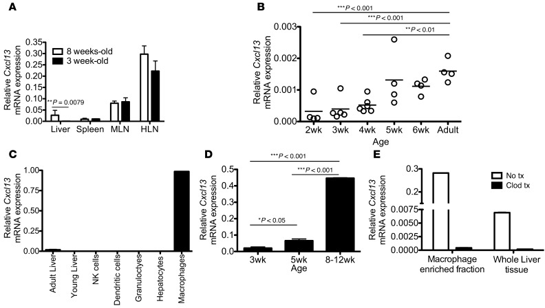
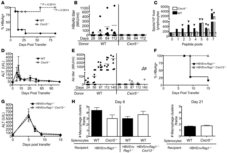
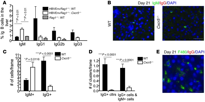
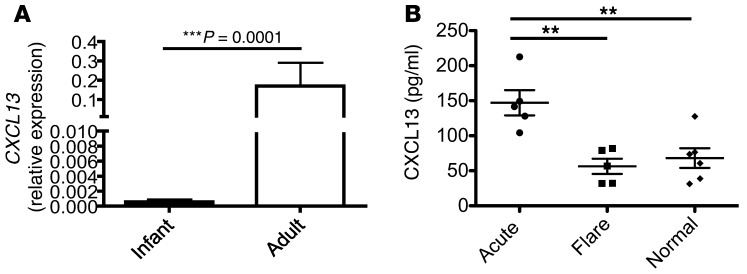
Hepatitis B virus (HBV) is a major human pathogen that causes immune-mediated hepatitis. Successful immunity to HBV is age dependent: viral clearance occurs in most adults, whereas neonates and young children usually develop chronic infection. Using a mouse model of HBV infection, we sought mechanisms underpinning the age-dependent outcome of HBV and demonstrated that hepatic macrophages facilitate lymphoid organization and immune priming within the adult liver and promote successful immunity. In contrast, lymphoid organization and immune priming was greatly diminished in the livers of young mice, and of macrophage-depleted adult mice, leading to abrogated HBV immunity. Furthermore, we found that CXCL13, which is involved in B lymphocyte trafficking and lymphoid architecture and development, is expressed in an age-dependent manner in both adult mouse and human hepatic macrophages and plays an integral role in facilitating an effective immune response against HBV. Taken together, these results identify some of the immunological mechanisms necessary for effective control of HBV.


Figures

Figure 1
Figure 1. HBV-specific immune responses that direct viral clearance are first detected in the liver.
(AL) HBV-specific T cell responses from lymphocytes isolated from the liver (A, E, and I), HLNs (B, F, and J), spleen (C, G, and K), and MLNs (D, H, and L) at days 3 (AD), 8 (EH), and 12 (IL) after adoptive transfer of adult WT syngeneic splenocytes into adult HBVEnvRag1–/– mice. Lymphocytes were stimulated with pools of HBV-envelope–derived peptides (15-mer peptides; pools of 12–14). IFN-γ was measured by ELISpot assay; representative data from 2 separate experiments are shown. Samples were pooled from n ≥ 3 mice. Pool 0 denotes no peptide; solid lines denote baseline IFN-γ levels; arrowheads denote a positive response ≥2× that of baseline. (M and N) Il21 mRNA expression levels from lymphocytes isolated from adult HBVEnvRag1–/– and Rag1–/– liver, spleen, MLNs, HLNs, and ILNs at 3 (M) and 8 (N) days after adoptive transfer of WT syngeneic splenocytes. Il21 levels relative to Gapdh were determined by real-time PCR. Data are representative of at least 2 independent experiments. **P = 0.0048, unpaired 2-tailed Student’s t test.
Figure 2
Figure 2. Adult mice show increased macrophage-associated lymphocyte organization, parenchymal lymphocyte clustering and IgG+ B cells compared to young mice.
(A) Representative images (original magnification, ×10) of macrophage (green; F4/80) and nuclei (DAPI; blue) staining of adult (top) and young (bottom) HBVEnvRag1–/– mouse liver tissue prior to adoptive transfer. (BE) Representative images (original magnification, ×10) of F4/80 (green), DAPI (blue), and CD4 (red; B), CD8 (red; C), B cells (red; B220; D) or DCs (red; CD11c; E) staining of adult and young HBVEnvRag1–/– liver tissue 8 days after adoptive transfer of adult WT splenocytes. Livers were frozen in OCT, sectioned at 6-μm thickness, and acetone fixed. (GI) 15 random frames per section of n ≥ 3 mice per group were scored by an unbiased pathologist for (G) number of macrophage clusters, defined as ≥4 macrophages adjacent to each other/frame; (H) number of CD4+, CD8+, B220+, or CD11c+ cells interacting with macrophage cluster/frame; and (I) number of parenchymal CD4+, CD8+, B220+ lymphocyte clusters/frame (≥2 cells within 15 μm). (F) Representative images (original magnification, ×10) of IgM (green), IgG (red; pooled IgG1 and IgG2b), and DAPI (blue) staining of adult and young liver tissue 21 days after adoptive transfer. (J) 15 random frames per section of n ≥ 3 mice per group were scored by an unbiased pathologist for number of IgM+ cells, IgG+ cells, and IgG clusters per frame (≥2 cells within 15 μm). Scale bars: 30 μm. *P < 0.05, **P < 0.01, ***P < 0.001, unpaired 2-tailed Student’s t test.
Figure 3
Figure 3. Macrophage depletion leads to an immune response that correlates with chronic HBV and impaired hepatic lymphocyte organization.
(A and B) HBVEnvRag1–/– mice were treated (tx) or not (untx) with clodronate liposome (Clod) on days –1, 2, 5, 8, and 11 and adoptively transferred on day 0 with WT splenocytes (n ≥ 5). (A) Plasma ALT. (B) Percent mice with circulating HBsAg. (C) HBV-specific T cell responses, identified by IFN-γ ELISpot, using liver-derived lymphocytes from clodronate-treated or untreated HBVEnvRag1–/– mice 4 months after transfer. Data are pooled from n ≥ 4 mice. Pool 0 denotes no peptide; solid and dotted lines denote baseline IFN-γ levels for untreated and clodronate-treated, respectively; arrowheads denote a positive response ≥2× that of baseline. (D) Il21 mRNA expression levels in liver-derived lymphocytes from untreated and clodronate-treated adult HBVEnvRag1–/– mice, untreated adult Rag1–/– mice, and untreated young HBVEnvRag1–/– mice 8 days after transfer, determined by RT-PCR (n ≥ 2). Data are representative of 3 (A, B, and D) or 2 (C) independent experiments. (EG) Images (original magnification, ×10) from untreated and clodronate-treated HBVEnvRag1–/– liver stained for (E) macrophages (green; F4/80) and nuclei (blue; DAPI) before adoptive transfer (day 0) or for F4/80 (green), DAPI (blue), and either CD4 (red; F) or B220 (red; G) 8 days after transfer. (H) 15 random frames from each section of n ≥ 3 mice per group were scored by an unbiased pathologist for number of parenchymal CD4+, CD8+, and B220+ lymphocyte clusters per frame (≥2 cells within 15 μm). Scale bars: 30 μm. **P < 0.01, unpaired 2-tailed Student’s t test.
Figure 4
Figure 4. Age-dependent hepatic CXCL13 expression is by macrophages and is susceptible to clodronate liposome treatment.
(A) Cxcl13 mRNA expression relative to Gapdh in 3- and 8-week-old Rag1–/– mouse liver, spleen, MLNs, and HLNs. (n ≥ 4). Data are representative of 2 independent experiments. (B) Relative Cxcl13 mRNA expression from liver of WT mice at the indicated ages. Data are representative of 2 independent experiments. (C) Cxcl13 mRNA expression levels in whole liver of adult and young Rag1–/– mice and in sorted cell populations from adult Rag1–/– liver. Cells were isolated using liver perfusion with DNAse and collagenase, followed by gradient separation and sorting for viability and the following parameters: NK cells (NK1.1+), DCs (NK1.1CD11c+CD11b), granulocytes/monocytes (NK1.1CD11b+F4/80). Macrophages were enriched by liver perfusion with DNase and collagenase followed by 30-minute incubation with pronase, isolated using a 25:50 Percoll gradient and sorted for CD45+CD11b+F4/80+. Hepatocytes were isolated and sorted by flow cytometry based on size and viability. (D) Cxcl13 mRNA expression in unsorted macrophage-enriched fractions from liver of Rag1–/– mice aged 3, 5, and 8–12 weeks (n = 3). (E) Relative CXCL13 levels in whole liver and macrophage-enriched fractions from adult Rag1–/– mice that were untreated or treated with clodronate 48 hours earlier (data are representative of 3 independent experiments). *P < 0.05, **P < 0.01, ***P < 0.001, Mann-Whitney test (A) or Tukey’s ANOVA multiple-comparison test (B and D).
Figure 5
Figure 5. Absence of CXCR5 on donor splenocytes or CXCL13 in recipient mice impairs the adaptive immune response to HBV.
(A) Percentage of mice with circulating HBsAg and (B) plasma HBsAb titer in HBVEnvRag1–/– recipients of WT or Cxcr5–/– adult splenocytes. (C) HBV-specific T cell responses 4 months after transfer, identified by IFN-γ ELISpot, using liver-derived lymphocytes from HBVEnvRag1–/– recipients of WT or Cxcr5–/– splenocytes. Data are pooled from n = 4 mice. Pool 0 denotes no peptide; solid and dotted lines denote baseline IFN-γ levels for WT and Cxcr5–/– recipient mice, respectively; arrowheads denote a positive response ≥2× that of baseline. (D) Plasma ALT levels in HBVEnvRag1–/– mice adoptively transferred with WT or Cxcr5–/– splenocytes. (E) HBsAb response in WT and Cxcr5–/– mice vaccinated with HBV vaccine (Merck) intramuscularly at days 0 and 56. (F) Percentage of mice with circulating HBsAg and (G) plasma ALT levels in HBVEnvRag1–/– or HBVEnvRag1–/–Cxcl13–/– mice adoptively transferred with WT splenocytes. Data are representative of 3 (A, B, and D) or 2 (C and EG) independent experiments. (H) Macrophage clustering in HBVEnvRag1–/– mouse liver tissue 8 and 21 days after adoptive transfer of WT or Cxcr5–/– splenocytes and in HBVEnvRag1–/– or HBVEnvRag1–/–Cxcl13–/– mouse liver tissue 8 days after adoptive transfer of WT splenocytes. **P = 0.0014, Fisher’s χ2 test.
Figure 6
Figure 6. Absence of CXCR5 on donor splenocytes alters hepatic IgM+ and IgG+ B cell number and B cell clustering.
(A) Frequency of B cells (plasma cells and plasmablasts) determined by FACS analysis on liver lymphocytes isolated 3 weeks after transfer of HBVEnvRag1–/– or Rag1–/– mice with WT or Cxcr5–/– splenocytes. Percentage of IgG1, IgG2b, and IgG3 B cell (B220loTCR-βCD44hi) and IgM B cell (B220loTCR-β) populations were identified in liver-derived lymphocytes by FACS (n = 3). (B) Representative images (original magnification, ×10) from liver tissue of HBVEnvRag1–/– mice 21 days after adoptive transfer of WT or Cxcr5–/– splenocytes, stained for IgM (green), IgG (red; IgG1 and IgG2b pooled) and DAPI (blue). (C and D) 15 random frames per section of n ≥ 3 mice per group were blinded and analyzed for (C) number of IgM+ and IgG+ cells and (D) number of IgG lymphocyte clusters (≥2 cells within 15 μm) and IgG+ cells associated with ≥1 IgM+ cell per frame. (E) Representative image (original magnification, ×40) from HBVEnvRag1–/– mouse liver stained for F4/80 (green), IgG (red; IgG1 and IgG2b pooled), and DAPI (blue) 21 days after adoptive transfer of WT syngeneic splenocytes. Scale bars: 30 μm (B); 10 μm (E). *P < 0.05, **P < 0.01, ***P < 0.001, Tukey’s ANOVA multiple-comparison test (A) or unpaired 2-tailed Student’s t test (C and D).
Figure 7
Figure 7. Adult human liver shows greater CXCL13 expression than infant liver, and plasma CXCL13 expression is significantly increased in patients who clear a-HBV.
(A) Fresh frozen paraffin-embedded liver biopsy samples from infants 6–12 weeks of age (n = 24) and adults (n = 9) was used for RNA extraction using RNeasy FFPE kit (Qiagen), and expression of CXCL13 relative to GAPDH was determined by RT-PCR. Infant liver tissue was obtained from liver biopsies performed to rule out a diagnosis of biliary atresia; of the 24 patients, 6 had biliary atresia, 6 had neonatal nonviral hepatitis, 10 had nonspecific liver disease diagnosis including cholestatsis and ductopenia, and 2 had an unknown diagnosis. Adult liver samples were obtained from donor livers prior to transplant. (B) Plasma was obtained from 5 patients with confirmed a-HBV infection at the time of active hepatitis and with confirmed subsequent viral clearance and HBsAb seroconversion, 5 patients with confirmed chronic HBV infection exhibiting a flare of disease, and 6 healthy volunteers and assayed for CXCL13 levels by ELISA. ***P < 0.001, Mann-Whitney test; **P = 0.0016, Tukey’s ANOVA multiple-comparison test.

Comment in

References

    1. Lee WM. Hepatitis B virus infection. N Engl J Med. 1997;337(24):1733–1745. doi: 10.1056/NEJM199712113372406. - DOI - PubMed
    1. Robinson WS. Principles and Practice of Infectious Diseases. 4th ed. London, United Kingdom: Churchill Livingstone; 1995.
    1. Ganem D, Prince AM. Hepatitis B virus infection--natural history and clinical consequences. N Engl J Med. 2004;350(11):1118–1129. doi: 10.1056/NEJMra031087. - DOI - PubMed
    1. Liang TJ. Hepatitis B: the virus and disease. Hepatology. 2009;49(5 suppl):S13–S21. doi: 10.1002/hep.22881. - DOI - PMC - PubMed
    1. Nebbia G, Peppa D, Maini MK. Hepatitis B infection: current concepts and future challenges. QJM. 2012;105(2):109–113. doi: 10.1093/qjmed/hcr270. - DOI - PMC - PubMed

Publication types

MeSH terms