Review
. 2013 Aug;61(8):394-400.
doi: 10.4103/0301-4738.116058.

Imaging modalities in keratoconus

Affiliations
Review

Imaging modalities in keratoconus

Himanshu Matalia et al. Indian J Ophthalmol. 2013 Aug.

Abstract

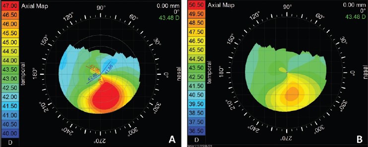
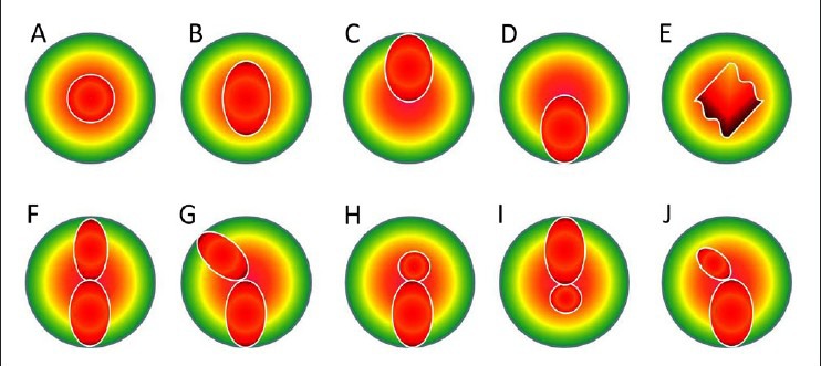
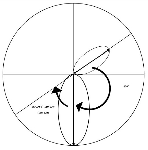
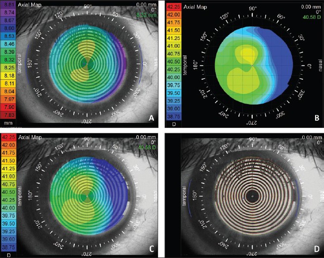
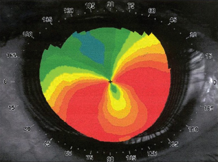
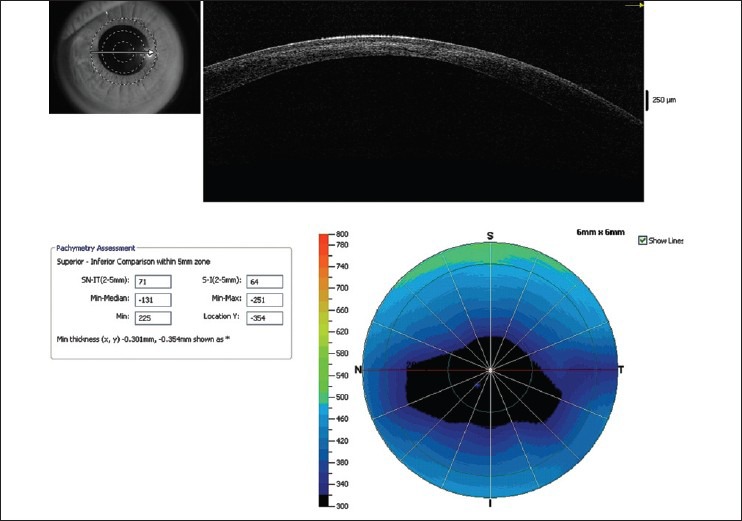
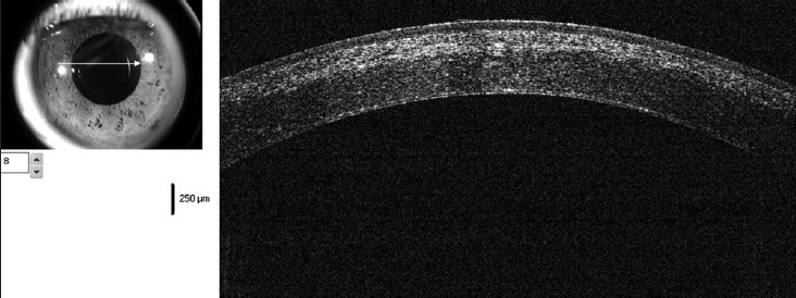
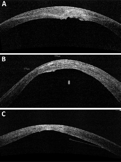
Diagnosis of keratoconus has greatly improved from simple clinical diagnosis with the advent of better diagnostic devices like corneal topographers based on placido disc, elevation based topographers and lately optical coherence tomography (OCT). These instruments are quite sensitive to pick up early keratoconus, which could help refractive surgeons to avoid serious complications like ectasia following keratorefractive surgeries. Each of these instruments has their advantages and disadvantages; in spite of that each one of them has its own place in the clinical practice. Currently, placido disc based topographers are the most commonly used topographers all over the world. There are many different companies making such devices, which follow the different techniques and color for the display. Due to these differences they are not directly comparable to each other. Various quantitative indices based on these topographers have been suggested and validated by different authors to aid in the diagnosis and quantification of keratoconus. OCT with its higher resolution and deeper penetration has created its place in the diagnostic armamentarium for keratoconus.


Conflict of interest statement

Conflict of Interest: None declared.

Figures

Figure 1
Figure 1
Topography of a same patient of keratoconus with different color steps, (a) having steps of 0.5 D and (b) having steps of 1.0 D, showing change in the pattern
Figure 2
Figure 2
Classification of various patterns on axial map of placido based topography. Top A, round; B, oval; C, superior steepening; D, inferior steepening; E, irregular; F, symmetric bow tie; G, symmetric bow tie with skewed radial axes; H, asymmetric bow tie with inferior steepening (AB/IS); I, asymmetric bow tie with superior steepening; J, asymmetric bow tie with skewed radial axes (AB/SRAX)
Figure 3
Figure 3
Calculation of SRAX: SRAX is calculated from 180-the angle between two steep axis above and below the horizontal meridian (smaller of the two angles). In this example two steep axis form 120° angle, hence SRAX = 60 (180−120)
Figure 4
Figure 4
Pseudokeratoconus: This figure shows the importance of misalignment of the normal eye mimicking a keratoconus. (a) Shows axial topography of a normal subject of with the rule astigmatism. (b) The same subject with the misalignment showing skewed radial axis mimicking keratoconus. (c) An overlay of the eye image and (d) mires overlay show apparent misalignment
Figure 5
Figure 5
Axial curvature map showing typical “crab claw”/“butterfly wing” pattern of pellucid marginal degeneration, which may mimic eccentric keratoconus. A careful slit-lamp examination and elevation based topography may help to differential it with keratoconus
Figure 6
Figure 6
Pachymetric map of Optovue RTVue optical coherence tomography (OCT) showing significant thinning in the paracentral cornea. Almost all quantitative indices exceed the cut off limits confirming the diagnosis of keratoconus. Line scan also shows hyperreflectivity in the anterior stroma secondary to mild scarring
Figure 7
Figure 7
OCT showing distinct demarcation line at the junction of cross-linked and non-cross-linked cornea at 3 weeks postoperative visit
Figure 8
Figure 8
(a-c) Various morphologies of Descemet's membrane (DM) irregularities seen in patients with keratoconus, suggestive of possible old hydrops. Presence of DM irregularities may affect choice of surgical procedure when considering intervention such as deep lamellar keratoplasty
Figure 9
Figure 9
Epithelial profile on OCT: Apical epithelial thinning over the apex of the cone in early ectasia can mask topographic changes on the anterior corneal surface. (a) Shows axial map of a forme fruste keratoconus showing minimal changes of keratoconus on the anterior surface, (b) shows the posterior elevation of the same eye showing significant elevation, (c) shows the total corneal pachymetry on Optovue RTVue OCT along with (d) epithelial thickness map, showing localized epithelial thinning masking the cone

References

    1. Krachmer JH, Feder RS, Belin MW. Keratoconus and related noninflammatory corneal thinning disorders. Surv Ophthalmol. 1984;28:293–322. - PubMed
    1. Rabinowitz YS. Keratoconus. Surv Ophthalmol. 1998;42:297–319. - PubMed
    1. Randleman JB, Russell B, Ward MA, Thompson KP, Stulting RD. Risk factors and prognosis for corneal ectasia after LASIK. Ophthalmology. 2003;110:267–75. - PubMed
    1. Li Y, Shekhar R, Huang D. Corneal pachymetry mapping with high-speed optical coherence tomography. Ophthalmology. 2006;113:792–9.e2. - PMC - PubMed
    1. Mazzotta C, Balestrazzi A, Traversi C, Baiocchi S, Caporossi T, Tommasi C, et al. Treatment of progressive keratoconus by riboflavin-UVA-induced cross-linking of corneal collagen: Ultrastructural analysis by Heidelberg Retinal Tomograph II in vivo confocal microscopy in humans. Cornea. 2007;26:390–7. - PubMed