. 2013 Oct;126(4):555-73.
doi: 10.1007/s00401-013-1160-3. Epub 2013 Aug 8.

Transfer of human α-synuclein from the olfactory bulb to interconnected brain regions in mice

Affiliations

Transfer of human α-synuclein from the olfactory bulb to interconnected brain regions in mice

Nolwen L Rey et al. Acta Neuropathol. 2013 Oct.

Abstract

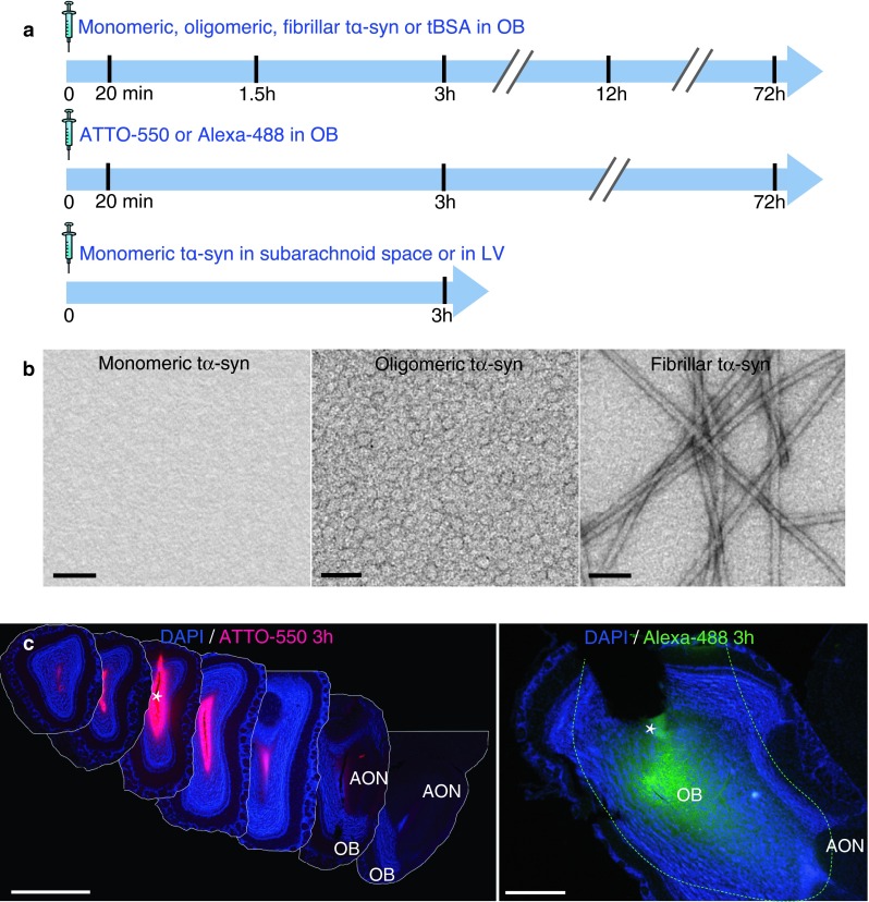
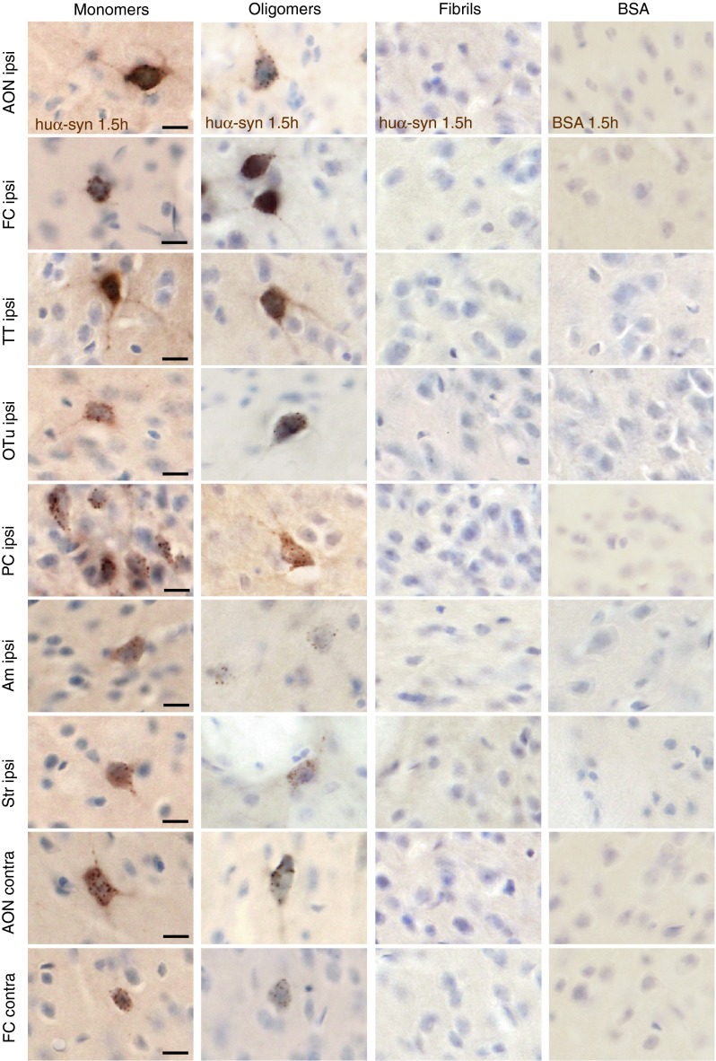
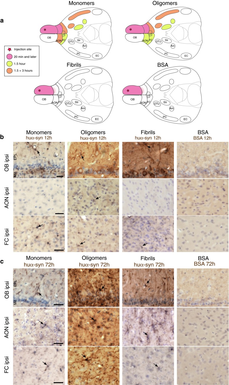
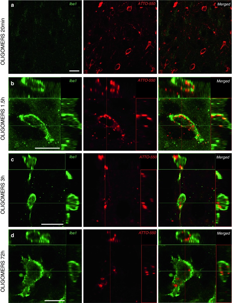
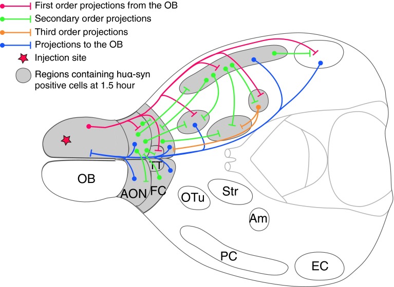
α-Synuclein (α-syn) is a protein prevalent in neural tissue and known to undergo axonal transport. Intracellular α-syn aggregates are a hallmark of Parkinson's disease (PD). Braak and collaborators have suggested that in people who are destined to eventually develop PD, α-syn aggregate pathology progresses following a stereotypic pattern, starting in the olfactory bulb (OB) and the gut. α-Synuclein aggregates are postulated to spread to interconnected brain regions over several years. Thus, propagation of the pathology via neural pathways can potentially explain how α-syn aggregates spread in PD. We have now studied if α-syn can transfer from the OB to other brain structures through neural connections, by injecting different molecular species of human α-syn (monomers, oligomers, fibrils) into the OB of wild-type mice. We found that non-fibrillar human α-syn is taken up very quickly by OB neurons. Within minutes to hours, it is also found in neurons in structures connected to the OB. Conversely, when we injected bovine serum albumin used as a control protein, we found that it does not diffuse beyond the OB, is rarely taken up by OB cells, and does not transfer to other structures. Taken together, our results show that OB cells readily take up α-syn, and that monomeric and oligomeric, but not fibrillar, forms of α-syn are rapidly transferred to interconnected structures within the timeframe we explored. Our results support the idea that α-syn can transfer along neural pathways and thereby contribute to the progression of the α-syn-related pathology.


Figures

Fig. 1
Fig. 1
Experimental design and controls of diffusion. a Experimental design. In wild-type mice, we stereotactically injected α-synuclein (α-syn) into the olfactory bulb (OB), the lateral ventricle (LV) or at the level of subarachnoid space dorsal to the OB. Different molecular species of human recombinant α-syn tagged with ATTO-550 and S-tag (tα-syn) were injected: monomeric, oligomeric, or fibrillar tα-syn; or bovine serum albumin tagged with ATTO-550 (tBSA); or unbound ATTO-550 or Alexa-488 alone as control. We killed mice 20 min, 1.5, 3, 12 or 72 h after injection for histology. b Characterization of recombinant tα-syn. Negatively stained transmission electron micrographs of monomeric, oligomeric and fibrillar tα-syn. Oligomer samples are homogeneous, and do not contain any α-syn fibrils. Scale bar represent 100 nm. c Photographs of the injected region in the OB 3 h after injection of unbound ATTO-550 or Alexa-488 (dissolved in PBS) into the OB of mice. The images illustrate how far the injected solution diffused in the neural tissue. Left panel is a montage of coronal sections of a brain injected with unbound ATTO-550 (inter-section interval = 450 μm). Three hours after injection, ATTO-550 diffused from the injection site to lateral layers of the OB, and until the very anterior part of the AON, but not further. Scale bar 1 mm. Right panel is a low magnification picture of a sagittal section of a brain injected with Alexa-488. At 3 h after injection, Alexa-488 diffused from the injection site to the posterior part of the OB, the accessory olfactory bulb (AOB) and until the very anterior part of the anterior olfactory nucleus (AON), but not further. Dashed line represent the limit of Alexa-488 diffusion. Scale bar represent 500 μm. The white asterisk marks the injection site
Fig. 2
Fig. 2
Tα-syn is taken up by OB cells less than 20 min after injection in the OB. Huα-syn- (stained with syn211 antibody) and BSA-staining in the OB, 20 min after injection of tα-syn, tBSA or ATTO-550 into the OB: a low magnification pictures of the ipsilateral OB (GL/Mit/GCL) (scale bar 50 μm), or high magnification pictures (scale bar 10 μm) of glomerular layer (GL) (b), mitral layer (Mit) (c), and granular cell layer (GCL) (d) of the ipsilateral OB. At 20 min after injection of monomeric, oligomeric and fibrillar tα-syn into the OB, we detected huα-syn-positive cells in various layers of the OB (GL, Mit, and GCL), but no BSA-positive cells in the OB injected with tBSA. A diffuse BSA-staining was observed in extracellular space. To control for specificity of our immunohistochemistry protocol, sections from groups that were injected with ATTO-550 into the OB were stained for huα-syn, and did not reveal any huα-syn-positive staining. A white arrow points examples of huα-syn-positive cells. e Uptake of injected α-syn at 20 min: number of huα-syn positive mitral cells in the ipsilateral OB at 20 min (H = 6.053, P < 0.04, post hoc test: monomers/fibrils P < 0.05). Scatter plots show data from individual mice
Fig. 3
Fig. 3
Tα-syn is localized within the somata of mitral cells in the OB 20 min after injection. We stained brain sections by immunofluorescence for Tuj1 (green), a neuronal marker, and for huα-syn or BSA. Tα-syn was identified both by its ATTO-550 fluorescent tag (red) and by the huα-syn (syn211 antibody) staining (blue). Similarly, in groups injected with BSA, the BSA was detected both by its ATTO-550 tag (red) and by BSA staining (blue). Confocal three-dimensional reconstructions (large panels) show ATTO-550 signal (red) colocalized with huα-syn staining (blue) within mitral cells (tuj1, green) in the OB of mice injected with monomers (a), oligomers (b) and fibrils (c), indicating that these cells contain huα-syn. We also occasionally detected BSA-positive punctae within Tuj1-positive mitral cells in mice injected with BSA (d). Smaller panels are confocal plans showing tuj1 (green), ATTO-550 (red), huα-syn or BSA (blue) staining, and merged pictures of green and red channels. Scale bars represent 10 μm in every panel. White arrowheads point to tα-syn punctae
Fig. 4
Fig. 4
Transfer of tα-syn to other structures 1.5 h after injection into the OB. Images illustrating huα-syn or BSA staining at high magnification (scale bar 10 μm) in various brain areas. We detected huα-syn-positive cells in various brain structures 1.5 h after the injection of monomeric and oligomeric tα-syn into the OB. We observed huα-syn-positive cells in the ipsi- and contralateral anterior olfactory nucleus (ipsi/contra AON), in ipsi- and contralateral frontal cortex (ipsi/contra FC), in ipsilateral tenia tecta (ipsi TT), olfactory tubercle (ipsi Otu), piriform cortex (ipsi PC), amygdala (ipsi Am) and striatum (ipsi Str). On the contrary, we detected huα-syn-positive cells only in the ipsilateral OB when we injected fibrillar tα-syn. tBSA injected as a control protein into the OB, was not detected in the brain, except in the injected OB where we observed only a diffuse staining in extracellular space, but no obvious BSA-positive cell
Fig. 5
Fig. 5
Quantifications of huα-syn-positive cells in different structures 1.5 h after injection into the OB. a Number of positive mitral cells in the ipsilateral OB at 90 min (H = 9.846, P < 0.01, post hoc test: monomers/fibrils P < 0.01). Positive cells in the ipsilateral anterior olfactory nucleus (iAON), ipsilateral frontal cortex (iFC) (b); ipsilateral tenia tecta (iTT), olfactory tubercle (iOTu), piriform cortex (iPC), and striatum (iStr) (c); ipsilateral amygdala (iAm) and contralateral tenia tecta (cTT), anterior olfactory nucleus (cAON) and frontal cortex (cFC) (d). e Time evolution of α-syn transfer and clearance in the ipsilateral piriform cortex. Scatter plots show data from individual mice. Black symbols represent animals that were excluded due technical issues during injection
Fig. 6
Fig. 6
Temporal evolution of tα-syn presence in the brain after injection in the OB. a Drawings representing schematic horizontal sections of the brain with the different regions exhibiting huα-syn-positive cells or a diffuse BSA-staining at different time points after protein injection in the OB. b BSA and huα-syn staining 12 h after injection in the OB. We detected huα-syn-positive cells in the ipsilateral OB, and FC after injection of monomeric, oligomeric and fibrillar tα-syn, as well as in the ipsilateral AON after injection of oligomers. On the contrary, we did not find BSA-positive cells in the brain 12 h after tBSA injection in the OB. Huα-syn-positive cells appeared to be either mitral/tufted cells, located in the external plexiform (EPL) and mitral cell (Mit) layers of the OB (in groups injected with oligomers or fibrils, grey arrows), or displayed the morphology of microglial cells (in groups injected with monomers, oligomers or fibrils; black arrows). Scale bar represent 25 μm. c Huα-syn- and BSA-staining 72 h after injection in the OB. Huα-syn-positive cells were detected in the ipsilateral OB, AON and FC after injection of monomers, oligomers and fibrils 72 h after injection, and those cells exhibit a microglia-like morphology. On the contrary, no BSA-positive cell was found in the brain 72 h after tBSA injection in the OB. Scale bar represents 25 μm
Fig. 7
Fig. 7
Tα-syn is localized within microglia 12 h after injection into the OB. Sections were stained by immunofluorescence for Iba1 (green), a microglial marker. Tα-syn and tBSA were identified by their ATTO-550 fluorescent tag (red). Confocal three-dimensional reconstructions show ATTO-550 signal (red) colocalized with Iba1 (green) within microglia in the OB of mice injected with monomers (a), oligomers (b) and fibrils (c), indicating that these microglial cells contain huα-syn. We also detected ATTO-550 signal in Iba1-positive cells in mice injected with the control protein tBSA (d). Scale bars represent 10 μm in each panel
Fig. 8
Fig. 8
Tα-syn is localized within microglia already 1.5 h after injection into the OB. Tα-syn was identified by its ATTO-550 fluorescent tag (red), and microglial cells by Iba1 staining (green) by confocal microscopy. At 20 min after injection of oligomers, no Iba1-positive cell containing ATTO-550 signal was detected (a). After 1.5 h, confocal three-dimensional reconstructions show ATTO-550 signal (red) colocalized with Iba1 (green) also within microglia in the OB of mice injected with oligomers at 1.5 h (b), 3 h (c) and 72 h (d) timepoints, indicating that these cells contain huα-syn. Scale bars represent 10 μm in each panel
Fig. 9
Fig. 9
Schematic diagram summarizing both the olfactory neural network and regions where huα-syn-positive cells are detected after injection into the OB. This schematic horizontal section of the brain illustrate some of the important regions connected to the OB. Red, green, yellow and blue pathways schematically represent connections within the olfactory system. Circular endpoint of pathways represents the cell body of a relay neuron and a strait end represents the axonal terminal. Structures filled with grey represent regions that displayed huα-syn-positive cells 1.5 h after injection of monomeric and oligomeric tα-syn. All those regions are directly, or indirectly connected to the OB by centrifugal or centripetal projections

References

    1. Angot E, Brundin P. Dissecting the potential molecular mechanisms underlying alpha-synuclein cell-to-cell transfer in Parkinson’s disease. Park Relat Disord. 2009;15(Suppl 3):S143–S147. doi: 10.1016/S1353-8020(09)70802-8. - DOI - PubMed
    1. Angot E, Steiner JA, Lema Tome CM, Ekstrom P, Mattsson B, Bjorklund A, Brundin P. Alpha-synuclein cell-to-cell transfer and seeding in grafted dopaminergic neurons in vivo. PLoS One. 2012;7(6):e39465. doi: 10.1371/journal.pone.0039465. - DOI - PMC - PubMed
    1. Ariesandi W, Chang CF, Chen TE, Chen YR. Temperature-dependent structural changes of Parkinson’s alpha-synuclein reveal the role of pre-existing oligomers in alpha-synuclein fibrillization. PLoS One. 2013;8(1):e53487. doi: 10.1371/journal.pone.0053487. - DOI - PMC - PubMed
    1. Bae EJ, Lee HJ, Rockenstein E, Ho DH, Park EB, Yang NY, Desplats P, Masliah E, Lee SJ. Antibody-aided clearance of extracellular alpha-synuclein prevents cell-to-cell aggregate transmission. J Neurosci. 2012;32(39):13454–13469. doi: 10.1523/JNEUROSCI.1292-12.2012. - DOI - PMC - PubMed
    1. Braak H, Del Tredici K. Poor and protracted myelination as a contributory factor to neurodegenerative disorders. Neurobiol Aging. 2004;25(1):19–23. doi: 10.1016/j.neurobiolaging.2003.04.001. - DOI - PubMed

Publication types

MeSH terms